Zooming in on the signals of cancer

This year, about 240,000 people in the U.S. will discover they have lung cancer. Some 200,000 of them will be diagnosed with non-small-cell lung cancer, which is the second leading cause of death after cardiovascular disease.
Georgia Tech researcher Ahmet Coskun is working to improve the odds for these patients in two recently published studies that are essentially focused on understanding why and how patients respond differently to disease and treatments.
“What we have learned is connectivity and communication between molecules and between cells is what really controls everything, regarding whether or not patients get healthy, or how they will respond to drugs,” said Coskun, an assistant professor in the Wallace H. Coulter Department of Biomedical Engineering at Georgia Tech and Emory University.
Published in the journals npj Precision Oncology and iScience, the studies detail the development of tools and techniques to deeply explore the tumor microenvironment at the subcellular level, utilizing the Coskun lab’s expertise in combining multiplex cellular imaging methods with artificial intelligence.
“We are developing a better grasp of cellular signaling and decision making, and how it is coordinated in the tumor microenvironment, which can lead to better personalized, precision treatments for these patients,” said Coskun, who is keenly interested in why some patients respond to groundbreaking immunotherapy drugs, and some don’t.
With that in mind, his team developed SpatialVizScore, a new method they describe in npj Precision Oncology, to deeply study tumor immunology in cancer tissues and help identify which patients are more likely to respond to an immunotherapy. It’s a significant upgrade to the current standard methodology used by cancer physicians and researchers, Immunoscore.

Read more →

Discovery broadens scope of use of CRISPR gene editing

A team of researchers at Northwestern University has devised a new platform for gene editing that could inform the future application of a near-limitless library of CRISPR-based therapeutics.
Using chemical design and synthesis, the team brought together the Nobel-prize winning technology with therapeutic technology born in their own lab to overcome a critical limitation of CRISPR. Specifically, the groundbreaking work provides a system to deliver the cargo required for generating the gene editing machine known as CRISPR-Cas9. The team developed a way to transform the Cas-9 protein into a spherical nucleic acid (SNA) and load it with critical components as required to access a broad range of tissue and cell types, as well as the intracellular compartments required for gene editing.
The research, published today in a paper titled, “CRISPR Spherical Nucleic Acids,” in the publication Journal of the American Chemical Society, and shows how CRISPR SNAs can be delivered across the cell membrane and into the nucleus while also retaining bioactivity and gene editing capabilities.
The work builds on a 25-year effort steered by nanotechnology pioneer Chad A. Mirkin, who led the study, to uncover the properties of SNAs and the factors that distinguish them from their well-known linear cousin, the blueprint of life. Mirkin is famed for his invention of SNAs, structures typically comprised of spherical nanoparticles densely covered with DNA or RNA, giving them chemical and physical properties radically different from those forms of nucleic acids found in nature.
Mirkin is the George B. Rathmann Professor of Chemistry in the Weinberg College of Arts and Sciences at Northwestern and director of the International Institute for Nanotechnology. Mirkin also is a professor of chemical and biological engineering, biomedical engineering and materials science and engineering in the McCormick School of Engineering and a professor of medicine at Northwestern University Feinberg School of Medicine.
Many classes of SNAs exist, with cores and shells of different chemical compositions and sizes, and SNAs are now being evaluated as potent therapeutics in six human clinical trials, including ones for debilitating diseases like glioblastoma multiforme (brain cancer) and a variety of skin cancers.
“These novel nanostructures provide a path for researchers to broaden the scope of CRISPR utility by dramatically expanding the types of cells and tissues that the CRISPR machinery can be delivered to,” said Mirkin. “We already know SNAs provide privileged access to the skin, the brain, the eyes, the immune system, the GI track, heart and lungs. When this type of access is coupled to one of the most important innovations in biomedical science in the last quarter-century, good things will follow.”
In this current research, Mirkin’s team used Cas9, a protein needed for gene editing, as the core of the structure, and attached DNA strands to its surface to make a new type of SNA. In addition, these SNAs were preloaded with RNA capable of performing gene editing and fused with peptides to control their ability to navigate compartmental barriers of the cell, thereby maximizing efficiency. These SNAs, like other classes of them, effectively enter cells without the use of transfection agents (which are often needed to deliver genetic materials into cells) and exhibit high gene editing efficiency between 32% and 47% across several human and mouse cell lines.
The research team included graduate student researchers Chi Huang, Zhenyu (Henry) Han and Michael Evangelopoulos. Their research was supported by the National Cancer Institute of the National Institutes of Health, the Sherman Fairchild Foundation Inc., Dr. John N. Nicholson Fellowship, and the Alexander S. Onassis Public Benefit Foundation.
Story Source:
Materials provided by Northwestern University. Original written by Win Reynolds. Note: Content may be edited for style and length.

Read more →

Researchers identify flu-fighting pathways and genes essential for influenza A immune defense

Researchers have identified the gene TDRD7 as a key regulator against influenza A virus (IAV), which causes respiratory tract infections in 5 to 20 percent of the human population. These findings could facilitate the development of novel therapeutic interventions against influenza virus infection. The study, led by the Icahn School of Medicine at Mount Sinai in collaboration with other institutions, was published in Science Advances on October 5.
IAV is responsible for 250,000-500,000 deaths per year worldwide. When IAV infects its host, an immunological response composed of a series of molecular processes begins. IAV can infect several different species, and physiological and genetic differences among these species can contribute to different host responses, although some responses are shared.
“Identifying key defense processes and key regulators in multiple species can facilitate the development of treatments for IAV in humans,” said Bin Zhang, PhD, Director of the Center for Transformative Disease Modeling, Willard T.C. Johnson Research Professor of Neurogenetics, and Professor of Genetics and Genomic Sciences at Icahn Mount Sinai, who led the study.
The study used RNA sequencing to analyze gene expression over time in cells and tissues collected from IAV-infected humans, ferrets, and mice, identifying multiple key defense processes specific to tissues and species. One gene found to play a key role in immunological defense mechanisms against IAV across all species was TDRD7, whichencodes a Tudor domain-containing protein, a type of protein shown to be involved in epigenetic regulation. In light of this discovery, the researchers conducted subsequent experiments inhibiting the function of TDRD7, resulting in an increase of virus replication in IAV-infected models.
“Identifying both common and species-specific responses to influenza is essential in developing effective therapies for the flu and can help inform future research of other respiratory infections, such as COVID-19,” said Christian Forst, PhD, Assistant Professor of Genetics and Genomic Sciences, and Microbiology, at Icahn Mount Sinai and a first author of the study.
Co-first authors on the research team also include Laura Martin-Sancho, PhD, Staff Scientist at Scripps Research, California, and Shashank Tripathi, PhD, Assistant Professor, Microbiology and Cell Biology, Centre for Infectious Disease Research, Indian Institute of Science, Bangalore.
Senior authors on the research team also include Adolfo García-Sastre, PhD, Irene and Dr. Arthur M. Fishberg Professor of Medicine and Director of the Global Health and Emerging Pathogens Institute at Icahn Mount Sinai in New York, and Elodie Ghedin, PhD, Chief of the Systems Genomics Section and Deputy Chief of the Laboratory of Parasitic Diseases at the National Institute of Allergy and Infectious Diseases, National Institutes of Health.
This study was supported by the National Institute of Allergy and Infectious Disease (NIAID) of the National Institutes of Health (NIH) under award numbers R21AI149013, U01AI111598, U19AI106754, U19AI135972, U19AI142733, and U19AI168631 and NIAID-funded Center for Research on Influenza Pathogenesis (CRIP) and Center of Excellence for Influenza Research and Response (CEIRR, contract #75N93021C00014).

Read more →

Researchers find link between immune cells' closest neighbors and survival time in patients with pancreatic cancer

Researchers from Johns Hopkins Medicine have discovered that the organization of different types of immune cells within pancreatic tumors is associated with how well patients with pancreatic cancer respond to treatment and how long they survive. The new findings, published Sept. 16 in Cancer Research, could eventually lead to new ways of treating pancreatic cancer, which has the highest mortality rate of all major cancers.
“Mapping the location of certain immune cells associated with a tumor could be a new biomarker to predict patient survival,” says Aleksander Popel, Ph.D., professor of biomedical engineering and director of the Systems Biology Laboratory at the Johns Hopkins University School of Medicine and member of the Johns Hopkins Sidney Kimmel Cancer Center. “We hope that our results will not only lead to a better fundamental understanding of cancer, but also the potential to provide prognostic guidance to clinicians treating pancreatic cancer.”
In 2022, the National Cancer Institute estimates that more than 62,000 Americans are expected to be diagnosed with pancreatic cancer and nearly 50,000 will die from the disease. On average, only about 10% of people with pancreatic cancer will survive for five years. Predicting which patients are most likely to respond to the few existing treatments is difficult; researchers have long been searching for more tools — cells, molecules or genes — that stratify pancreatic cancer patients by survival.
In recent years, scientists studying many types of cancer have discovered the importance of the noncancerous cells, molecules and blood vessels surrounding tumors — called the tumor microenvironment. Part of this tumor microenvironment is immune cells; some have the ability to target a tumor for destruction while others help the tumor evade the immune system.
In previous research on pancreatic cancer, researchers tallied up how many immune cells are present in the tumor microenvironment and found no association with patient outcomes, but Popel and Johns Hopkins graduate student Haoyang Mi hypothesized that the physical arrangement of immune cells might be more important than the total number.
In the new study, Popel, Mi and collaborators at Oregon Health & Science University used a method called multiplexed immunohistochemistry to pinpoint the locations of 27 different immune molecules in surgically resected tumors from 45 people with pancreatic ductal adenocarcinoma — the most common form of pancreatic cancer. The patients were 52% women, a median of 63.5 years old, and had all stages of cancer, with 41% of participants’ cancer spread to at least four lymph nodes.

Read more →

Stem cell patch surgery to mend spina bifida in the womb

Published28 minutes agoSharecloseShare pageCopy linkAbout sharingImage source, Christopher NelsonBy Michelle RobertsDigital health editorUS doctors say they have successfully carried out surgery on babies in the womb to repair harmful spine defects using a special, therapeutic stem cell patch method. Experts hope the pioneering research at UC Davis Health could help others with spina bifida – when the spinal cord and spine do not develop properly. Three of the babies in the trial have now been born. The team will monitor them for at least six years. Without treatment, spina bifida can sometimes lead to a range of lifelong issues, including problems with mobility because of nerve damage. In extreme cases, the spinal canal remains open and exposed. If the defect is not closed to protect it shortly before or after birth, it can cause total paralysis of the legs. Surgeons have already used keyhole surgery on babies in the womb to mend the gap. Now the US team have gone a step further, fitting a graft or implant to bridge the repair. It is a patch that contains immature cells, called stem cells, that can grow with the baby. The researchers say they have already had very promising results in animals with this technique. They have tried it in baby sheep and a pair of English bulldog puppies – Darla and Spanky – to refine the process. Baby Robbie is one of the first humans to have the treatment. Her mum, Emily, says it was a lifeline that they could not refuse. “We didn’t know about spina bifida until the diagnosis. We are so thankful that we got to be a part of this. We are giving our daughter the very best chance at a bright future,” she said.Their surgeon, Diana Farmer, says the operation went off without a hitch: “Mother and foetus did great!” Emily recalls the day Robbie was born more than a year ago.”One of my first fears was that I wouldn’t be able to see her, but they brought her over to me. I got to see her toes wiggle for the first time. It was so reassuring and a little bit out of this world.”The UC Davis team plan to treat about 35 babies as part of their trial. More studies and follow-up are needed to assess how well the treatment works. Robbie and the other babies will have check-ups to see their progress with skills such as walking and potty training. It is not known what causes spina bifida but a number of things can increase the risk of a baby developing the condition.Not having enough folic acid (vitamin B9) during pregnancy is one of the most important ones.Prof Neena Modi, an expert in neonatal medicine at Imperial College London in the UK, stressed the importance of women taking supplements around conception and in pregnancy – a cheap and easy intervention that can help prevent cases of spina bifida. More on this storyKeyhole surgery repairs baby spine in womb17 May 2019Surgery in the womb: ‘I’ve done the best for her’2 May 2021Related Internet LinksSpina bifida – NHSThe CuRe Trial – UC Davis HealthThe BBC is not responsible for the content of external sites.

Read more →

Teenagers Keep Vaping Despite Crackdowns on E-Cigarettes

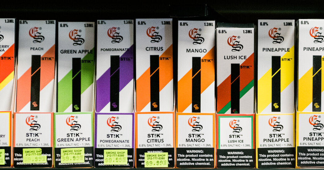
While use among youths has fallen since the peaks of 2018-19, resumption of in-school classes this year shows students still have access to flavored, disposable vapes.High school students resumed taking the annual National Youth Tobacco Survey in school this year and 14 percent of them reported using e-cigarettes, underscoring how an upstart industry is dodging regulators’ efforts to spare a generation from nicotine addiction.The number shows a slight change from 11 percent last year, but researchers cautioned against drawing comparisons to 2021’s survey, which was conducted differently because it took place when many schools were closed during the pandemic. The latest results were released by the Centers for Disease Control and Prevention on Thursday.Though the age-old force of peer pressure may still be encouraging use, the percent of high school students who reported vaping in the last 30 days was still far lower than record-high levels reached in 2019 of nearly 28 percent.Overall, the survey found that 2.5 million middle and high school students, or about 9 percent, used e-cigarettes in the last 30 days. That puts their overall rate of use several times higher than that of adults, which is estimated at about 3 percent.In the survey, which was conducted from January through May of this year, high school students reported strongly favoring fruit- and candy-flavored vapes. Some mentioned PuffBar, Vuse and Juul as their favorite brand among those on the survey’s list.Read More on Smoking, Vaping and E-CigarettesThe Business of Addiction: McKinsey’s ties to opioid makers are well known, but for decades the consulting giant worked with Big Tobacco and has also advised Juul.Youth Vaping Settlement: Juul tentatively agreed to pay $438.5 million to settle an investigation by nearly three dozen states over marketing and sales practices that they contend set off the teen vaping crisis.Ban Suspension: The Food and Drug Administration ordered Juul to stop selling e-cigarettes on the U.S. market, though it later suspended the ban citing “scientific issues” that warrant a review of the decision.Nicotine Levels: The F.D.A. is planning to require tobacco companies to slash the amount of nicotine in their cigarettes. But experts warn that drastic cuts could prove disruptive and fuel underground markets.But many said their favored e-cigarette brand was not one of the 13 listed. That finding highlights how nimble the industry has been in stamping an array of brand names on vapes with flavors like strawberry ice cream and fresh vanilla that are largely made in China and shipped from warehouses to corner stores and into e-commerce.“What that shows is that playing Whac-A-Mole with a few products is not going to solve the problem,” said Vince Willmore, a spokesman for the Campaign for Tobacco-Free Kids. “As long as any flavored products are still on the market, kids are going to shift to them. To solve the problem, you have to clear the market of all flavored products.”One stark finding was that one in four of the high school students who were e-cigarette users reported vaping every day. Groups opposed to e-cigarettes and tobacco products were particularly troubled by one other result that reflected the highest frequency-of-use to date: Nearly half of the high school students who were vaping said they were doing so 20 to 30 days a month.“That’s a real signal of addiction and setting up young people for a lifetime of addiction which they don’t want, they didn’t choose and they don’t like,” said Robin Koval, president of the Truth Initiative, a nonprofit organization aimed at eliminating youth tobacco use.The Food and Drug Administration considers e-cigarettes to be generally beneficial, because they can provide an alternative to adult users of traditional cigarettes, which coat the lungs in tar. The agency’s hope for health gains, though, has existed in the shadow of a youth vaping crisis that exploded in 2018-19, prompting an outcry from parents, schools, lawmakers and public health experts.The F.D.A. began to crack down on vape makers in 2019, banning many flavors and ordering manufacturers to apply for marketing authorization to keep their products on the market — an ongoing process. That effort has been challenged by e-cigarette makers who saw a loophole in making e-cigarettes with synthetic nicotine and jumped into the market with blueberry, kiwi and candy-flavored vapes.This spring Congress gave the F.D.A. the authority to rein in those devices. The agency said it was reviewing about one million applications to sell synthetic nicotine products. In July, the agency gained authority to remove unauthorized nontobacco products from the market but has said it needs to move methodically as it enforces the law.The consequences for teens who develop a nicotine addiction are just beginning to be understood. Dr. Rose Marie Robertson, science and medicine officer with the American Heart Association, said scientists were seeing toxic effects from the inhaled flavoring ingredients. She said researchers were also detecting signs of e-cigarette use on the heart and lungs.“It took us 40 years to show that women would develop lung cancer more readily if they smoked,” Dr. Robertson said. “The fact that we’re seeing any effects at an early stage is very worrisome.”The persistent rate of e-cigarette use among teenagers also concerns experts who were thrilled to see youth cigarette smoking rates fall steadily for years.“To have decades of progress wiped away by e-cigarettes has been astonishing to us who’ve been there all along,” Dr. Robertson said.

Read more →

Eye-opening discovery about adult brain's ability to recover vision

A discovery about how some visually impaired adults could start to see offers a new vision of the brain’s possibilities. The finding that the adult brain has the potential to partially recover from inherited blindness comes from a collaboration between researchers in the University of California, Irvine School of Biological Sciences and the School of Medicine. Their paper appears in Current Biology.
The team was examining treatment for Leber congenital amaurosis, known as LCA. The term refers to a group of inherited retinal diseases distinguished by severe visual impairment at birth. The condition, which stems from mutations in any of over two dozen genes, causes degeneration or dysfunction in the retina’s photoreceptors.
Administering chemical compounds that target the retina, called synthetic retinoids, can restore a notable amount of vision in children with LCA. The UCI team wanted to find out if the treatment could make a difference for adults who have the condition.
“Frankly, we were blown away by how much the treatment rescued brain circuits involved in vision,” said Sunil Gandhi, professor of neurobiology and behavior and the corresponding author. Gandhi is a fellow of UCI’s Center for the Neurobiology of Learning and Memory and a member of the Center for Translational Vision Research. “Seeing involves more than intact and functioning retinae. It starts in the eye, which sends signals throughout the brain. It’s in the central circuits of the brain where visual perception actually arises.” Until now, scientists believed that the brain must receive those signals in childhood so that central circuits could wire themselves correctly.
Working with rodent models of LCA, the collaborators were surprised by what they found. “The central visual pathway signaling was significantly restored in adults, especially the circuits that deal with information coming from both eyes,” Gandhi said. “Immediately after the treatment, the signals coming from the opposite-side eye, which is the dominant pathway in the mouse, activated two times more neurons in the brain. What was even more mind-blowing was that the signals coming from the same-side eye pathway activated five-fold more neurons in the brain after the treatment and this impressive effect was long-lasting. The restoration of visual function at the level of the brain was much greater than expected from the improvements we saw at the level of the retinae. The fact that this treatment works so well in the central visual pathway in adulthood supports a new concept, which is that there is latent potential for vision that is just waiting to be triggered.”
The finding opens exciting research possibilities. “Whenever you have a discovery that breaks with your expectations about the possibility for the brain to adapt and rewire, it teaches you a broader concept,” Gandhi said. “This new paradigm could aid in the development of retinoid therapies to more completely rescue the central visual pathway of adults with this condition.”
Gandhi and first author Carey Huh, PhD, who initiated the project, teamed with Krzysztof Palczewski, Distinguished Professor of ophthalmology. Palczewski, director of the Center for Translational Vision Research, is renowned for his work on retinoids and the visual cycle. Philip Kiser, associate professor of physiology and biophysics, an expert on visual cycle biochemistry, helped lead the group. Kiser, who holds a joint appointment in ophthalmology, is a member, Center for Translational Vision Research.
The research was funded by the National Institutes of Health, the Department of Veterans Affairs and the Research to Prevent Blindness foundation.
Story Source:
Materials provided by University of California – Irvine. Note: Content may be edited for style and length.

Read more →

Fluorescence imaging system lays groundwork for better tumor removal

Researchers have developed a low-cost, simple imaging system that uses tumor-targeting fluorescent molecules to determine the depth of tumor cells in the body. The portable system could eventually help surgeons distinguish between healthy and cancerous tissue with higher precision when removing a tumor.
Doctors can use fluorescent molecules during tumor resection to make cancer cells light up so that the surgeon can see if any cancerous tissue remains. However, the equipment necessary for this technique isn’t widely accessible and typically doesn’t provide quantitative information about how deep within the tissue the cancer cells reside. Having access to depth information would help surgeons to remove a full healthy layer of tissue around the tumor, which has been shown to provide the best possible outcomes for patients.
“The few commercial systems that do provide quantitative depth information are large and expensive, limiting use outside of large medical centers,” said research team leader Christine M. O’Brien from Samuel Achilefu’s Lab at Washington University School of Medicine in St. Louis. “Our group built upon prior work in this field to develop a low-cost, simple system that can quickly determine the depth of tumor cells using near-infrared (NIR) fluorescent probes.”
The researchers describe their new system in the Optica Publishing Group journal Biomedical Optics Express. The portable and easy-to-use system could be used in low-resource clinical centers, which could help minimize health disparities.
“Systems like this could be used in the future to improve surgical outcomes of patients undergoing tumor removal,” said O’Brien. “It would also prevent the need to wait for pathology results before confirming whether cancer cells are still present after tumor removal.”
Lighting up cancer
Research has shown that surgical treatments for cancer tend to be most successful if surgeons remove not only the tumor but also a healthy layer of tissue completely surrounding it. However, this can be difficult because it’s hard to pinpoint the margins between where the tumor ends and healthy tissue begins. In addition, the optimal thickness of the healthy layer depends on the tumor type and location.

Read more →

Promising medication for sleep apnea

Targeting a condition suffered by nearly a billion people worldwide, a new study from Flinders University has shown a drug previously used to treat depression can reduce obstructive sleep apnoea (OSA) severity.
While not yet identifying a cure, the authors say the study opens up further avenues for the development of future drug treatments targeted at the huge number of people unable to tolerate current sleep apnoea therapies, such as continuous positive airway pressure (CPAP) machines.
“Obstructive sleep apnoea can be a debilitating disease, causing poor quality sleep at night and sleepiness during the day,” says study lead author Dr Thomas Altree from FHRMI: Sleep Health (formerly the Adelaide Institute for Sleep Health).
“It affects millions of Australians and causes major impacts on health and productivity.
“Recent research found a combination of the medicines reboxetine and oxybutynin, which were both previously used for unrelated conditions, could be an effective treatment for obstructive sleep apnoea but can cause side effects.
“We wanted to see if reboxetine on its own could be effective and assess exactly how it changes breathing during sleep.”
The team ran a double blind, placebo controlled, randomised, multicentre cross-over trial with collaborators at the Woolcock Institute in Sydney (following a gold standard for drug trials) with 16 people who had OSA. They tested single doses of reboxetine compared to a combination of reboxetine and oxybutynin or placebo.

Read more →

Dodging death: Surprise discovery redirects efforts for treatment of liver disease

WEHI researchers have revealed that common liver diseases are not driven by inflammatory cell death as previously thought, resolving a long-standing controversy in gastroenterology and pointing to a new direction for treatment.
The research team studied liver diseases that affect billions worldwide, including non-alcoholic fatty liver disease and hepatitis B, to understand what drives their progression.
Their unexpected finding — that liver cells are unable to undergo an inflammatory form of cell death called ‘necroptosis’ — resolves crucial unanswered questions in the field and will help guide the development of new therapeutic interventions.
At a glance WEHI researchers have for the first time revealed that an important type of liver cells cannot undergo necroptosis, eliminating this type of cell death as a driver of common liver diseases The surprise findings define the role and relevance of necroptosis in non-cancerous liver diseases, which affect billions of people worldwide The results will help to inform new strategies for the development of treatments for these liver diseasesThe findings, published in Gastroenterology, offer clarity on the heavily debated role of necroptosis in the progression of liver pathologies, and provide fundamental insights to guide future pre-clinical and clinical studies in a new direction.
The study was led by chief investigator Dr Marcel Doerflinger, former WEHI PhD researcher Dr Simon Preston and principal investigator Professor Marc Pellegrini, in collaboration with researchers from the Peter Doherty Institute for Infection and Immunity and the University of Queensland.

Read more →