Maharashtra: India village goes offline daily to help people talk
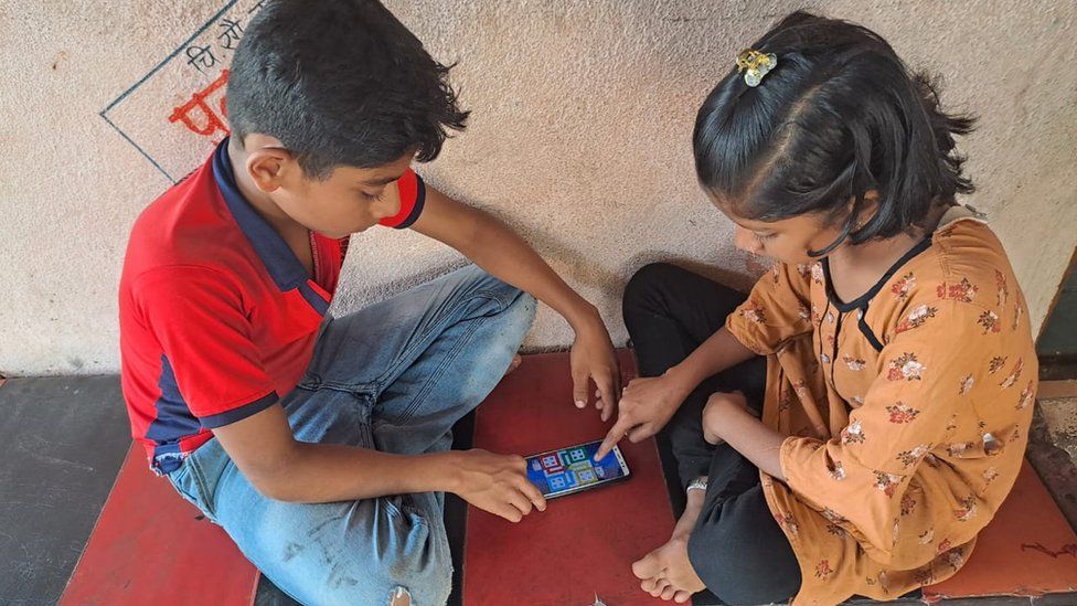
Published8 hours agoSharecloseShare pageCopy linkAbout sharingBy Imran QureshiBBC HindiA village in India’s Maharashtra state has declared “independence” from two modern-day addictions – television and mobile internet. At least, for a couple of hours every day.A siren goes off at 7pm every evening in Vadgaon village in Sangli district, an indication to all residents to switch off their TV sets and mobile phones.The two instruments of “addiction” can be switched on when the village council sounds the siren again at 8.30pm.”We decided at the village meeting on 14 August – the eve of India’s Independence Day – that we needed to stop this addiction,” Vijay Mohite, president of the village council, told BBC Hindi. “From the next day, all television sets and mobiles were shut down when the siren went off.” Vadgaon has a population of about 3,000 people, made up largely of farmers and sugar mill workers.Mr Mohite said children had become dependent on TV and mobile phones for online classes during the Covid-19 pandemic. As educational institutions reopened this year, children returned to regular classes in schools and colleges.”But they returned [from class] to either play on their mobile phones or sit and watch television,” he said, adding that many adults were also spending too much time on their devices and not talking to each other. Vandana Mohite (not related to Vijay Mohite) said she was finding it difficult to supervise her two children “because they would be focused completely on playing with the phone or watching TV”.”Since this new norm began, it is far easier for my husband to return home from work and help them study and I can peacefully do my work in the kitchen,” she added. But it was not easy for the village council to get everyone to agree with the idea of a digital detox.Mr Mohite said initially, when the council discussed the issue and a proposal was taken to the villagers, men scoffed at the idea. The council then gathered the village women, who were open enough to admit that they could get drawn into watching a lot of TV serials and agreed that the entire village should shut down television and mobiles for a few hours. Another meeting of the council was held and it was decided that a siren would be installed above the village temple. Why India has world’s cheapest mobile dataThe forgotten ‘father of internet’ in IndiaThe decision was not easy to implement. As the siren went off, council staff and groups of villagers had to go around, urging people to switch off their TVs and mobile phones. “[Now], the decision has finally been implemented fully across the village,” Mr Mohite says. But, does briefly switching off your TV and phone help?It can, says Dr Manoj Kumar Sharma, professor of clinical psychology at the National Institute of Mental Health and Neurosciences (Nimhans).”Covid has increased preference for online activities or time spent on online activities,” he says. A study conducted by Dr Sharma and his colleagues among 682 adults (495 female and 187 male) between July and December 2020, showed that “problematic internet use” was a rapidly emerging phenomenon among adolescents and young adults. It is one of the most critical challenges that has emerged from increased internet usage. “The risk for problematic usage increases with excessive non-productive usage of the internet, which can cause psychological stress,” the study found. “It has the potential to harm many aspects of adolescent life.”Adolescents predisposed to psychological stress or those experiencing stress were likely to use the internet in its many forms to escape temporarily from unpleasant emotional states, it added. This could lead to them skipping in-person social interactions, social get-togethers, family interactions and extracurricular events to gradually become isolated.Why India is the world leader of internet shutdownsConscious digital fasting as a family to engage in quality-based activities is a cornerstone for decreasing dependence on online activities, Dr Sharma says.”You need to talk to children and ensure they have physical or offline leisure activities as well as adequate sleep and food intake,” he says.Dilip Mohite, a sugarcane farmer who has three school-going sons, says he can see the difference the decision has made. “The children were just not concentrating on their studies before,” he says. “Now, there is normal conversation [at home, even] among the adults.” Read more India stories from the BBC:India facing a pandemic of resistant superbugsIndian political stalwart Mulayam Singh Yadav diesAmitabh Bachchan as you’ve never seen him beforeWhy Indians are fleeing halfway around the worldTiger that killed nine people in India shot dead Gambia police probe deaths linked to Indian syrup India student alleges rape in school by seniors

Read more →
‘Exploited’ foreign doctors worry about risk to UK patients

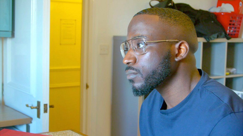
Published9 hours agoSharecloseShare pageCopy linkAbout sharingBy Paul Kenyon & Anna MeiselFile on 4Doctors recruited from some of the world’s poorest countries to work in UK hospitals say they’re being exploited – and believe they’re so overworked they fear putting patients’ health at risk.A BBC investigation has found evidence that doctors from Nigeria are being recruited by a British healthcare company and expected to work in private hospitals under conditions not allowed in the National Health Service.The British Medical Association (BMA) has described the situation as “shocking” and says the sector needs to be brought in line with NHS working practices.The BBC has spoken to several foreign medics – including a young Nigerian doctor who worked at the private Nuffield Health Leeds Hospital in 2021.Augustine Enekwechi says his hours were extreme – on-call 24 hours a day for a week at a time – and that he was unable to leave the hospital grounds. He says working there felt like being in “a prison”. The tiredness was so intense, he says, there were times he worried he couldn’t properly function. “I knew that working tired puts the patients at risk and puts myself also at risk, as well for litigation,” he says. “I felt powerless… helpless, you know, constant stress and thinking something could go wrong.”Nuffield Health disputes those working hours, saying its doctors are offered regular breaks, time off between shifts, and the ability to swap shifts if needed. The company adds that “the health and wellbeing of patients and hospital team members” is its priority. Augustine was hired out to the Nuffield Health Leeds Hospital from a private company – NES Healthcare. It specialises in employing doctors from overseas, many from Nigeria, and using them as Resident Medical Officers, or RMOs – live-in doctors found mainly in the private sector.Augustine says he was so excited to be offered a job that he barely looked at the NES contract. In fact it opted him out of legislation that protects UK workers from excessive working hours – the Working Time Directive – and left him vulnerable to a range of punishing salary deductions.Listen to File on 4 – Paul Kenyon investigates the ‘brain drain’ of doctors from developing countries to work in the UKAugustine is far from alone. The BMA and the front line lobbying group the Doctors’ Association has given the BBC’s File on 4 and Newsnight exclusive access to the findings of a questionnaire put to 188 Resident Medical Officers. Most of the doctors were employed by NES but some were with other employers. It found that 92% had been recruited from Africa and most – 81% – were from Nigeria. The majority complained about excessive working hours and unfair salary deductions.For years now, the World Health Organization (WHO) has warned against the “active recruitment” of doctors and nurses from developing countries with severe shortages of medical personnel. The WHO has compiled a list of 47 such countries – most of them in Africa. The UK government has incorporated that list into its own code of practice – calling it the “red list”. In effect, it makes Nigeria a no-go destination for British medical recruiters.So how did the doctors come to be working in the UK in the first place? We travelled to Nigeria and witnessed another troubling side to this story.In an exam hall in Lagos, the country’s biggest city, we found hundreds of doctors queuing to take what’s called a Professional and Linguistic Assessments Board test – or PLAB 1. The paper is set by the General Medical Council in London and is the first step required by the British medical authorities to secure a licence to work in the UK. The doctors we spoke to said they were attracted by the potential of higher salaries and better working conditions. The event was being overseen by staff from the British Council – an organisation sponsored by the Foreign Office.The GMC also offers the exams in several other red-list countries – Ghana, Sudan, Pakistan and Bangladesh. Both the GMC and British Council deny they are involved in “active recruitment” and say they’re simply helping provide a service for doctors wanting to come to the UK independently – something that is allowed under the guidelines.In Augustine’s case, he was studying for the second part of those PLAB exams in the UK, when he was approached by NES Healthcare and later offered visa sponsorship and a potential job.While that does appear to have been “active recruitment” – NES says it wasn’t, because it is not a recruitment agency and, as such, only engages with doctors from overseas once they’ve already committed to practising in the UK. But the Department of Health and Social Care told us the UK code of practice did apply to NES – so the company was in breach of it.We spoke to several African doctors recruited in this way by NES. They all had similar stories about what the terms and conditions of their contracts meant in reality, once they had been hired out to private UK hospitals.Dr Femi Johnson was sent to a different hospital to Augustine, but says he was also expected to work 14 to 16-hour days and then be on call overnight. “I was burnt out,” he says. “I was tired, I needed sleep. It’s not humanly possible to do that every day for seven days.”But when he needed a break because he was too exhausted to continue, NES were entitled to deduct money from his salary. The company says that is to cover the cost of finding a replacement doctor, but Femi says it leaves NES doctors in a terrible dilemma.”In situations like that, I always make that internal discussion with my inner self – ‘Femi are you doing right by yourself and are you doing right by the patient?'” he tells us. “Unfortunately, I haven’t always been able to answer that question.” Some NES doctors have received help from Dr Jenny Vaughan from the Doctors’ Association. She receives many complaints from Resident Medical Officers and says the UK healthcare system has developed into two tiers – one for NHS doctors, the other for international recruits working in the private sector.NHS doctors can only be scheduled to work up to 48 hours, and if they request, up to 72 hours a week.”No doctor in the NHS does more than four nights consecutively because we know that it’s frankly not safe,” says Dr Vaughan.”This is a slave-type work with… excess hours, the like of which we thought had been gone 30 years ago. “It is not acceptable for patients for patient-safety reasons. It is not acceptable for doctors. ”We took our findings to the BMA – and its deputy chair, Emma Runswick. She told us the situation was a “disgrace to UK medicine”. “Our international colleagues have come a long way to the UK, and have found conditions so exploitative it beggars belief.” NES Healthcare told us that our “feedback about doctors’ experiences” with the company was “extremely positive”. It says it provides doctors “with a safe and supportive route to pursue their career choice in the National Health Service, and in the UK healthcare system more generally, and that their work is of “great benefit to the British public.”

Read more →

After stroke in an infant's brain, right side of brain compensates for loss of language in left side

A clinical study conducted by researchers at Georgetown University Medical Center found that, for children who had a major stroke to the left hemisphere of their brain within days of their birth, the infant’s brain was ‘plastic’ enough for the right hemisphere to acquire the language abilities ordinarily handled by the left side while also maintaining its own language abilities as well.
The left hemisphere of the brain is normally responsible for sentence processing (understanding words and sentences as we listen to speech). The right hemisphere of the brain is normally responsible for processing the emotion of the voice — is it happy or sad, angry or calm. This study sought to answer the question “what happens when one of the hemispheres is injured at birth?”
The findings appear in PNAS the week of October 10, 2022.
The participants in this study developed normally during pregnancy. But around birth they had a significant stroke, one that would have debilitating outcomes in adults. In infants, a stroke is much rarer but does happen in roughly one out of every four thousand births.
The researchers studied perinatal arterial ischemic stroke, a type of brain injury occurring around the time of birth in which blood flow is cut off to a part of the brain by a blood clot. The same type of stroke occurs much more commonly in adults. Previous studies of brain injury in infants have included several types of brain injury, but the focus in this study on a specific type of injury enabled the authors to find more consistent effects than in previous work.
“Our most important conclusion is that plasticity in the brain, specifically the ability to reorganize language to the opposite side of the brain, is definitely possible early in life,” says Elissa Newport, Ph.D., director of the Center for Brain Plasticity and Recovery at Georgetown Medical Center, professor in the departments of Neurology and Rehabilitation Medicine and first author of this study. “However, this early plasticity for language is restricted to one brain region. The brain is not able to reorganize injured functions just anywhere as more dramatic reorganization is not possible even in early life. This gives us great insights into the regions we might be able to focus on for potential breakthroughs in developing techniques for recovery in adults as well.”
The investigators recruited people from across the United States who all had medium to large strokes to the cortex region of their left hemisphere around the time of birth. To assess long-term outcomes in their language abilities, participants were given language tests at 9 to 26 years of age and were compared to their close-in-age healthy siblings. They were also scanned in an MRI to reveal which brain areas were involved in sentence comprehension.
The participants and their healthy siblings all completed the language tasks almost perfectly. The major difference was that the stroke participants processed sentences on the right side of the brain while their siblings processed sentences on the left side. The stroke participants showed a very consistent pattern of language activation in the right hemisphere, regardless of the extent or location of damage from the stroke to the left hemisphere. Only one of the 15 participants, who had the smallest stroke, did not show clear right hemisphere dominant activation.
“It is also notable that many years after their strokes our participants are all such highly functioning adults. Some are honor students and others are working toward or have gotten their master’s degrees,” says Newport. “Their achievements are remarkable, especially since some of their parents had been told when they were born that their strokes would produce life-long impairments.”
In future studies the researchers hope to gain a better understanding of why the left hemisphere routinely becomes dominant in healthy brains but consistently loses out to the right hemisphere when there is a significant left-hemisphere stroke. An additional question of special interest — and clinical importance — is why left hemisphere language can successfully reorganize to the right hemisphere if injuries occur very early in life but not later. Research on stroke recovery and sentence processing in adults suggests that plasticity narrows with age, something that Newport hopes to study as it could be of great benefit, and potential therapeutic interest, to adult stroke survivors.
Story Source:
Materials provided by Georgetown University Medical Center. Note: Content may be edited for style and length.

Read more →

Common approach to demystify black box AI not ready for prime time

Artificial intelligence models that interpret medical images hold the promise to enhance clinicians’ ability to make accurate and timely diagnoses, while also lessening workload by allowing busy physicians to focus on critical cases and delegate rote tasks to AI.
But AI models that lack transparency about how and why a diagnosis is made can be problematic. This opaque reasoning — also known “black box” AI — can diminish clinician trust in the reliability of the AI tool and thus discourage its use. This lack of transparency could also mislead clinicians into over-trusting the tool’s interpretation.
In the realm of medical imaging, one way to create more understandable AI models and to demystify AI decision-making have been saliency assessments — an approach that uses heat maps to pinpoint whether the tool is correctly focusing only on the relevant pieces of a given image or homing in on irrelevant parts of it.
Heat maps work by highlighting areas on an image that influenced the AI model’s interpretation. This could help human physicians see whether the AI model focuses on the same areas as they do or is mistakenly focusing on irrelevant spots on an image.
But a new study, published in Nature Machine Intelligence on Oct. 10, shows that for all their promise, saliency heat maps may not be yet ready for prime time.
The analysis, led by Harvard Medical School investigator Pranav Rajpurkar, Matthew Lungren of Stanford, and Adriel Saporta of New York University, quantified the validity of seven widely used saliency methods to determine how reliably and accurately they could identify pathologies associated with 10 conditions commonly diagnosed on X-ray, such as lung lesions, pleural effusion, edema, or enlarged heart structures. To ascertain performance, the researchers compared the tools’ performance against human expert judgment.

Read more →

Turning the spotlight on cells in tissues so RNA can tell their story

A new advance overcomes present limitations in spatial transcriptomics with a DNA nanotechnology-driven method called ‘Light-Seq.’ Light-Seq allows researchers to ‘geotag’ the full repertoire of RNA sequences with unique DNA barcodes exclusive to a few cells of interest. These target cells are selected using light under a microscope via a fast and effective photocrosslinking process, and their RNAs made available to next-generation sequencing with the help of a new DNA nanotechnology-driven technique. This entire process can then be repeated for different cell populations in the same sample.
Under the microscope, researchers often observe different cell types organizing themselves in peculiar patterns within tissues, or sometimes a rare cell type that stands out by occupying a unique position, exhibiting an unusual shape, or expressing a specific biomarker molecule. To determine the deeper meaning of their observations, they have developed approaches to also access cells’ gene expression patterns (transcriptomes) by analyzing the gene-derived RNA molecules present within them, which they can match with cells’ shapes, spatial positions, and molecular biomarkers.
However, these “spatial transcriptomics” approaches still only capture a fraction of a cell’s total RNA molecules, and cannot deliver the depth and quality of analysis provided by single-cell sequencing methods, which were developed to investigate the transcriptomes of individual cells isolated from tissues or biofluids via next-generation sequencing (NGS) techniques. Nor do they allow researchers to only home in on specific cells based on their location in a tissue, which would greatly facilitate the pursuit of disjointed cell populations, or rare, difficult-to-isolate cells like rare brain cells with unique functions, or immune cells that invade tumors. In addition, because the original tissue environment is disrupted, many spatial transcriptomics and all single-cell sequencing methods prevent researchers from revisiting their samples to perform follow-up analysis, and they are costly because they require specialized instruments or reagents.
A new advance made at the Wyss Institute for Biologically Inspired Engineering at Harvard University now overcomes these limitations with a DNA nanotechnology-driven method called “Light-Seq.” Light-Seq allows researchers to “geotag” the full repertoire of RNA sequences with unique DNA barcodes exclusive to a few cells of interest. These target cells are selected using light under a microscope via a fast and effective photocrosslinking process.
With the help of a new DNA nanotechnology, the barcoded RNA sequences are then translated into coherent DNA strands, which can then be collected from the tissue sample and identified using NGS. The Light-Seq process can be repeated with different barcodes for different cell populations within the same sample, which is left intact for follow-up analysis. With a performance comparable to single-cell sequencing methods, it significantly broadens the depth and scope of investigations possible on a tissue sample. The method is published in Nature Methods[BB1] .
“Light-Seq’s unique combination of features fills an unmet need: the ability to perform imaging-informed, spatially prescribed, deep-sequencing analysis of hard, if not impossible-to-isolate cell populations or rare cell types in preserved tissues, with one-to-one correspondence of their highly refined gene expression state with spatial, morphological, and potentially disease-relevant features,” said Peng Yin, Ph.D., one of four corresponding authors and a Core Faculty member at the Wyss Institute, where his group developed Light-Seq. “It thus has potential to fast-forward the biological discovery process in various biomedical research areas.” Yin is also a Professor of Systems Biology at Harvard Medical School (HMS).

Read more →

New survey: 91% of parents say their family is less stressed when they eat together

Chronic, constant stress can increase lifetime risk of heart disease and stroke, but a new survey from the American Heart Association, a global force for longer, healthier lives for all, reveals regular mealtime with others could be a simple solution to help manage stress.
Of the 1,000 U.S. adults nationwide surveyed in September 2022 for the American Heart Association’s Healthy for GoodTM movement by Wakefield Research, the vast majority (84%) say they wish they could share a meal more often with loved ones, and nearly all parents report lower levels of stress among their family when they regularly connect over a meal.
To make mealtime togetherness a little easier and help people claim the heart mind and body benefits that go with it, the American Heart Association will share practical and budget-friendly meal tips each Tuesday through December. People can follow #TogetherTuesday on social media or text 2gether to 51555 to get tips sent directly to their phone.
“Sharing meals with others is a great way to reduces stress, boost self-esteem and improve social connection, particularly for kids,” said Erin Michos, M.D, M.H.S, American Heart Association volunteer, associate director of preventive cardiology at Johns Hopkins and a co-author of the American Heart Association’s statement on Psychological Health, Well-being, and the Mind-Heart-Body Connection. “Chronic, constant stress can also increase your lifetime risk of heart disease and stroke, so it is important for people to find ways to reduce and manage stress as much as possible, as soon as possible.”
Connecting with friends, family, coworkers and neighbors benefits people beyond stress relief. In fact, the survey found 67% of people say sharing a meal remind them of the importance of connecting with other people, and 54% say it reminds them to slow down and take a break.
Those surveyed say they are more likely (59%) to make healthier food choices when eating with other people but have difficulty aligning schedules with their friends or family to do so, according to the survey. Overall, respondents reported eating alone about half of the time.
“We know it’s not always as easy as it sounds to get people together at mealtime. Like other healthy habits, give yourself permission to start small and build from there,” Michos said. “Set a goal to gather friends, family or coworkers for one more meal together each week. If you can’t get together in person, think about how you can share a meal together over the phone or a computer.”
The American Heart Association’s survey also identified the majority (65%) of adults say they are at least somewhat stressed and more than a quarter (27%) are extremely or very stressed. Nearly 7 in 10 (69%) of survey respondents who are employed full or part-time said they would feel less stressed at work if they had more time to take a break and share a meal with a co-worker.
Story Source:
Materials provided by American Heart Association. Note: Content may be edited for style and length.

Read more →

Home sensors can detect opioid withdrawal signs at night

Some smart home technology could help curb opioid overdose. A Washington State University pilot study showed that a set of noninvasive home sensors could provide accurate information about overnight restlessness and sleep problems for people recovering from opioid use disorder.
Disrupted sleep is a major complaint of people trying to quit highly addictive opioids. Although methadone is effective at reducing cravings and withdrawal, it is often prescribed once daily and adjusting for the proper dosage can take time. In the meantime, treatments can wear off during the night, returning withdrawal symptoms and pain — which increases the risk of resuming drug use and accidental overdose.
In the study, detailed in the journal Pain Management Nursing, WSU researchers found the home sensors matched other monitoring methods for sleep disturbance about 89% of the time.
“One day when people go home from addiction treatment, we could send them to a smart home sensor environment, so we can know remotely if they’re struggling to sleep and getting up and down a lot,” said lead author Marian Wilson, a WSU nursing professor. “We know that poor sleep is a trigger for substance use and could lead to unintended overdose.”
Wilson and her colleagues designed the study to see if they could create a system that could detect problems such as sleep apnea and other breathing issues as well as physical movement indicating an inability to sleep well. This information could then potentially help alert healthcare workers of the problems and prompt medication changes or supportive interventions. The findings also simply validated patients’ concerns.
“Our study confirms what people with opioid use disorder have been saying — their sleep can be restless and disturbed. We need to appreciate that people may be suffering,” said Wilson. “There’s a misconception that substance use is all about that euphoric ‘high.’ By the time people get into a methadone treatment program, they’re usually just trying to feel normal.”
For the study, which was conducted at WSU’s Sleep and Performance Center, researchers collected about 16 hours of data over two nights for four people in methadone treatment programs. The sensors were placed on a wall, overhead and on the door to monitor movements. They compared the data collected by the unattached, camera-less sensors to information collected by observers via video and polysomnography, the most common sleep study method. Polysomnography involves attaching monitoring wires and equipment to the patient to collect measurements like oxygen saturation and leg movement.
The researchers found that the home sensors captured a lot of the same information as the systems with wires and cameras.
“This tells us that maybe in the future, we don’t need to have the person come into the sleep lab to be observed,” said Wilson. “The home sensors would be a very unobtrusive way to monitor somebody with no cameras, no videos, just the sensors that tell us about their movements.”
The next step for this research would be to test the sensor concept in the home environment instead of a lab, which would involve placing sensors in the homes of patients who are being discharged from an in-patient treatment program or starting an outpatient medication treatment program.
In addition to Wilson, co-authors on this study include Roschelle Fritz of the WSU College of Nursing, Myles Finlay of WSU Elson S. Floyd College of Medicine and Diane Cook of the WSU School of Electrical Engineering and Computer Science. This research received support from the National Institutes of Health.
Story Source:
Materials provided by Washington State University. Original written by Sara Zaske. Note: Content may be edited for style and length.

Read more →

New antibiotic comes from a pathogenic bacterium in potatoes

The growing threat of antimicrobial resistance has led researchers to search for new compounds everywhere. This week in mBio, a multinational team of researchers in Europe report the discovery of a new antifungal antibiotic named solanimycin. The compound, initially isolated from a pathogenic bacterium that infects potatoes, appears to be produced by a broad spectrum of related plant pathogenic bacteria.
Solanimycin acts against a wide range of fungi known to infect and wreak havoc on agricultural crops, according to the researchers. In lab studies, the compound also acted against Candida albicans, a fungus that occurs naturally in the body but can cause dangerous infections. The results suggest that solanimycin, and related compounds, could be useful in both agricultural and clinical settings.
Soil microbes, especially from the Actinobacteria phylum, produce most therapeutic antibiotics used today. The new discovery suggests plant-based microorganisms are worth a closer look, especially as crops develop resistance to existing treatments, says microbiologist Rita Monson, Ph.D., at the University of Cambridge. She co-led the study with molecular microbiologist Miguel Matilla, Ph.D., at the Spanish Research Council’s Estación Experimental del Zaidín, in Granada.
“We have to look more expansively across much more of the microbial populations available to us,” Monson said.
The pathogenic potato bacterium Dickeya solani, which produces solanimycin, was first identified more than 15 years ago. Researchers in the lab of molecular microbiologist George Salmond, Ph.D., at the University of Cambridge, began investigating its antibiotic potential about a decade ago.
“These strains emerged rapidly, and now they are widely distributed,” said Matilla.

Read more →

Researchers find tumor microbiome interactions may identify new approaches for pancreatic cancer treatment

Investigators from Rutgers Cancer Institute of New Jersey, the state’s leading cancer center and only National Cancer Institute-Designated Comprehensive Cancer Center, together with RWJBarnabas Health, examined the microbiome of pancreatic tumors and identified particular microorganisms at single cell resolution that are associated with inflammation and with poor survival. According to the researchers, these microorganisms may be new targets for earlier diagnosis or treatment of pancreatic cancer, which is the fourth leading cause of cancer death for both men and women in the United States. The findings are published in the online version of Cancer Cell.
Microbes are living things that are too small to be seen with the naked eye. We have more microbes living in our body than the total number of human cells, and can be found in organs like the pancreas, which at one time was considered microbe-free. Subhajyoti De, PhD, principal investigator at Rutgers Cancer Institute and senior author of the study along with graduate student Bassel Ghaddar, a student in the MD/PhD program at Rutgers Robert Wood Johnson Medical School, began exploring if there are microbes residing in pancreatic tumors, and if they have consequences for cancer progression or treatment. However, studying microbes in tumors is difficult, in part since every patient is different, and because microbial footprints are too subtle to detect reliably.
To explore further, the researchers teamed up with Martin Blaser, MD, Henry Rutgers Chair of the Human Microbiome at Rutgers University and microbiome expert. The investigators developed a genomic approach called SAHMI (Single-cell analysis of Host-Microbiome Interactions) to identify microorganisms associated with individual human cells. Sifting through millions of RNA sequences using sophisticated software, they identified which ones likely represent human genes, and which ones are microbial in origin. “This new technique allowed us to identify tumor-associated microbes and measure the activity of the host cells at the same time, which is a significant technical advance, and the results were stunning,” notes Dr. De, who is also an associate professor of cancer systems biology at Rutgers Robert Wood Johnson Medical School.
Studying two independent groups of pancreatic tumors, the team found that some had bacteria that associated with specific cell-types within the tumor, which were essentially absent in normal pancreatic tissues. These bacteria were predominantly located within tumor cells, and their abundance correlated with cancer-related cell activities. The specific signatures of the microbes that were found predicted particularly aggressive cancer progression and poor prognosis.
The microbial footprints within the pancreatic tumors raised the question of whether the immune cells that were present were responding to the cancer or to the microbes. The study findings suggested that the immune responses were mostly responding to the microbes in the tumor and not to the cancer cells. “Our observations provide a new view about why pancreatic cancers are so difficult to treat,” notes Dr. Blaser, who is also a research member at Rutgers Cancer Institute and professor of epidemiology and biostatistics at Rutgers School of Public Health. “But better understanding these interactions may identify new approaches for therapies.”
Along with Drs. De, Ghaddar, and Blaser, other investigators include: Antara Biswas, PhD, Center for Systems and Computational Biology, Rutgers Cancer Institute; Chris Harris, PhD, Department of Surgery, University of Rochester Medical Center; M. Bishr Omary, PhD, Center for Advanced Biotechnology and Medicine, Rutgers University and Darren R. Carpizo, MD, PhD, Department of Surgery, University of Rochester Medical Center.
Story Source:
Materials provided by Rutgers University. Note: Content may be edited for style and length.

Read more →

Pockets of resistance found in survey of pathogen diversity

Unparalleled insights into the secret life of Streptococcus pneumoniae, the bacterium responsible for hundreds of thousands of infant deaths each year, have been revealed by new research from the Wellcome Sanger Institute, the University of Oxford, the Shoklo Malaria Research Unit and Imperial College London.
The study, published today (10 October) in Nature Microbiology, details the genetic diversity of the pathogen within individual infants, including hidden multidrug-resistant and virulent strains. The findings suggest that the detection of such resistant variants is only possible using a population deep sequencing (PDS) approach and that their presence may be due to individuals being treated with antibiotics.
The research highlights the potential for PDS to improve our understanding of pathogens like S. pneumoniae and to inform treatment strategies when antimicrobial resistance is a concern.
Streptococcus pneumoniae, also known as the pneumococcus, is a bacterial pathogen that causes diseases ranging from ear infections through to pneumonia, septicaemia and meningitis. It is responsible for around nine million global infections annually, with elderly adults and children particularly susceptible. More than 300,000 children die from pneumococcal infection each year, mainly in low- and middle-income countries (LMICs).
Not all individuals colonised by S. pneumoniae will become ill. The bacteriumis carried without symptoms by up to 60 per cent of children and by a smaller percentage of adults. Children are generally colonised by common strains, and this allows our immune systems to recognise and guard against them. As adults, we tend to be colonised by rarer strains that we may not have encountered before.
In this new study, researchers at the Wellcome Sanger Institute, the University of Oxford and Imperial College London set out to document the diversity of S. pneumoniae in children and adults.

Read more →