New tech aims to reduce racial disparities in blood measurements

A team led by a University of Texas at Arlington bioengineering professor and an Austin businessman has published key findings in the British Medical Journal Innovations that illustrate how a new device measures hemoglobin more accurately in individuals with darker skin pigmentations.
George Alexandrakis, UT Arlington bioengineering professor, and Dr. Vinoop Daggubati of Shani Biotechnologies LLC conducted a clinical study at UT Arlington with 16 healthy volunteers and measured their hemoglobin and oxygen content using the newly developed technology. The team compared the results to those obtained using a commercially available pulse-oximeter for accuracy and variability.
Racial disparities in hemoglobin and blood oxygen measurements are an urgent public health issue. Currently available devices are inaccurate in people with dark skin. The U.S. Food and Drug Administration has issued a safety communication and organized an advisory committee meeting on Nov. 1, 2022, to discuss this issue at length.
The findings from the UTA team’s research are encouraging, and the new technology has massive potential to address this clinical unmet need. Alexandrakis said their intent is to develop a wearable device, such as a watch or a monitor, that would read the blood through the skin.
Most currently available methods for monitoring hemoglobin require blood samples and expensive equipment. The available noninvasive spectroscopic methods have a high degree of variability and often are inaccurate in people of color due to differences in skin melanin. There is a significant unmet need for a reliable, noninvasive device to estimate hemoglobin, irrespective of skin color.
Currently available pulse-oximeters use red-infrared light and are based on technology first designed more than 50 years ago. In contrast, the team’s device relies on the spectroscopic properties of hemoglobin in the blue-green light spectra.
“We have used the green-blue light and have successfully tested the device in preclinical and clinical studies,” Daggubati said. “Our group has addressed the issues around shorter wavelength, scattering of light and the impact of skin melanin. The scientific community should open its mind to the concept of green light for these measurements. The Shani device has huge potential to eliminate this racial disparity.”
Story Source:
Materials provided by University of Texas at Arlington. Note: Content may be edited for style and length.
Read more →

Cellular housekeeping process implicated in fatal neurological disorder

Huntington’s disease, a fatal, inherited neurodegenerative condition, is caused by a genetic error present at birth, though its symptoms often don’t begin until middle adulthood. Scientists at Washington University School of Medicine in St. Louis have been trying to understand how the aging process triggers the onset of symptoms, with the expectation that such knowledge could point to treatments that delay or prevent neurodegeneration.
To that end, a new study from Washington University indicates that as patients age, the disease gradually impairs an important cellular housekeeping process called autophagy, which is responsible for eliminating waste from cells. This housekeeping is significant in Huntington’s because a buildup of waste in a specific kind of neuron leads to such cells’ untimely deaths.
The researchers also showed that enhancing the autophagy pathway in such neurons that were created from skin cells of Huntington’s patients protects those cells from dying.
“Our study reveals how aging triggers a loss of the crucial process of autophagy — and hints at how we might try to restore this important function, with the aim of delaying or even preventing Huntington’s disease,” said senior author Andrew S. Yoo, PhD, a Washington University professor of developmental biology.
The study, published Oct. 27 in the journal Nature Neuroscience, also may offer clues to understanding cognitive decline in aging generally.
Huntington’s disease destroys a specific type of brain cell called medium spiny neurons, the loss of which causes involuntary muscle movements, impaired mental health and cognitive decline. Patients typically live about 20 years after signs of the disease first appear.

Read more →

Stem cell grafts and rehabilitation combined boost spinal cord injury results

In recent years, researchers have made measurable progress, using animal models, to promote tissue regeneration in spinal cord injuries (SCI) through implanted neural stem cells or grafts. Other efforts have shown that intensive physical rehabilitation can improve function after SCI by promoting greater or new roles for undamaged or spared cells and neural circuits.
In a new paper, published August 22, 2022 in the journal JCI Insight, researchers at University of California San Diego School of Medicine address the question of whether rehabilitation can augment functional outcomes when combined with pro-regenerative therapies, such as stem cell grafting.
Using a rat model, researchers induced a cervical lesion that impaired the animals’ ability to grasp with its forelimbs. There were four groups: animals who underwent the lesion alone; animals who received a subsequent grafting of neural stem cells designed to grow and connect with existing nerves; animals who received rehabilitation only; and animals who received both stem cell therapy and rehabilitation.
Rehabilitation therapy for some animals began one month after initial injury, a time point that approximates when most human patients are admitted to SCI rehabilitation centers. Rehabilitation consisted of daily activities that rewarded them with food pellets if they performed grasping skills.
The researchers found that rehabilitation enhanced regeneration of injured corticospinal axons at the lesion site in rats, and that a combination of rehabilitation and grafting produced significant recovery in forelimb grasping when both treatments occurred one month after injury.
“These new findings indicate that rehabilitation plays a critically important role in amplifying functional recovery when combined with a pro-regenerative therapy, such as a neural stem cell transplant,” said first author Paul Lu, PhD, associate adjunct professor of neuroscience at UC San Diego School of Medicine and research health science specialist at the Veterans Administration San Diego Healthcare System.
“Indeed, we found a surprisingly potent benefit of intensive physical rehabilitation when administered as a daily regimen that substantially exceeds what humans are now provided after SCI.”
Senior author Mark H. Tuszynski, MD, PhD, professor of neurosciences and director of the Translational Neuroscience Institute at UC San Diego School of Medicine, and colleagues have long worked to address the complex challenges of repairing SCIs and restoring function.
In 2020, for example, they reported on the observed benefits of neural stem cell grafts in mice and in 2019, described 3D-printed implantable scaffolding that would promote nerve cell growth.
Spinal cord injuries remain a largely unresolved medical challenge. Nearly 18,000 people in the United States suffer SCIs each year, with another 294,000 persons living with an SCI, usually involving some degree of permanent paralysis or diminished physical function, such as bladder control or difficulty breathing.
“There is a great unmet need to improve regenerative therapies after SCI,” said Tuszynski. “We hope that our findings point the way to a new potential combination treatment consisting of neural stem cell grafts plus rehabilitation, a strategy that we hope to move to human clinical trials over the next two years.”
Co-authors include: Camila Marques De Freria, Lori Graham, Amanda N. Tran and Dena Yassin, all at UC San Diego; Ashley Villarta, Veterans Administration Medical Center, San Diego; J. Russell Huie and Adam R. Ferguson, UC San Francisco.
Story Source:
Materials provided by University of California – San Diego. Original written by Scott LaFee. Note: Content may be edited for style and length.

Read more →

Study finds persistent disparities in access to prenatal care among pregnant people based on citizenship status and education level

Immigrant pregnant people faced persistent inequities in obtaining timely prenatal care as compared with native-born pregnant people in the United States over an eight-year period, a factor that could be contributing to longstanding health disadvantages, according to an analysis published in JAMA Network Open on October 28.
The study, led by researchers from the Icahn School of Medicine at Mount Sinai and the University at Albany, SUNY, covered the period between 2011 and 2019, a time when access to prenatal care (defined by the study as health care provided to pregnant people within the first trimester) was increased in states that expanded Medicaid as part of the Affordable Care Act.
While access to timely prenatal care increased overall after Medicaid expansion, disparities between immigrant and U.S.-born pregnant people grew larger among those of Hispanic descent. In states that expanded Medicaid, 76.3 percent of immigrant Hispanic pregnant people received timely prenatal care after expansion versus about 81.1 percent of U.S.-born Hispanic pregnant people.
About one in every four births in the United States is to a person who is an immigrant or a non-U.S. citizen, according to the Centers for Disease Control and Prevention. Some immigrants are excluded from safety-net programs including Medicaid, which provides health care to low-income U.S. citizens.
“Our study shows that immigrant exclusions to Medicaid eligibility may exacerbate disparities,” said co-corresponding author Teresa Janevic, PhD, MPH, Associate Professor of Obstetrics, Gynecology and Reproductive Science, Population Health Science and Policy, and Global Health and Health Systems Design at Icahn Mount Sinai. “Prompt and appropriate access to prenatal care is important because it benefits mothers and infants beyond pregnancy and birth. We know insurance coverage prior to pregnancy results in an earlier start to prenatal care; therefore, Medicaid coverage before pregnancy is an important lever to improve timely prenatal care.”
The cross-sectional analysis used data from the National Center for Health Statistics and reviewed prenatal care among more than 6 million pregnant people pre- and post-Medicaid expansion in 16 states, including 400,000 immigrant pregnant people. The researchers also factored in characteristics including age, number of children, education level, race, and ethnicity.
Dr. Janevic said the findings could inform current policy discussions around maternal health equity, demonstrating that certain restrictions on Medicaid coverage based on immigration status contribute to disparities between native-born and immigrant people seeking prenatal care in the United States. The study also encourages health care workers and policymakers to address how citizen-based exclusions to health care and social benefits contribute to structural racism faced by immigrant communities, she said.
“Non-citizens face many obstacles to obtaining health coverage both during and outside of pregnancy,” said co-corresponding author Ashley M. Fox, PhD, MA, Associate Professor of Public Administration and Policy at University at Albany, SUNY. “The pathways available for immigrants to access care are often complex and vary based on qualifying status, time in the country, and state or locality of residence. Recent policy changes that have extended Medicaid eligibility, both prior to and during the COVID-19 pandemic, have often excluded immigrants either inadvertently or advertently.”

Read more →

Identity theft the secret of the cat parasite's success

The parasite Toxoplasma is carried by a large portion of the global human population. Now a study led by researchers at Stockholm University shows how this microscopic parasite so successfully spreads in the body, for example to the brain. The parasite infects immune cells and hijacks their identity. The study is published in the scientific journal Cell Host & Microbe.
In order to fight infections, the various roles of immune cells in the body are very strictly regulated. Scientists have long wondered how Toxoplasma manages to infect so many people and animal species and spread so efficiently.
“We have now discovered a protein that the parasite uses to reprogram the immune system,” says Arne ten Hoeve, researcher at the Department of Molecular Biosciences, Wenner-Gren Institute at Stockholm University.
The study shows that the parasite injects the protein into the nucleus of the immune cell and thus changes the cell’s identity. The parasite tricks the immune cell into thinking it is another type of cell. This changes the gene expression and behavior of the immune cell. Toxoplasma causes infected cells which normally should not travel in the body to move very quickly and in this way the parasite spreads to different organs.
The phenomenon has been described as Toxoplasma turning immune cells into Trojan horses or wandering “zombies” that spread the parasite. The newly published study provides a molecular explanation for the phenomenon, and also shows that the parasite is much more targeted in its spread than previously thought.
“It is astonishing that the parasite succeeds in hijacking the identity of the immune cells in such a clever way. We believe that the findings can explain why Toxoplasma spreads so efficiently in the body when it infects humans and animals,” says Professor Antonio Barragan, who led the study, which was carried out in collaboration with researchers from France and the USA.
About the parasite Toxoplasma and the disease toxoplasmosis:
Toxoplasmosis is probably the most common parasitic infection in humans globally. Toxoplasma also infects many animal species (zoonosis), including our pets. The WHO has estimated that at least 30% of the world’s human population is a carrier of the parasite. Studies indicate that 15-20% of the Swedish population carry the parasite (the vast majority without knowing it). The incidence is higher in several other European countries.
Felines, not just domestic cats, have a special place in the life cycle of Toxoplasma: it is only in the cat’s intestine that sexual reproduction takes place. In other hosts, for example humans, dogs or birds, reproduction takes place by the parasite dividing.
Toxoplasma is spread through food and contact with cats. In nature, the parasite spreads preferentially from rodents to cats to rodents and so forth. The parasites are “sleeping” in the rodent’s brain and when the cat eats the mouse, they multiply in the cat’s intestine and come out via the feces. The parasite ends up in the vegetation and when the rodent eats the vegetation it becomes infected. Humans become infected through meat consumption or through contact with cats, specifically cat feces.
The parasite causes the disease toxoplasmosis. When a person is infected for the first time, mild flu-like symptoms occur that can resemble a cold or a flu. After the first infection phase, the parasite transitions to a “sleeping” stage in the brain and begins a chronic silent infection that can last for decades or for life. The chronic infection usually causes no symptoms in healthy individuals. Toxoplasma can, however, cause a life-threatening brain infection (encephalitis) in people with a weakened immune system (HIV, organ transplant recipients, after chemotherapy) and can be dangerous to the fetus during pregnancy. Eye infections can occur in otherwise healthy individuals.
Story Source:
Materials provided by Stockholm University. Note: Content may be edited for style and length.

Read more →

Researchers reveal why shingles can lead to stroke

Scientists investigating why people who have had shingles are at a higher risk of stroke, now believe the answer lies within lipid vesicles called exosomes that shuttle proteins and genetic information between cells, according to new research from the University of Colorado Anschutz Medical Campus,
The study, published today in The Journal of Infectious Diseases, details the mechanisms behind the link between shingles and strokes.
“Most people know about the painful rash associated with shingles, but they may not know that the risk of stroke is elevated for a year after infection,” said the study’s lead author Andrew Bubak, PhD, assistant research professor in the Department of Neurology at the University of Colorado School of Medicine. “Importantly, the rash is often completely healed and individuals feel normal but nonetheless are walking around with this significant elevation in stroke risk.”
Herpes zoster (HZ) or shingles is caused by the varicella zoster virus which causes chicken pox. The virus lingers in the ganglionic neurons and can reactivate causing excruciating pain. But researchers have found that shingles can also increase the risk of stroke especially for those under age 40 where the shingles vaccine is not typically recommended.
The risk is greatest in people with the rashes on their faces, perhaps due to the proximity to the brain.
To better understand how this works, Bubak and his team began looking more closely at exosomes.

Read more →

Large stroke trial finds intensive blood pressure lowering after clot removal worsens recovery

The results of the trial, stopped early due to the significance of the findings, were presented in a late-breaking session at the World Stroke Congress and simultaneously published in The Lancet.
Professor Craig Anderson, Director of Global Brain Health at The George Institute for Global Health, said the rapid emergence of this effect suggested the more aggressive approach was compromising the return of blood flow to the affected area.
“Our study provides a strong indication that this increasingly common treatment strategy should now be avoided in clinical practice,” he said.
Around 85 percent of strokes are ischaemic strokes, caused by the loss of blood flow to an area of the brain due to a blockage in a blood vessel, leading to a loss of neurological function.
Endovascular thrombectomy is an increasingly used non-surgical treatment for ischaemic stroke, in which microcatheters or thin tubes visible under X-rays are inserted into the blood clot to dissolve it.
“A potential downside of this now widely used and effective treatment is that the rapid return of blood supply to an area that has been deprived of oxygen for a while can cause tissue damage known as reperfusion injury,” said Professor Anderson.

Read more →

A stem cell protein facilitates relapse of pediatric brain tumors

The malignant brain tumour type medulloblastoma can become resistant to therapy which can cause relapse. Researchers at Uppsala University have discovered a certain protein that makes tumour cells resting and insensitive to radiation treatment. The research group hopes that the results could eventually lead to better treatments for children that have the highest risk to develop relapses.
Medulloblastoma is the most common form of malignant brain tumour in children, which often can be cured with operation, radiation and chemotherapy. But a distinct type of medulloblastoma has an increased risk of becoming resistant to the treatment. This will cause recurrence that is strongly connected to increased mortality.
That certain cancer cells can avoid treatment suggests there are biologic factors inside cancer cells that can enhance their resistance towards for example radiation. In the present study, the research team has shown that certain cells within the tumour bulk did not divide as often as other cancer cells did, which made them less sensitive to irradiation. They also found an accumulation of a specific protein, SOX9, in recurrent samples from patients that had been operated before and after they developed a relapse. They then suspected that SOX9 was involved in the recurrence process.
“We found that a stem cell protein called SOX9 was present with high activity in rare cells in the tumours and that cells with high activity of SOX9 were resting. SOX9 protected the resting cancer cells against radiation by temporary inhibiting another protein that otherwise promotes cell division. In experiments where we knocked out SOX9 with the CRISPR/Cas9 genetic scissors, tumour cells lost their capability to relapse, which suggests that SOX9 is important for this process,” says Fredrik Swartling at Uppsala University, who led the study.
In animal models that resembled relapse in patients, SOX9 gave rise to increased migration and metastases of cancer cells in areas within the spinal cord. The cancer cells also became reprogrammed to avoid recognition by the immune system. These are contributing factors that might facilitate relapse.
The researchers further examined how substances that inhibited SOX9 influenced the development of relapses in animal models. Using bioinformatic analyses they discovered a few drugs that are used for other treatments that unexpectedly had a suppressing effect on SOX9 in relapses.
“We hope that our discovery could lead to more specific treatments against those SOX9 positive, slow dividing cancer cells. Eventually, it might improve the possibilities to treat children with medulloblastoma who have the highest risk of developing relapses,” says Anna Borgenvik a postdoc in the research group who performed the treatment studies based on the bioinformatic analyses developed by Holger Weishaupt, a researcher in the team.
The study was made in collaboration with the Preclinical Cancer Treatment Center, a SciLifeLab and Uppsala University sponsored pilot facility.
Story Source:
Materials provided by Uppsala University. Original written by Linda Koffmar. Note: Content may be edited for style and length.

Read more →

Sticky cell fingers help keep breast tumors contained

Researchers at Turku University and Åbo Akademi University, Finland, have identified that finger-like cellular extensions called filopodia contribute to building a barrier surrounding breast tumours.
At the early stage of breast cancer malignant cells are imprisoned by a tissue barrier called a basement membrane that stops them from disseminating into other parts of the body. This early disease stage is typically not life-threatening, as surgery can remove the tumour. However, breast cancer can become lethal if it spreads and forms metastases.
To escape and spread, tumour cells first need to break through their most proximal barrier, the basement membrane. Researchers at University of Turku and Åbo Akademi University have discovered that cellular structures called filopodia help preserve the basement membrane surrounding the tumour, blocking their escape.
“These results are very surprising as we previously thought that these cancer cells’ sticky fingers were only used to invade nearby tissues. Now we find that these structures can also help contain the tumour,” says InFLAMES group leader Professor Johanna Ivaska, University of Turku.
These sticky fingers are generated by a protein called Myosin-10. The research teams led by Professor Ivaska, Docent at the Institute of Biomedicine at the University of Turku Dr. Emilia Peuhu, and InFLAMES group leader, Associate Professor of Cell Biology Dr. Guillaume Jacquemet from Åbo Akademi University, found that cancer cells lacking Myosin-10 cannot build and maintain their surrounding barrier, the basement membrane. This makes it easier for cancer cells to escape.
“Remove Myosin-10, and the tumours are clearly more aggressive. Their basement membrane is almost completely gone and they spread more freely to the surrounding tissue,” says Dr. Peuhu.
For several years, the Ivaska and Jacquemet teams have focused their efforts on understanding how cancer cells use filopodia to move and invade surrounding tissue. Their previous findings highlight that filopodia are used by cancer cells to disseminate once they escape the primary tumour. Now the teams found that filopodia have an opposite role at the early stage of the disease.
“We’ve been looking into developing anti-filopodia strategies to treat cancers, but our new results clearly emphasize that targeting filopodia or Myosin-10 too early could actually make things worse,” says Dr. Jacquemet.
The teams and their collaborators are now assessing how filopodia regulate basement membranes’ assembly.
InFLAMES Flagship is a joint initiative of University of Turku and Åbo Akademi University, Finland. The goal of the Flagship is to integrate the immunological and immunology-related research activities to develop and exploit new diagnostic and therapeutic tools for personalised medicine. InFLAMES is funded by Academy of Finland.
Story Source:
Materials provided by University of Turku. Note: Content may be edited for style and length.

Read more →

Croydon Hospital: Patient tells PM to pay NHS staff more

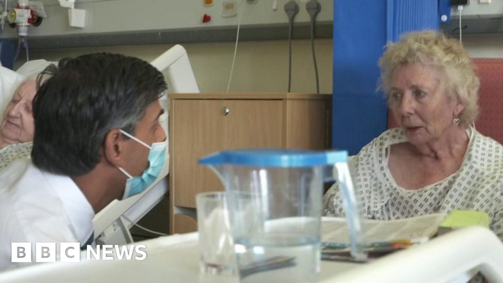
Rishi Sunak has been told he needed to “try harder” over plans to give pay rises to hospital staff.Visiting Croydon University Hospital, the prime minister spoke to in-patients, including Catherine Poole, aged 77.After telling him he needed to pay medical staff more, she rejected his answer over his efforts, and told him he was “not trying”.

Read more →