Staying warm: What does an unheated room do to your body?
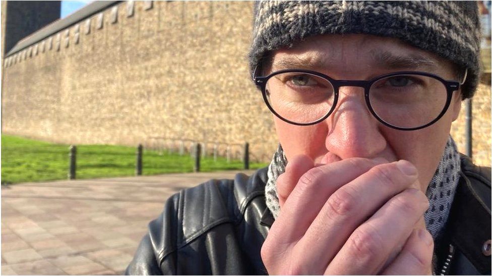
Published2 days agoSharecloseShare pageCopy linkAbout sharingImage source, James GallagherBy James GallagherInside Health presenter, BBC Radio 4Mention deadly cold and I think of polar explorers with icicles dangling from their beards and mountaineers tackling the heights of Everest; of fingers turning black with frostbite and the chilling clutch of hypothermia. So I was sceptical when I was asked to take part in a cold experiment that took place at just 10 degrees Celsius. Yes, 10C.To me that’s mild, nowhere near freezing and certainly no Arctic blast. Surely we’d have to go much colder before putting a strain on the body? I was wrong.”It sounds mild, but it is a real physiological challenge,” Prof Damian Bailey, from the University of South Wales, tells me. He’s invited me to his laboratory to explore the impact of cold homes on our bodies and why such seemingly mild temperatures can become deadly. “Ten degrees is the average temperature that people will be living in, if they can’t afford to heat their homes,” said Prof Bailey.And as I was about to find out, 10C has a profound impact on the heart, lungs and brain. Listen to Inside Health podcast: How can a cool homes affect your health?I’m led into the environmental chamber in the corner of the laboratory – it’s all shiny metal walls and thick, heavy doors. In this air-tight room, scientists can precisely set the temperature, humidity and oxygen levels. I’m hit with a blast of warm 21C air. The plan is to start at 21C, drop the temperature down to 10C and chart how my body responds to the chill. First, I am wired up to countless state-of-the-art gizmos for the most in-depth analysis my body has ever faced.My chest, arms and legs are dotted with monitors to track my body temperature, heart rate and blood pressure.”You will look like something out of Star Wars,” says Prof Bailey as another sensor and trailing cable is attached to my body. A headset is fitted to monitor the blood flow in my brain just as the first beads of sweat breakout on my brow; an ultrasound inspects the carotid arteries in my neck (hearing the rhythmic whoosh of blood going to my brain is oddly reassuring) and I breathe into a huge tube that analyses the air I exhale. Image source, James GallagherThe measurements are done. The scientists know how my body performs in a pleasant 21C. So the fans kick in and a cool breeze gradually lowers the temperature in the chamber. “Your brain is tasting your blood as we speak and it’s tasting the temperature and the brain is now sending signals to the rest of your body,” Prof Bailey tells me. The goal is to keep my core – that’s my major organs including my heart and liver – at around 37C.I was still unaware of the profound changes happening inside my body, but there were already clues on the outside. By the time the room has dropped to 18C I was no longer sweating and the hairs on my arms were starting to stand up to help insulate my body. “Science tells us that 18 degrees is the tipping point… the body is now working to defend that core temperature,” Prof Bailey shouts over the droning fans.Next my fingers turn white and they feel cold. The blood vessels in my hands are being closed off – known as vasoconstriction – in order to keep my warm blood for my critical organs.This would happen even more quickly if I were a different gender. “Women do tend to feel the cold more, because of hormones (oestrogen) their blood vessels in their hands and feet are more likely to constrict… and that makes us feel cold,” says Dr Clare Eglin from the University of Portsmouth.My first shiver kicks in at 11.5C as my muscles begin to shake to generate heat.At 10C the fans shut down. I’m feeling uncomfortable, but not freezing as we repeated all the bodily measurements again at the lower temperature and it soon became clear I was wrong to doubt that 10C would affect me. “The body is working jolly hard at 10 degrees,” says Prof Bailey.What shocks me is the change in blood flow to the brain and how much longer it takes me to complete a shape-sorting game.I wouldn’t want to be trying to do school homework in a cold room or to have this compound something like dementia.”You’re delivering less blood to the brain, so there’s less oxygen and less glucose [sugar] getting into the brain and the downside of that is it’s having a negative impact on your mental gymnastics,” Prof Bailey says.But my body is achieving its main goal of keeping my core body temperature stable – it’s just having to do more work. I’m pumping warm blood around my body more intensely with my heart beating faster and blood pressure also shooting up. “That increasing blood pressure is a risk factor for a stroke, it’s a risk factor for a heart attack,” Prof Bailey tells me.The blood itself is also changing “so it becomes a bit like treacle”, says Prof Bailey, and this thicker gloopier blood also adds to the risk of a dangerous blockage.It’s why heart attacks and strokes are more common in the winter. Image source, James GallagherFortunately, I started off with “fabulous vasculature”, Prof Bailey tells me, but these internal changes are a risk to those who already have poor heart health and the elderly. “The evidence clearly suggests that cold is more deadly than the heat, there are a higher number of deaths caused through cold snaps than there are through the heat snaps,” says Prof Bailey.”So I really do think that more recognition needs to be paid for the dangers associated with cold.”Cold favours viruses tooThe cold also lends a helping hand to many infections that thrive in the winter months such as flu. Pneumonia, when there is inflammation in the lungs because of an infection, is more common after cold weather. It is easier for viruses to spread because we’re more likely to meet up indoors with the windows shut and no fresh air to blow viruses away. Cold also makes it easier for viruses to survive outside the body and cold air contains less virus-trapping moisture. Dry air allows viruses to travel further distances, says Prof Akiko Iwasaki, an immunobiologist from Yale University. She has also performed experiments showing breathing in cold air affects how the immune system works in the nose.Prof Iwasaki tells me: “At these cooler temperatures, your immune response becomes less active and this can allow virus to grow better within your nose.”What can you actually do about it?In an ideal world we’d all heat the room we’re in to at least 18C. When that is not possible, Prof Bailey says “it’s like preparing for a mountaineering expedition”.His tips are:focus on clothes that provide good insulation such as those made of woolgloves and warm socks are more important than a hat (but a woolly hat will help too)switch foods to a higher carbohydrate diet generate more body heat by moving around and not just sitting in a chair and watching TV.This video can not be playedTo play this video you need to enable JavaScript in your browser.Follow James on Twitter. Inside Health was produced by Gerry Holt. More from Inside HealthCovid: Have I dodged it and what does it mean?Multiple sclerosis: Is a virus we all have causing MS?Long Covid: ‘I’ve had long Covid for two years now’

Read more →

What does an unheated room do to your body?

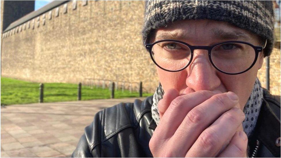
Published19 November 2022Shareclose panelShare pageCopy linkAbout sharingImage source, James GallagherBy James GallagherInside Health presenter, BBC Radio 4Mention deadly cold and I think of polar explorers with icicles dangling from their beards and mountaineers tackling the heights of Everest; of fingers turning black with frostbite and the chilling clutch of hypothermia. So I was sceptical when I was asked to take part in a cold experiment that took place at just 10 degrees Celsius. Yes, 10C.To me that’s mild, nowhere near freezing and certainly no Arctic blast. Surely we’d have to go much colder before putting a strain on the body? I was wrong.”It sounds mild, but it is a real physiological challenge,” Prof Damian Bailey, from the University of South Wales, tells me. He’s invited me to his laboratory to explore the impact of cold homes on our bodies and why such seemingly mild temperatures can become deadly. “Ten degrees is the average temperature that people will be living in, if they can’t afford to heat their homes,” said Prof Bailey.And as I was about to find out, 10C has a profound impact on the heart, lungs and brain. Listen to Inside Health podcast: How can a cold home affect your health?I’m led into the environmental chamber in the corner of the laboratory – it’s all shiny metal walls and thick, heavy doors. In this air-tight room, scientists can precisely set the temperature, humidity and oxygen levels. I’m hit with a blast of warm 21C air. The plan is to start at 21C, drop the temperature down to 10C and chart how my body responds to the chill. First, I am wired up to countless state-of-the-art gizmos for the most in-depth analysis my body has ever faced.My chest, arms and legs are dotted with monitors to track my body temperature, heart rate and blood pressure.”You will look like something out of Star Wars,” says Prof Bailey as another sensor and trailing cable is attached to my body. A headset is fitted to monitor the blood flow in my brain just as the first beads of sweat breakout on my brow; an ultrasound inspects the carotid arteries in my neck (hearing the rhythmic whoosh of blood going to my brain is oddly reassuring) and I breathe into a huge tube that analyses the air I exhale. Image source, James GallagherThe measurements are done. The scientists know how my body performs in a pleasant 21C. So the fans kick in and a cool breeze gradually lowers the temperature in the chamber. “Your brain is tasting your blood as we speak and it’s tasting the temperature and the brain is now sending signals to the rest of your body,” Prof Bailey tells me. The goal is to keep my core – that’s my major organs including my heart and liver – at around 37C.I was still unaware of the profound changes happening inside my body, but there were already clues on the outside. By the time the room has dropped to 18C I was no longer sweating and the hairs on my arms were starting to stand up to help insulate my body. “Science tells us that 18 degrees is the tipping point… the body is now working to defend that core temperature,” Prof Bailey shouts over the droning fans.Next my fingers turn white and they feel cold. The blood vessels in my hands are being closed off – known as vasoconstriction – in order to keep my warm blood for my critical organs.This would happen even more quickly if I were a different sex. “Women do tend to feel the cold more, because of hormones (oestrogen) their blood vessels in their hands and feet are more likely to constrict… and that makes us feel cold,” says Dr Clare Eglin from the University of Portsmouth.My first shiver kicks in at 11.5C as my muscles begin to shake to generate heat.At 10C the fans shut down. I’m feeling uncomfortable, but not freezing as we repeated all the bodily measurements again at the lower temperature and it soon became clear I was wrong to doubt that 10C would affect me. “The body is working jolly hard at 10 degrees,” says Prof Bailey.What shocks me is the change in blood flow to the brain and how much longer it takes me to complete a shape-sorting game.I wouldn’t want to be trying to do school homework in a cold room or to have this compound something like dementia.”You’re delivering less blood to the brain, so there’s less oxygen and less glucose [sugar] getting into the brain and the downside of that is it’s having a negative impact on your mental gymnastics,” Prof Bailey says.But my body is achieving its main goal of keeping my core body temperature stable – it’s just having to do more work. I’m pumping warm blood around my body more intensely with my heart beating faster and blood pressure also shooting up. “That increasing blood pressure is a risk factor for a stroke, it’s a risk factor for a heart attack,” Prof Bailey tells me.The blood itself is also changing “so it becomes a bit like treacle”, says Prof Bailey, and this thicker gloopier blood also adds to the risk of a dangerous blockage.It’s why heart attacks and strokes are more common in the winter,

Read more →

FDA Approves a Drug That Can Delay Type 1 Diabetes

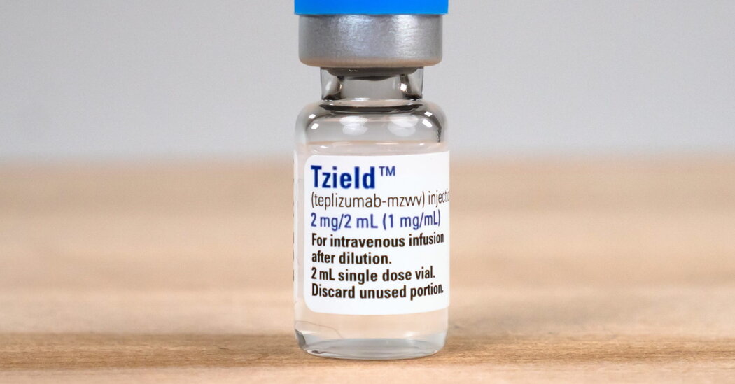
The therapy will cost nearly $200,000. But it’s the first treatment in 100 years to alter the course of the disease, which usually begins in adolescence.The Food and Drug Administration has approved the first treatment that can delay — possibly for years — the onset of Type 1 diabetes, a disease that often emerges in teenagers.The new drug, teplizumab, is made by Provention Bio, which will partner with Sanofi to market the drug in the United States under the brand name Tzield. In an investor call on Friday, Provention said the drug would cost $13,850 a vial or $193,900 for the 14-day treatment. The company said teplizumab should be available by the end of the year.The drug, which the F.D.A. approved on Thursday, does not cure or prevent Type 1 diabetes. Instead, it postpones its onset by an average of two years and, for some lucky patients, much longer — the longest so far is 11 years, said Dr. Kevan Herold of Yale, a principal investigator in trials of the drug.The only other treatment for the disease — insulin — was discovered 100 years ago and does not affect the course of the disease. It just replaces what is missing.Teplizumab will be used to treat patients at high risk for Type 1 diabetes who have antibodies that indicate an immune attack on their pancreas and whose glucose tolerance is not normal. Treatment involves a 14-day infusion of the drug, a monoclonal antibody that blocks T cells, preventing them from attacking the insulin-producing cells of the pancreas.“Talk to anyone who has Type 1 diabetes and any day you are not burdened by measuring blood sugar four times a day and injecting yourself with insulin is a glorious day,” said Dr. Mark S. Anderson, director of the diabetes center at the University of California, San Francisco, and a researcher for the pivotal clinical trial that led to the treatment’s approval. Dr. Anderson has been a paid consultant for Provention in the past.Dr. John Buse, a diabetes expert at the University of North Carolina who was not involved in the study, called the approval “really exciting” and said it would “turn the world of Type 1 diabetes on its head.”“There has always been a notion that screening would be a good idea,” he said. But medical experts “have never really promoted it to detect Type 1 diabetes.”It will not be easy — no screening test is. But in this case, very few who are screened will have this rare but dire disease, which affects just four in 1,000 in the general population, or 1.4 million Americans.Type 1 diabetes typically emerges in adolescence when patients suddenly are tired all the time, urinating frequently, drinking copious amounts of water, and losing weight.With a diabetes diagnosis, their lives are completely changed. They have to measure their blood sugar and take insulin for the rest of their lives. Every time they eat a meal, they have to calculate how much insulin they need. If they take too much, they can pass out or have a seizure or even end up in intensive care.They also face the specter of complications — eye disease that can lead to blindness, kidney failure, heart disease and stroke. Without good control of blood glucose, complications can set in as early as five years after diagnosis, Dr. Anderson said.The new treatment, Dr. Anderson said, “opens the door,” much in the way that the first immunotherapy for cancer was a breakthrough to a new era of treatment about a decade ago. He expects that as immunotherapy for diabetes improves, the disease may be halted before it can take hold.The new drug is not a treatment for the much more common type of diabetes, Type 2, in which the pancreas makes insulin but the body’s cells do not respond to it.The story of the new treatment dates back to the 1980s and involves determined researchers who pursued the idea as company after company — four in total before Provention — got interested but then ended up dropping the drug for a variety of reasons.Dr. Jeffrey A. Bluestone, an academic until recently who is now chief executive of Sonoma Biotherapeutics, said he and his colleague, Dr. Herold, who has consulted for Provention and other companies, spent 20 years “trying to keep the drug alive.”“Scientists were passionate about it,” said Dr. Bluestone, who has consulted for Provention and has been a company board member. “Guys at companies were really passionate about it.” But for reasons unrelated to its potential, it kept being dropped.Dr. Herold said he vividly remembered trips that he and Dr. Bluestone and a French researcher, Lucienne Chatenoud of Paris Descartes University, made to drug companies “begging them to pick this thing up.”At one point, Dr. Bluestone actually took the antibody, developed at that time by Ortho Pharmaceuticals, and made a clinical batch of the drug in the lab. Dr. Herold tested it in a small study of people who were newly diagnosed with Type 1 diabetes.The treatment prolonged the period in which they made some insulin, but eventually all got diabetes.In 2011, Dr. Bluestone and Dr. Herold proposed a different sort of study. They would treat people who were at high risk of diabetes but who had not yet developed it. It was a bold move, Dr. Bluestone noted. “Other than vaccines, there aren’t many drugs given before diagnosis,” he said.To find those people, the researchers worked with a group of clinical trial sites, TrialNet, that were supported by the National Institutes of Health. Dr. Herold is now the group’s chairman. TrialNet investigators screened 200,000 people who were immediate family members of people with Type 1 diabetes, looking for antibodies indicating an immune attack on the pancreas and abnormal glucose metabolism.The result was a study, published in The New England Journal of Medicine, that led to Thursday’s approval.Now that the drug is approved, the challenge will be to find people who could benefit. Screening only people with immediate relatives who have diabetes will miss 85 percent of patients.The JDRF, a nonprofit group that advocates for people with Type 1 diabetes and supports research, which, along with the N.I.H., funded the trial that led to the drug’s approval, wants antibody screening tests to become part of routine pediatric care.“Most families say diagnosis is a bolt out of the blue,” said Aaron Kowalski, chief executive of the JDRF. And most patients, he added, are very sick when they first are diagnosed.The group has conducted blood tests to look for antibodies in Germany and in parts of Colorado. And it has offered an at-home test people can order, underwritten by the foundation. But, Dr. Kowalski said, “we want pediatric offices to do it.”Testing also offers another opportunity, he said. It turns out that antibodies indicating an immune attack often occur when people are as young as 5 or 6 years old, although most do not develop diabetes until they are teenagers.Now, he said, his hope is to treat people even earlier, as soon as those antibodies emerge. Clinicians and the F.D.A. had previously objected to treating before the disease was clearly underway, asking, “How can you give an immunotherapy if they are normal?” Dr. Kowalski said.But, he said, the antibodies tell a different story.“They do have diabetes,” he said, although not according to the usual definition of the disease. “It just hasn’t unmasked itself yet. We need to help them save their beta cells,” the insulin-secreting cells of the pancreas.Dr. Herold is cautious. If someone has antibodies but their pancreas is not actively being attacked, the treatment may not help.“It’s hard to stop something that isn’t happening,” he said.Dr. Bluestone and Dr. Herold wonder if giving a second round of treatment could improve results even further.For now, Dr. Bluestone would like to see the treatment used to help younger patients than were in the trial. It is approved for patients at least 8 years old. “But the disease affects a lot of patients who are younger than 8,” he said.Although he and others are excited about the possibility of some day preventing the disease entirely, there is an immediate challenge for diabetes experts.“The most important thing right now is finding the potential patients,” Dr. Bluestone said.

Read more →

Toxins force construction of 'roads to nowhere'

Toxins released by a type of bacteria that cause diarrheal disease hijack cell processes and force important proteins to assemble into “roads to nowhere,” redirecting the proteins away from other jobs that are key to proper cell function, a new study has found.
The affected proteins are known as actins, which are highly abundant and have multiple roles that include helping every cell unite its contents, maintain its shape, divide and migrate. Actins assemble into thread-like filaments to do certain work inside cells.
Researchers found that two toxins produced by the Vibrio genus of bacteria cause actins to start joining together into these filaments — which could be thought of as cellular highways on which cargo is delivered — at the wrong location inside cells, and headed in the wrong direction.
“Growing in the wrong direction is a totally new function that was not previously known and was not thought to be possible for actin filaments inside the cell,” said senior author Dmitri Kudryashov, associate professor of chemistry and biochemistry at The Ohio State University. “A large fraction of actin in the cell is consumed in formation of the ‘highways’ where they are not needed, so the cell resources are wasted and cannot be used to satisfy the cell’s basic needs.”
The research is published today (Nov. 18, 2022) in the journal Science Advances.
These disruptive toxins are called VopF and VopL, and are produced by two strains of Vibrio bacteria living in seawater: V. cholerae and V. parahaemolyticus, both of which can contaminate oysters and other shellfish that, when eaten raw, make people sick.

Read more →

New study identifies connection between diabetes medications, multiple sclerosis

A new University of Arizona Health Sciences study found that people older than 45 whose Type 2 diabetes was treated with anti-hyperglycemic medications had an increased risk of multiple sclerosis, particularly among women, while anti-hyperglycemic exposure in people younger than 45 reduced that risk.
“Our findings reinforce the need for a precision medicine approach to preventing MS in these vulnerable populations,” said lead researcher Kathleen Rodgers, PhD, associate director of translational neuroscience at the Center for Innovation in Brain Science.
Multiple sclerosis (MS) is an unpredictable autoimmune neurological disorder that affects the central nervous system and leads to severe physical and cognitive disability. It is estimated that nearly 1 million adults in the U.S. and more than 2.8 million worldwide are living with MS.
For people with Type 2 diabetes, there is mounting evidence linking metabolic disorders and MS through a common driver of increased autoimmunity. This brings into question the impact of anti-hyperglycemic therapeutics used to treat Type 2 diabetes, including insulin, on the incidence of MS.
“Previous research has shown a neuroprotective effect of anti-hyperglycemic medications in Alzheimer’s disease and other related dementias,” Dr. Rodgers said. “For MS, we wanted to further examine age and sex differences, particularly among men and women under 45 with Type 2 diabetes.”
They found that men older than 45 years old had a slightly significant increase of MS risk and women older than 45 years exhibited a significant increase in MS incidence after anti-hyperglycemic exposure. In addition to age differences, the risk analysis by drug class showed that exposure to insulin in patients older than 45 years old was associated with a greater increased risk compared with other therapies.
In patients younger than 45, anti-hyperglycemic exposure was protective against the development of MS.
The study utilized a U.S.-based insurance claims database of 151 million participants to identify more than 5 million patients with a diagnosis of Type 2 diabetes and either early-onset or late-onset MS. Researchers segmented the data by age — patients diagnosed with Type 2 diabetes prior to or after age 45 — and sex to decode the factors driving MS risk in both populations, especially in women over 45 years of age.
The paper, “Age and sex differences on anti-hyperglycemic medication exposure and risk of newly diagnosed multiple sclerosis in propensity score-matched type 2 diabetics,” was published recently in the journal Heliyon.
Co-authors from the Center for Innovation in Brian Science include Roberta Diaz Brinton, PhD, director and Regents Professor; Francesca Vitali, PhD, research assistant professor of neurology; Georgina Torrandell-Haro, doctoral candidate and graduate research assistant; and Gregory Branigan, PhD, third-year medical student in the UArizona College of Medicine — Tucson’s MD-PhD program.
This research was supported in part by the National Institute on Aging (P01AG026572, T32AG061897, R37AG053589) and the National Institute of Neurological Disorders and Stroke (R25NS107185), both divisions of the National Institutes of Health.
Story Source:
Materials provided by University of Arizona Health Sciences. Note: Content may be edited for style and length.

Read more →

Can pharmacotherapies prevent alcohol use disorder in people with PTSD?

Survivors of abuse and trauma are vastly more likely than other people to develop alcohol use disorder (AUD); according to some estimates, as many as three-quarters of people with post-traumatic stress disorder (PTSD) report drinking problems.
Now, Scripps Research scientists have identified a class of drugs that might break this link. In animal models of PTSD, the drug decreased alcohol preference and intake as well as other behaviors associated with PTSD, including aggression, excessive fear and hyperarousal. The findings were published in Neuropsychopharmacology on November 18, 2022.
“The overlap of PTSD and AUD is a major problem,” says co-senior author Marisa Roberto, PhD, the Schimmel Family Chair of Molecular Medicine and a professor of Neuroscience at Scripps Research. “We’ve shown that there is potential to alleviate both disorders by targeting brain pathways that they share.”
According to the U.S. Department of Veterans Affairs National Center for PTSD, about 12 million adults in the U.S. have PTSD during a given year. Men and women who have PTSD at any point in their lives are more than twice as likely as other people to have alcohol abuse or dependence. Moreover, people who suffer from both PTSD and AUD are at a higher risk of suicidal thoughts and extreme aggression compared to those with either disorder alone.
Researchers have known that FKBP5, a protein found in the brain, plays a role in both disorders. The FKBP5 gene is responsible for lifting the brakes on the brain’s stress response pathways, and its genetic variants are associated with increased risk of AUD and PTSD. In animals, higher levels of FKBP5 have been linked to both stress exposure and alcohol exposure.
In the new study, co-first authors Bryan Cruz, PhD, and Valentina Vozella, PhD, and additional colleagues studied rats with symptoms similar to comorbid human PTSD and AUD. Like people with the disorders, the animals drink more alcohol than average, are irritable and fearful, and exhibit anxiety and sleep disturbances, the team showed. The researchers treated the animals with either of two drugs known to target FKBP5: benztropine (Cogentin®), which is FDA-approved to treat Parkinson’s disease and targets a number of molecules in the brain, or SAFit2, an experimental compound designed specifically for blocking FKBP5.
They found that benztropine reduced alcohol preference in stressed male and female animals, as well as aggressive behavior in the females. SAFit2 reduced alcohol drinking in stressed males, and decreased levels of extreme fear in both male and females. Neither drug impacted sleep.
“The results may have varied between male and female animals because of reproductive hormones,” says Cruz. “There is new literature suggesting that the activity of these kinds of compounds varies in females throughout the estrous cycle.”
The team says that the fact that benztropine is already FDA-approved suggests the potential for repurposing it in people with PTSD.
“We think FKBP5 inhibitors might be useful in preventing AUD after the onset of PTSD,” adds co-senior author Eric Zorrilla, PhD, associate professor in the Department of Molecular Medicine. “More work is needed to determine whether these compounds also can prevent the recurrent relapse that hampers recovery.”
Support for this study was provided by The National Institute on Alcohol Abuse and Alcoholism (AA027700, AA028879, AA013498, P60 AA006420, AA017447, AA021491, AA029841, AA015566, K99 AA026638 and T32 AA007456), the Schimmel Family Endowed Chair, and the Department of Defense (DoD).

Read more →

Dietary change starves cancer cells, overcoming treatment resistance

A dietary change could be a key to enhancing colon cancer treatment, a new study from the University of Michigan Rogel Cancer Center finds.
Cancer cells need nutrients to survive and grow. One of the most important nutrient sensing molecules in a cell is called mTORC1. Often called a master regulator of cell growth, it allows cells to sense different nutrients and thereby grow and proliferate. When nutrients are limited, cells dial down nutrient sensing cascade and turn off mTORC1.
While mTORC1 is known to be hyperactive in colon cancer, the key question is whether colon tumors hijack nutrient sensing pathways to fire up the master regulator.
“In colon cancer, when you decrease the nutrients available in the tumors, the cells don’t know what to do. Without the nutrients to grow, they undergo a kind of crisis, which leads to massive cell death,” said senior author Yatrik M. Shah, Ph.D., Horace W. Davenport Collegiate Professor of Physiology at Michigan Medicine.
Researchers found in cells and in mice that a low-protein diet blocked the nutrient signaling pathway that fires up a master regulator of cancer growth. Results are published in Gastroenterology.
The regulator, mTORC1, controls how cells use nutritional signals to grow and multiply. It’s highly active in cancers with certain mutations and is known to cause cancer to become resistant to standard treatments. A low-protein diet, and specifically a reduction in two key amino acids, changed the nutritional signals through a complex called GATOR.

Read more →

Scientists produce 'DNA virus vaccine' to fight DNA viruses

Rutgers scientists have developed a new approach to stopping viral infections: a so-called live-attenuated, replication-defective DNA virus vaccine that uses a compound known as centanamycin to generate an altered virus for vaccine development.
The method was tested to produce a weakened or “attenuated” version of a mouse cytomegalovirus, a common virus, that has been altered so it can’t reproduce or replicate inside the cell. A replication-defective DNA virus is incapable of replicating its genome, its essential genetic matter. As a result, it is unable to produce an infectious progeny virus in infected cells, and thus restricted primarily to the site of inoculation.
When the weakened viral particles are injected into animals, the researchers said, they stimulate a specific host’s immune system to recognize the invading live virus particles as foreign, causing the virus to be eliminated whenever it is detected.
The new approach, published in Cell Reports Methods, has been shown to effectively shut down viral infections in lab animals.
“We have found that this method is safe; the attenuated virus infects certain cells without proliferating beyond that, and alerts the host to produce specific neutralizing antibodies against it,” said Dabbu Jaijyan, a researcher in the Department of Microbiology at Rutgers New Jersey Medical School and an author of the study. “We see this as a novel method that we hope will accelerate vaccine development for many untreated viral infections in humans and animals.”
The method is called a live-attenuated DNA virus vaccine because it specifically targets DNA viruses — viruses such as cytomegalovirus, chicken pox and herpes simplex that reproduce by making copies of their DNA molecules — and uses an altered DNA virus to fight against them. Developing a method that can quickly and easily generate replication-defective live-attenuated viruses, the researchers said, will accelerate vaccine development for diseases caused by DNA viruses.

Read more →

Brain organoids reveal in detail the harms of prenatal alcohol exposure

Alcohol consumption during pregnancy poses significant peril to the healthy development of the unborn child. There is no known safe amount of alcohol during pregnancy.
The consequences of prenatal alcohol exposure (PAE) are reflected in the different diagnoses that emerge under the umbrella of fetal alcohol spectrum disorders. At one end of the spectrum, growth deficits and physical differences define fetal alcohol syndrome (FAS), but in most cases, irreversible brain damage leads to behavior and learning challenges even without a physical impact. Experts estimate that 1.1 to 5 percent of U.S. schoolchildren — as many as 1 in 20 — may be affected by PAE, with some percentage experiencing FAS.
Although the clinical effects of fetal alcohol spectrum disorders are well documented, the precise molecular effects on the human fetal cerebral cortex are not fully understood. In a new study, published November 16, 2022 in Molecular Psychiatry, researchers at University of California San Diego School of Medicine used human brain organoids to more specifically document how alcohol exposure impairs the development and functioning of new brain cells.
“The findings underscore the broad threat of alcohol exposure to the fetal brain. The harm inflicted is profound and extensive,” said Alysson R. Muotri, PhD, professor in the Departments of Pediatrics and Cellular and Molecular Medicine at UC San Diego School of Medicine.
Muotri is co-corresponding author of the study with Cleber A. Trujillo, a former project scientist in Muotri’s lab and now associate director of stem cell biology at Massachusetts-based Vesalius Therapeutics.
Using human induced pluripotent stem cells, Muotri and colleagues created three-dimensional brain organoids that develop similarly to human fetal corticogenesis — the formation of the outer layers of the brain that house many high-level functions, such as reasoning, conscious thought, emotional control and speech.
Alcohol exposure at different points of fetal brain development resulted in different but invariably negative effects, from fundamental dysfunction of cellular processes, to faulty construction of brain architecture and inadequate creation of support cells (gliogenesis) and connections between brain cells (synaptogenesis).
The researchers followed up by conducting electrophysiology recordings to monitor electrical activity patterns in the cortical organoids, documenting and confirming impaired cortical organoid function.
The authors said the findings improve upon previous studies using animal models.
“They overcome the suboptimal recapitulation of non-human models,” said co-author Miguel Del Campo, MD, PhD, associate professor at UC San Diego School of Medicine and medical geneticist at Rady Children’s Hospital-San Diego. “In fact, they show organoids are a valuable model for better assessing, more fully and deeply, the effects of alcohol exposure on the developing human brain.”
Co-author Kenneth L. Jones, MD, professor of pediatrics at UC San Diego School of Medicine, elaborated: “That is crucial because we can better see what prominent growth and signaling pathways are disrupted and perhaps discover new targets to therapeutically impede or prevent the neuropathology of prenatal alcohol exposure. The good news is that some of these alterations were reversed using specific experimental drugs.”
Co-authors include: Jason W. Adams, Priscilla D. Negraes, Justin Truong, Timothy Tran, Ryan Szeto, Carmen Teodorof and Stephen A. Spector, all at UC San Diego.
Story Source:
Materials provided by University of California – San Diego. Original written by Scott LaFee and Nicole Mlynaryk. Note: Content may be edited for style and length.

Read more →