Measuring organ development

Organs in the human body have complex networks of fluid-filled tubes and loops. They come in different shapes and their three-dimensional structures are differently connected to each other, depending on the organ. During the development of an embryo, organs develop their shape and tissue architecture out of a simple group of cells. Due to a lack of concepts and tools, it is challenging to understand how shape and the complex tissue network arise during organ development. Metrics for organ development have now been defined for the first time by scientists from the Max Planck Institute of Molecular Cell Biology and Genetics (MPI-CBG) and the MPI for the Physics of Complex Systems (MPI-PKS), both in Dresden, as well as the Research Institute of Molecular Pathology (IMP) in Vienna. In their study, the international team of researchers provide the necessary tools to transform the field of organoids — miniature organs — into an engineering discipline to develop model systems for human development.
The collective interaction of cells leads to the shaping of an organism during development. The different organs feature various geometries and differently connected three-dimensional structures that determine the function of fluid-filled tubes and loops in organs. An example is the branched network architecture of the kidney, which supports the efficient filtration of blood. Observing embryonic development in a living system is hard, which is why there are so few concepts that describe how the networks of fluid-filled tubes and loops develop. While past studies have shown how cell mechanics induce local shape changes during the development of an organism, it is not clear how the connectivity of tissues emerges. By combining imaging and theory, the researcher Keisuke Ishihara started to work on this question first in the group of Jan Brugues at the MPI-CBG and MPI-PKS. He later continued his work in the group of Elly Tanaka at the IMP. Together with his colleague Arghyadip Mukherjee, formerly a researcher in the group of Frank Jülicher at MPI-PKS, and Jan Brugués, Keisuke used organoids derived from mouse embryonic stem cells that form a complex network of epithelia, which line organs and function as a barrier. “I still remember the exciting moment when I found that some organoids had transformed into tissues with multiple buds that looked like a bunch of grapes. Describing the change in the three-dimensional architecture during development proved to be challenging, though,” remembers Keisuke and adds, “I found that this organoid system generates astonishing internal structures with many loops or passages, resembling a toy ball with holes.”
Studying the development of tissues in organoids has several advantages: they can be observed with advanced microscopy methods, making it possible to see dynamic changes deep inside the tissue. They can be generated in large numbers and the environment can be controlled to influence development. The researchers were able to study the shape, number, and connectivity of the epithelium. They tracked the changes in the internal structure of organoids over time. Keisuke continues, “We discovered that tissue connectivity emerges from two different processes: either two separate epithelia fuse or a single epithelium self-fuses by fusing its two ends, and thereby creating a doughnut shaped loop.” The researchers suggest, based on theory of epithelial surfaces, that the inflexibility of epithelia is a key parameter that controls epithelial fusion and in turn the development of tissue connectivity.
The supervisors of the study, Jan Brugues, Frank Jülicher, and Elly Tanaka conclude, “We hope that our findings will lead to a fresh view of complex tissue architectures and the interplay between shape and network connectivity in organ development. Our experimental and analysis framework will help the organoid community to characterise and engineer self-organising tissues that mimic human organs. By revealing how cellular factors influence organ development, these results may also be useful for developmental cell biologists who are interested in organisational principles.”
Story Source:
Materials provided by Max Planck Institute of Molecular Cell Biology and Genetics (MPI-CBG). Note: Content may be edited for style and length.


Read more →

Local alcohol availability related to child maltreatment

The number of stores selling alcohol in a neighborhood is linked to cases of child abuse and neglect in the same area, a new study suggests.
A study in Sacramento, California, found that having one more off-premises alcohol outlet — those selling alcohol to be consumed elsewhere — in a census tract was related to 13.5% more substantiated cases of child abuse and neglect in that area in a year and 10.5% entries into foster care.
That was after taking into account a variety of other factors that could impact child maltreatment numbers in the neighborhood.
The results show the importance of alcohol availability in creating conditions that may lead to child abuse, said Bridget Freisthler, lead author of the study and professor of social work at The Ohio State University.
“The relationship between the alcohol environment and child abuse and neglect is complex,” Freisthler said.
“Individual interventions to reduce substance use will not completely solve the problem without addressing the issue of alcohol supply.”
The study was published online recently in the journal Alcoholism: Clinical and Experimental Research.

Read more →

Legs of sprinters: Highly muscular, still maneuverable

A comprehensive knowledge of human biomechanics is useful for training athletes more effectively. From a biomechanical perspective, sprinting is challenging in comparison to walking and endurance running, making it an intriguing area of research. In addition, a detailed biomechanical understanding of sprinter limbs can be insightful for more effective training of sprinters and for advancing our understanding of human motor performance in general.
Previous studies have revealed that sprinters have inhomogeneous muscular development, with well-developed hip flexors, extensors, and muscles located in the proximal thigh. The muscularity in sprinters corresponds well with the musculoskeletal demands for sprinting, which can be a positive aspect in force-related factors. At the same time, however, the morphological differences would involve differences in the mass distribution and, therefore, maneuverability. More muscular legs would lead to a larger mass and, in turn, to a general speculation of the trade-off between greater torque exertion ability and greater difficulty in moving the lower limb.
This raises an interesting question: How do sprinters balance muscular strength and brisk movements? Contextually, not much is known regarding the maneuverability of sprinter legs from an inertial viewpoint. This knowledge gap caught the attention of a research team led by Professor Yasuo Kawakami from the Faculty of Sport Sciences, Waseda University, who, along with his colleagues Hoshizora Ichinose from Nihon University and Dr. Natsuki Sado from University of Tsukuba, investigated athlete leg morphology from an inertial perspective.
“As former athletes, we have been interested in the relevance of the human body in sports. Athletes train hard, but in many cases, they do not know how their body responds specifically to training. We wanted to address this and help in the development of effective training strategies,” says Kawakami, explaining the motivation behind their study.
In their study, the researchers analyzed fat- and water-separated MRI scans of 11 male sprinters, all trained athletes with careers spanning more than 7 years, and 12 male non-sprinters, individuals who had not undergone resistance training or played sports within the previous two years at the time of the study. By comparing the MRI characteristics, they found that the sprinters had a higher relative mass of the lower limbs than did the non-sprinters. This increase in muscularity, in turn, aided power production during running. These findings have been published in Medicine & Science in Sports & Exercise. To understand the trade-off between joint torque and segment maneuverability, the researchers further computed the moment of inertia of the lower limbs around the hip. This comparison revealed that the moment of inertia did not differ between sprinters and non-sprinters, thereby suggesting that the increased musculature does not compromise the maneuverability of the lower limbs in sprinters.
On comparing the different parts of the lower limbs between sprinters and non-sprinters, the researchers found that while the relative mass of the thighs differed between sprinters and non-sprinters, the relative masses of the shank and foot did not vary. The researchers thus conclude that these top-heavy, bottom-light characteristics of sprinter limbs help them in executing athletic movements with ease, judging from an inertial perspective.
This novel study sheds light on the unique characteristics of the lower limbs of sprinters. The findings will be useful for the development of new training strategies. Based on their findings, what do the researchers suggest athletes and trainers do for more effective training? Kawakami comments, “Sprinters can focus on training their lower limb muscles without worrying about the increased mass associated with sprint-induced lower limb muscularity and the resulting difficulty of moving due to higher moment of inertia. This strategy will help in balancing running power and limb movements.”
Story Source:
Materials provided by Waseda University. Note: Content may be edited for style and length.
Read more →

Long-term effects of COVID-19 in diabetics

Can a COVID-19 infection have long-term health effects on people with diabetes, including advancing their risk for heart disease?
College of Medicine researcher Dr. Dinender Singla believes that the genetic makeup of patients with diabetes or those predisposed to the disease makes them more prone to post-COVID inflammatory conditions that impact the heart and brain.
“We believe that COVID-19 can alter a person’s genetic makeup which can enhance the proliferation of disease and cause further deterioration in diabetes and associated heart disease,” explained Dr. Singla, who is the AdventHealth Chair of Cardiovascular Science at the College of Medicine.
Dr. Singla has spent much of his research career studying heart failure, diabetes and inflammation. In a recent article published in the American Journal of Physiology-Heart and Circulatory Physiology, he examined the mechanisms and possible effects of COVID-19 on patients with high-risk diabetes and the virus’ potential to advance the disease, leading to inflammation and heart failure.
“Our thinking is COVID-19 could have three major long-term effects on patients, Dr. Singla noted. “One is cognitive dysfunction, which can lead to Alzheimer’s disease. Second, it can enhance diabetes in pre-diabetic patients or pre-diabetic conditions. Third, it can exacerbate complications of diabetes such as cardiomyopathy or muscle dysfunction.”
Dr. Singla theorizes that some diabetic patients who were infected with COVID-19 may have developed a different cellular composition in their blood compared to diabetic patients who never had COVID. The next step in his research is to analyze specific cellular differences in diabetics with and without a COVID infection.
“Our goal is to look into whether there is a difference in blood composition or variations in cytokines — proteins that affect communications between cells — compared to the non-COVID diabetic patients,” Dr. Singla said. “If any differences are noted, then we would need to examine what kind of diseases they could potentially cause or enhance in those patients.”
COVID-19 has affected more than 600 million people worldwide, and because vaccines have made the virus not as alarming today as it was two years ago, Dr. Singla said there are still many unanswered questions about COVID’s long-term impact on health.
“For example, if someone was genetically predisposed to developing heart disease or Alzheimer’s disease, if that person is affected by COVID-19, will that person develop heart disease or Alzheimer’s earlier than they were predisposed to?” Dr. Singla said. “Also how severe will their disease be and will it be different in people who contracted or did not have COVID-19?”
Dr. Singla said he is currently working on securing funding to explore the unanswered questions left in the wake of the virus.
“We want to know will diabetes be present in patients infected with COVID-19 10 or 20 years from now?” Dr. Singla said. “Will they develop a special type of cardiomyopathy or diabetic muscle pain and will those diseases be much more in enhanced? Having this information will allow us to be one step ahead in developing therapeutics and treatments to manage any variations of diseases that may occur.”
Story Source:
Materials provided by University of Central Florida. Original written by Christin Senior. Note: Content may be edited for style and length.

Read more →

Exercise can reduce severity of breast cancer treatment side effects

Breast cancer is the most common form of the disease among women; in Australia, one in eight women will be diagnosed with breast cancer by the age of 85.
Radiotherapy has emerged as an important component of breast cancer treatment but can lead to cancer-related fatigue and negatively impact patients’ health-related quality of life including their emotional, physical and social wellbeing.
However, latest research by Edith Cowan University (ECU) has revealed exercise may make radiotherapy more tolerable for patients.
ECU’s Exercise Medicine Research Institute included 89 women in the study, with 43 completing a home-based 12-week program, consisting of a weekly exercise regime of one to two resistance training sessions and an accumulated 30-40 minutes of aerobic exercise.
The remaining patients were a control group who did not participate in the exercise program.
Researchers found patients who exercised recovered from cancer-related fatigue quicker during and after radiotherapy compared to the control group and saw a significant increase in health-related quality of life post radiotherapy.

Read more →

Chris Hemsworth: Alzheimer’s risk prompts actor to take acting break

Published23 hours agoSharecloseShare pageCopy linkAbout sharingImage source, Getty ImagesBy Steven McIntoshEntertainment reporterActor Chris Hemsworth says he is taking a break from acting after learning he has a heightened risk of developing Alzheimer’s disease.The Thor star made the discovery after undergoing tests as part of his Disney+ documentary series Limitless.He told Vanity Fair the tests confirmed his “biggest fear”, adding he will now be trying to take “preventative steps”.Alzheimer’s is the most common form of dementia and can cause memory problems, confusion and communication issues.Hemsworth learned that he has two copies of the gene ApoE4, one from his mother and one from his father, making him between eight and 10 times more likely to develop the disease than those without both copies of the gene.About 2 to 3 per cent of the population carries two copies of the gene.”It’s not like I’ve been handed my resignation,” Hemsworth said, but added the news “really triggered something in me to want to take some time off”.”If you look at Alzheimer’s prevention, the benefit of preventative steps is that it affects the rest of your life,” he said. “It’s all about sleep management, stress management, nutrition, movement, fitness. It’s all kind of the same tools that need to be applied in a consistent way.”The Limitless series sees Hemsworth test his body and explore ways to live longer and healthier.He explained that he had not been diagnosed with Alzheimer’s disease, but had been warned of the heightened risk. “It’s not a pre-deterministic gene, but it is a strong indication,” he said. “Ten years ago, I think it was more thought of as determinant.”Image source, Getty ImagesHemsworth said the original plan for the series would have seen him receive all his genetic test results live to camera – but series creator Darren Aronofsky told him privately once they got the results.The Marvel star was subsequently given the option of removing any references to Alzheimer’s from the show, but decided to include his genetic risk of Alzheimer’s to improve awareness and understanding.”My concern was I just didn’t want to manipulate it and over-dramatise it, and make it into some sort of hokey grab at empathy or whatever for entertainment,” he said.Hemsworth also confirmed to the magazine his grandfather has also been diagnosed with Alzheimer’s.The actor will soon be seen in George Miller’s forthcoming Mad Max sequel Furiosa, which concluded filming earlier this month. The 39-year-old said he would take a break from acting after finishing the publicity tour for Limitless, along with his other contracted work.He will be going home to Byron Bay in Australia to spend time with his partner, actor Elsa Pataky, and their three children.More on this storyAlzheimer’s research takes ‘leap forward’4 AprilAlzheimer’s-slowing drug labelled historic28 September

Read more →

Changing the lives of Senegalese people disabled by clubfoot

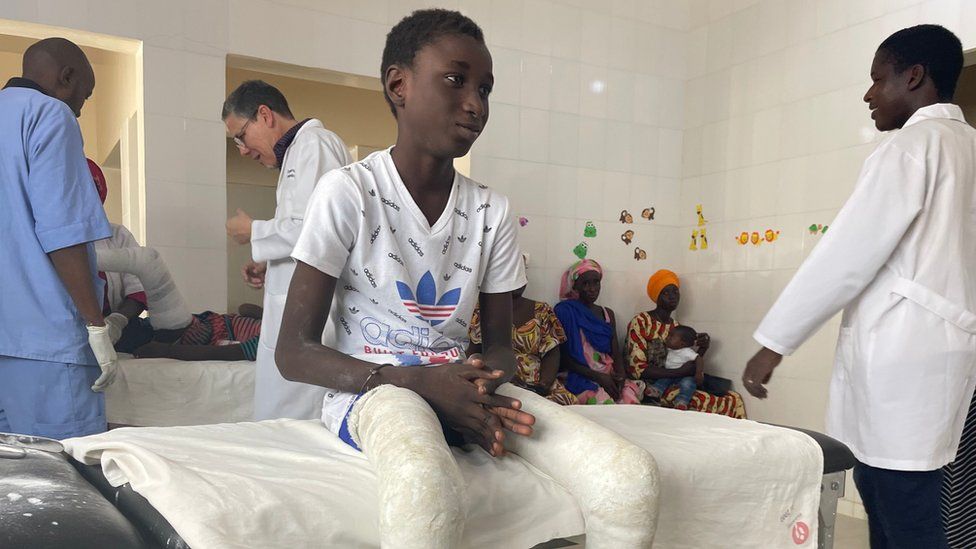
Published7 hours agoSharecloseShare pageCopy linkAbout sharingBy Naomi GrimleyGlobal health correspondent, DakarAs Africa holds its first ever conference on clubfoot – something about one in 800 people across the world are born with – the BBC visits a health clinic in Senegal to witness a transformative treatment which is turning lives around. Earlier this year, 14-year-old Serigne was reluctant to ever leave the house. He could walk very slowly, but – born with both his feet pointing inwards – he was just too ashamed. Some would make fun of the way he walked, others were afraid and would call him names. But now, less than six months on, his feet are transformed and his dream of playing football for Senegal seems at least possible. Serigne has clubfoot, also known as talipes, and today is another step in the journey to correct that. Every year 200,000 babies are born with clubfoot. According to the charity MiracleFeet, of the nearly 10 million people alive today who were born with it, as many as eight million have never received treatment. Senegal has been a relatively late adopter of Ponseti – a pioneering method of correcting clubfoot named after the Spanish doctor who invented it – compared with some other African countries. Malawi and Uganda, for example, were ahead of many Western countries in recognising its potential. But this means Senegalese medics have perfected the original concept and are now using it to treat teenagers or even adults who thought they had passed the age for any intervention.Serigne’s mother is beaming at her son’s progress. She knows better than anyone the cruel nature of this disability. “One time he even refused to leave home for three days. It caused me so much hurt inside. But now we’re happy.” As part of the Ponseti method, a new plaster cast is put on every week to gradually manipulate the foot into a better position. Because his case is so severe, he has had 20 casts in as many weeks, but the average is between five and eight. As with most cases, towards the end of his treatment a minor operation to release his Achilles tendon has been required, but otherwise this treatment is non-invasive. Better still, it is low cost, doesn’t need to be done by highly-trained surgeons, and is extremely effective – 90% of children receiving help under the age of two with routine cases will be left with good foot function, whereas surgery can lead to complications. The change is truly astonishing when you look at the video footage of Serigne’s shuffle before treatment began. A few days ago, Serigne had his final cast put on. After that, he will need some physiotherapy to help him learn to bear weight on his legs, and he must initially wear short leg braces to keep his feet in position, but his future is very promising. Fatou, another child being treated in this Dakar clinic, has travelled 400 miles (644km) to get the same treatment for her right foot. Today is her 13th birthday but the day is starting with the removal of her fourth cast. The nurses saw through the plaster and then, in a cloud of dust, prise it off her leg. She winces a little, but the clinic’s head nurse, Nicanor Manga, reassures her that some pain is normal and it will be worth it in the end. “We believe that her foot will be corrected and that later on she will be able to walk properly,” he says. “Really, it’s a joy for us,” he smiles. “When we lift the cast off and see the good progress, we’re really happy.”Fatou explains that because she lives in such a remote part of the country, she never expected to get her foot corrected. So she is happy to be patient.School has been hard – watching other children run around and jump – whereas Fatou has been forced to rely on crutches. “Everything I longed to do, I couldn’t. But once I’m cured, I’ll be able to do anything I want.” That includes working towards achieving her new dream of becoming a doctor, so positive has been her experience here.But not everyone is so fortunate. Take 32 year-old Oumou Barry, for example. When we visit her in Lac Rose, a suburb of the Senegalese capital, she shows us her left foot, which is bulky and misshapen. She hobbles on the side of her foot and shooting pain means she can’t walk far. Her life has been marked by the stigma of clubfoot, and she wipes away tears as she recalls her childhood.”I used to ask my mother why I was like this? She would answer me: ‘It’s God’s will’ and ‘You were born like this.'” Oumou has experienced exactly the kind of fatalism that stops many families in Senegal from seeking treatment.She says her toughest time was during her teens, when her friends began wearing heels, but she had to stick to sandals. In the end she dropped out of school – no longer able to cope with the bullying. Even away from spiteful classmates, life was tough. She had to give up on a sewing course when she discovered that operating the sewing machine pedal was just too painful.Oumou has a husband and two children so her life isn’t as isolated as some who have been ostracised because of clubfoot, but she still feels the limitations of a very visible disability which has left her unemployed. “I hold on to nothing concrete,” she weeps. Unfortunately, due to her age and a botched surgery, Oumou is not able to receive Ponseti treatment. Some adults can respond well to Ponseti, but it is more complicated because their feet are less supple. In most cases, the exact causes of congenital clubfoot are still not well understood, although boys are twice as likely than girls to be born with it. The vast majority of global cases are in low and middle income countries, due to high birth rates, and it’s in poorer countries that the condition is most likely to go uncorrected.One woman who has dedicated her life to making sure fewer people reach adulthood with clubfoot is Aisha Mballo. When we meet her, she is walking confidently through a market in the town of Thies, a big picture book under her arm. The book explains clubfoot and how the Ponseti method can correct it. She has come here to seek out cases of clubfoot which might otherwise go undetected. Aisha says her own feet were “completely turned backwards” until, in the 1990s when she was in her mid-teens, she was sent to the US for complex surgery. Now she volunteers as an ambassador for the charity MiracleFeet, championing early screening for newborns. “If people recognise the condition and get their kids out of hiding, we can get them to hospital for treatment.”Rosalind Owen of the Global Clubfoot Initiative, who has studied the perception of clubfoot in Africa, says that in traditional communities there are “huge problems with stigma”. “People think you were cursed, or had witchcraft against you, or perhaps that your parents did wrong.” This can include, she says, a belief that the mother committed adultery and this is the punishment. Still, fewer than 20% of clubfoot cases are receiving treatment at birth in Senegal, so there’s a lot of room for improvement. Before leaving Thies, we drop in on another clinic to see a two-week-old baby girl getting a new plaster cast.The doctors and nurses are winding bandages around her tiny legs and then smoothing down wet plaster on top.This is the ideal time to correct clubfoot.In the waiting room, a two-year-old boy, possibly a budding footballer, is kicking a juice carton around. He’s a little unsteady – with one foot still turned inward – but at least he’s getting the treatment he needs.Photography and additional reporting by Gabriella O’Donnell, Nick Loomis and Borso TallRelated Internet LinksClub foot – NHS ChoicesThe BBC is not responsible for the content of external sites.

Read more →