Sinonasal cancer: AI facilitates breakthrough in diagnostics

Researchers at LMU and Charité hospital in Berlin have developed a method for classifying difficult-to-diagnose nasal cavity tumors.
Although tumors in the nasal cavity and the paranasal sinus are confined to a small space, they encompass a very broad spectrum with many tumor types. As they often do not exhibit any specific pattern or appearance, they are difficult to diagnose. This applies especially to so-called sinonasal undifferentiated carcinomas (SNUCs).
Now a team led by Dr. Philipp Jurmeister and Prof. Frederick Klauschen from the Institute of Pathology at LMU and Prof. David Capper from Charité University Hospital as well as the German Cancer Consortium (DKTK) ), partner sites Munich and Berlin, has achieved a decisive improvement in diagnostics. The team developed an AI tool that reliably distinguishes tumors on the basis of chemical DNA modifications and assigns the SNUCs, which the methods available before now have been unable to distinguish, to four clearly distinct groups. This breakthrough could open up new opportunities for targeted therapies.
Tumor-specific DNA modifications
Chemical modifications in DNA play a vital role in the regulation of gene activity. This includes DNA methylation, whereby an extra methyl group is added to the DNA building blocks. In earlier studies, the scientists had already demonstrated that the methylation pattern of the genome is specific for different tumor types, because it can be traced back to the tumor’s cell of origin.
“On this basis, we’ve now recorded the DNA methylation patterns of almost 400 tumors in the nasal cavity and paranasal sinus,” says Capper. Thanks to an extensive international collaboration, the researchers managed to compile such a large number of samples even though these tumors are rare and comprise only about four percent of all malignant tumors in the nose and throat area.
Four tumor groups with different prognoses
For the analysis of the DNA methylation data, the researchers have developed an AI model that assigns the tumors to different classes. “Due to the large volumes of data involved, machine learning methods are indispensable,” says Jurmeister. “To actually recognize patterns, we had to evaluate several thousand methylation positions in our study.” This revealed that SNUCs can be classified into four groups, which also differ in terms of further molecular characteristics.
Furthermore, these results are clinically relevant, as the various groups have different prognoses. “One group takes a surprisingly good course, for example, even though the tumors look very aggressive under the microscope,” says Klauschen. “Whereas another group has a poor prognosis.” On the basis of the molecular characteristics of the groups, researchers may also be able to develop targeted new therapy approaches in the future.
Story Source:
Materials provided by Ludwig-Maximilians-Universität München. Note: Content may be edited for style and length.
Read more →

New material discovered that helps diabetic wounds heal quickly

Scientists have discovered a new material that can be applied to diabetic wounds to heal them faster with just one application.
Researchers from the University of Nottingham have discovered a new class of polymer that can provide instructions to both immune and non-immune cells to aid healing in hard-to-treat diabetic wounds. The findings have been published in Advanced Materials.
Wound healing is a complex biological process that involves various cell types working together, with a cell type called fibroblasts playing a critical role in forming new tissue required for healing. Diabetes can disrupt these processes in cells making wound healing slow and difficult to treat. This can lead to infection and in extreme cases the need for amputation.
Experts from the School of Life Sciences and Pharmacy screened 315 different polymer surfaces, examining the different chemical make-up of each until they identified a polymer type that actively drives fibroblasts and immune cells to promote healing. A team from the School of Engineering made small particles that are decorated with this polymer on their surface. These particles could be directly applied to the wound area.
A polymer is a chemical compound made up of molecules bonded together in long, repeating chains. This structure gives polymers unique properties that can be tailored for different uses. Using polymer microparticles the team showed how this new material, when delivered to a wound on an animal model, produces three times more fibroblast activity over a period of up to 96 hours and achieved more than 80% wound closure.
This new polymer could be applied as a coating to standard wound dressings to provide a fast and effective treatment.
Professor Amir Ghaemmaghami from the School of Life Sciences at the University of Nottingham is one of the lead authors on the study and says: “This research is a significant step towards being able to create a new, low cost, effective treatment for diabetic wounds. The results we saw were achieved in just one application, which could be transformative for patients whose current treatment often involves repeated treatments delivered by trained health professionals.”
Professor Morgan Alexander from the School of Pharmacy at the University of Nottingham added: “We have shown the medical potential of novel polymers in previous work; our bacterial biofilm resistant materials are used on urinary catheters in the NHS, showing how this can prevent infection by changing the bacterial cell behaviour at the polymer surface. These polymers also have the potential to be easily applied to dressings, and we are already working with industry partners to develop ways to help wound healing in this way.”
Story Source:
Materials provided by University of Nottingham. Note: Content may be edited for style and length.

Read more →

The brain's immune cells can be triggered to slow down Alzheimer's disease

The brain’s big-eating immune cells can slow down the progression of Alzheimer’s disease. This is shown by a study that is now published in Nature Aging.
The brain’s own immune cells are called microglia and are found in the central nervous system. They are big eaters that kill viruses, damaged cells and infectious agents they come across. It has long been known that microglial cells can be activated in different ways in several neurological diseases such as Alzheimer’s and Parkinson’s diseases. Depending on how they are activated, they can both drive and slow disease development. Researchers from Lund University and Karolinska Institutet have now shown that a certain type of activation of the microglial cells triggers inflammatory protective mechanisms in the immune system:
“Most people probably think that inflammation in the brain is something bad and that you should inhibit the inflammatory system in case of illness. But inflammation doesn’t just have to be negative,” says Joana B. Pereira, researcher at Lund University and Karolinska Institutet who is first author of the study.
One of the proteins that sits on the surface of microglial cells is TREM2. When an unusual mutation occurs in this protein, the risk of developing Alzheimer’s increases. However, when the protein is activated, it can instead be protective. Namely, the TREM2 receptor seems to sense residual products of disintegrating cells in the brain, causing it to be triggered. When TREM2 is activated in people with Alzheimer’s, the researchers have found that less of the thread-like structures formed by the protein tau accumulate in the brain cells.
“This in turn means that the development of the disease is slower and the deterioration of the patient’s cognitive abilities is slowed down,” says Oskar Hansson, professor of neurology at Lund University and senior physician at Skåne University Hospital.
In some animal studies, it has been previously observed that microglial cells can eat tau proteins and thus clean up what is abnormal in the brain. Oskar Hansson believes that this could be behind what is also happening in this research study, which is conducted in humans. Oskar Hansson also thinks that the results of the study are particularly interesting, given that several pharmaceutical companies are now developing antibodies that can activate TREM2 in particular, and he hopes for a future treatment method for Alzheimer’s disease.
“In addition to trying to find therapies to reduce the proteins beta-amyloid and tau, I see this as a third treatment principle. Perhaps in the future patients can receive a cocktail of drugs that, in addition to reducing beta-amyloid, also boost TREM2 antibodies and thus slow down the course of the disease,” concludes Oskar Hansson.
Story Source:
Materials provided by Lund University. Note: Content may be edited for style and length.

Read more →

New tools map seizures, improve epilepsy treatment

Two new models could solve a problem that’s long frustrated millions of people with epilepsy and the doctors who treat them: how to find precisely where seizures originate to treat exactly that part of the brain.
By helping surgeons decide if and where to operate, the tools developed by Johns Hopkins University researchers and newly detailed in the journal Brain, could help patients avoid risky and often-ineffective surgeries as well as prolonged hospital stays.
“These are underserved patients,” said Sridevi V. Sarma, associate director of Johns Hopkins Institute of Computational Medicine and head of the Neuromedical Control Systems Lab. “We want surgeries to go well, but we also want to prevent surgeries that may never go well.”
Using equations based on machine learning and calculus to reveal patterns in brain activity, the models identify where seizures begin in the brain. And they do it in just minutes.
Typically patients spend five to 14 days hospitalized with electrodes stuck to their heads, while doctors hope that they’ll have a seizure so that surgeons can map the brain, pinpoint the trouble spot, and plan how to remove it.
“This is a new paradigm,” said Joon-Yi Kang, a neurologist at Johns Hopkins Hospital, who co-authored the studies. “We’re getting more insights into specific brain networks. We’re not waiting around for seizures to happen.”
More than 65 million people in the world have epilepsy, a condition makes them three times more likely to die. While most patients respond to medication, about 30% have drug-resistant epilepsy. Two treatment options are available to them: a device implanted into the brain to stop seizures with stimulation, or surgery to remove or disconnect brain regions where seizures originate.

Read more →

New study provides evidence for three-year interval for multi-target stool DNA screening for those at average risk of colon cancer

A scientific study exploring the appropriate interval for colorectal cancer screening via non-invasive multi-target stool DNA testing for individuals with average risk for the disease reported finding no colorectal cancers three years after an initial negative multi-target stool DNA test. These results suggest that at least a three-year interval between screenings using this method is clinically appropriate.
The results of this multicenter study, led by Regenstrief Institute Research Scientist and Indiana University School of Medicine faculty member Thomas F. Imperiale, M.D., were determined via repeating the multi-target stool DNA test at year three, with test results confirmed via colonoscopy, the gold standard test for detecting colon cancers.
While no cancers were found by screening at the three-year interval, 63 advanced precancerous lesions were identified in the 591 study participants. The study authors note that because the test targets cancer and high risk pre-cancerous polyps and is not designed to identify non-advanced lesions, some found at three years may have become advanced since the previous multi-target stool DNA screening.
“This study provides screening age range adults and their clinicians with evidence-based information that they need on multi-target stool DNA screening frequency,” said Dr. Imperiale. “However, due to the constraints imposed by the pandemic when individuals and healthcare systems postponed or canceled appointments, the number of participants dwindled over the three years, and further study of the test interval is required. A three-year interval may be the most appropriate, but it is possible that a longer interval may work as well or better.”
A study of 10,000 patients led by Dr. Imperiale, published in the New England Journal of Medicine in 2014, reported that multi-target stool DNA testing, which requires a single stool sample expelled from the body directly into a container, detects 92.3 percent of colon cancers.
The ability of a test to detect disease — known as sensitivity — is the most important characteristic for cancer screening tests because the primary role of these tests is to “rule out” disease — in this case, a cancer of the colon or rectum. In 2021, national guidelines lowered the age recommended to begin screening for colon cancer from 50 to 45.
“Colon cancer screening tests like the multi-target stool DNA or the less sensitive annual fecal immunochemical test [FIT] are efficient ways to screen the population, especially those at the low-risk end of the average risk population, which includes most individuals in the younger age range of screening,” said Dr. Imperiale. “Using at-home tests encourages those who appreciate their ease of use and lower hassle factor to be screened. And it doesn’t simply benefit the individual, it benefits others who are high risk and are best screened with colonoscopy.”
Approximately seven or eight out of 10 individuals who fall within the range of those for whom colorectal cancer screening is recommended by national guidelines are considered to be at average risk for the disease.
Colon cancer is the third most common cancer in the U.S. and has a significant death rate for both men and women.
Dr. Imperiale focuses on developing and testing screening strategies based on individual risk factors and assessing diagnostics and therapeutics. Both the 2022 and the 2014 studies on multi-target stool DNA testing were funded by Exact Sciences Corporation, manufacturer of the multi-target stool DNA screening test.
Story Source:
Materials provided by Regenstrief Institute. Note: Content may be edited for style and length.

Read more →

Common veterinary drugs show effectiveness against bed bugs

Two common drugs used by veterinarians to combat parasites may be effective against bed bugs, with one showing especially strong potential, according to a new study from North Carolina State University that examined the drugs in the context of controlling resurgent bed bug populations on poultry farms.
Fluralaner and ivermectin, which are used to kill fleas and ticks on household pets like dogs and cats, among other uses, were tested for their effectiveness in killing bed bugs. In a collaboration between entomologists and veterinary scientists from NC State’s College of Veterinary Medicine, researchers tested bed bug mortality rates in different experiments: after the pests consumed blood mixed with the drugs on the lab bench and after bed bugs bit and fed off chickens that had either ingested or received topical treatment with the drugs.
Fluralaner is a relatively new, longer-lasting anti-parasitic drug used mostly for companion animals; however, Europe and Australia have approved its use for the poultry industry. Besides household pet uses, ivermectin effectively serves anti-parasitic uses in human populations, particularly in Africa, as well as in larger animals.
Both drugs showed powerful efficacy on the lab bench, killing most bed bugs, although fluralaner performed much more effectively on bed bugs that showed resistance to common insecticides.
“The drugs affect receptors in the insect’s nervous system,” said Coby Schal, Blanton J. Whitmire Distinguished Professor of Entomology at NC State and the corresponding author of a paper describing the work.
Fluralener was highly effective at killing bed bugs that fed on chickens dosed with the drug. Ivermectin, meanwhile, was ineffective against bed bugs that fed on dosed chickens.

Read more →

Toward early detection of pathological social withdrawal, Hikikomori

Kyushu University researchers have developed a new ‘Hikikomori Questionnaire’ in an effort to detect the condition at an earlier stage. Preliminary results show that ‘isolation’ is a possible factor that can distinguish between non-hikikomori and pre-hikikomori individuals, providing possible validation of the new questionnaire as a tool for early detection and treatment.
Hikikomori is a complex pathological condition where an individual withdraws from society and remains at home almost every day for more than six months. Although it may be viewed as a condition unique to Japan, hikikomori has been reported globally from across Asia, to Europe and North America.
“Hikikomori was first defined in 1998. As we studied the condition, we found that it is a very complex pathology caused by an overlap of physical, societal, and psychological conditions,” explains Takahiro A. Kato of Kyushu University’s Faculty of Medical Sciences and first author of the study. “Its growing international recognition has put hikikomori into the purview of many researchers and medical professionals, especially on the heels of the years long COVID-19 pandemic. Just this year, Hikikomori was recognized in the revised edition of the DSM-5.”
Kato and his team have been working on measures to evaluate, identify, and treat hikikomori patients, to the point of opening the world’s first outpatient clinic for hikikomori individuals in 2013. In 2018, the team developed the ‘Hikikomori Questionnaire,’ or HQ-25, that was design to assess whether individuals under social withdrawal after six months are symptomatic of hikikomori.
“This questionnaire allowed us to identify symptomatic individuals with hikikomori. As our work progressed, we found that we needed an assessment tool that could evaluate symptomatic individuals at an earlier stage to help detect and potentially prevent hikikomori,” continues Kato.
The new Hikikomori Questionnaire, or HQ-25M — made in collaboration with Nihon University and Oregon Health and Science University — is composed of 25 questions that evaluate the three subfactors of socialization, isolation, and emotional support on a scale to 0-4, 4 being ‘strongly agree.’
For example, questions such as ‘I feel uncomfortable around other people’ gauge socialization, while ‘there are few people I can discuss important issues with’ covers emotional support.
The pilot test of the new questionnaire, reported in Psychiatry and Clinical Neurosciences, was conducted with 762 Japanese individuals. The questionnaire first asked about the individual’s social withdrawal status in the prior month in order to categorize participants into hikikomori, non-hikikomori, and pre-hikikomori groups. The team also added a questionnaire that evaluates the individual’s psychological distress during the same month.
“We analyzed the data to see any comparable differences between the different category groups,” explains Kato. “Multiple models showed us that hikikomori groups scored significantly higher on all metrics compared to non- and pre-hikikomori.”
Interestingly, between the pre- and non-hikikomori respondents, of all the three subfactors that were measured, the isolation subfactor was the only one that showed a significant difference in scores.
While still preliminary, the team is pleased with their initial findings and plans to use them to improve their questionnaire and data collecting.
“These initial findings are promising and show that our questionnaire may be a good tool for early detection of hikikomori,” concludes Kato. “Nonetheless, we have to work on expanding and diversifying our sample size and fine tune our questions. Moreover, since the hikikomori pathology is being reported around the world, we must work with researchers and patients outside of Japan.”
Story Source:
Materials provided by Kyushu University. Note: Content may be edited for style and length.

Read more →

Speeding up treatment for pregnancy-related hypertension

An initiative developed by Cedars-Sinai investigators improves the timeliness of treatment for women with severe pregnancy-related hypertension, one of the leading causes of pregnancy-related death.
Under the new treatment protocol, detailed in The Joint Commission Journal on Quality and Patient Safety, nearly 95% of patients were treated within 30 minutes of confirmed severe hypertension. Speeding up treatment reduces the risk of maternal stroke and other morbidity, compared to the current national standard of treating pregnant hypertension patients within 30 to 60 minutes of confirmed diagnosis.
Investigators also found that while Black, Asian and Hispanic women were more likely than white women to experience severe pregnancy-related hypertension, race and ethnicity did not play a role in the timeliness of treatment.
“We are constantly looking to develop strategies to decrease both severe maternal morbidity and mortality, while identifying ways of narrowing the racial disparity gap,” said John Ozimek, DO, the director of Labor & Delivery and the Maternal-Fetal Care Unit at Cedars-Sinai and first author of the study. “One of the ways that we can minimize, or at least decrease risk of complication from severe hypertension, is by recognizing and treating it quickly.”
Hypertensive disorders of pregnancy include chronic hypertension, gestational hypertension, preeclampsia-eclampsia, and chronic hypertension with superimposed preeclampsia. In the U.S., all remain a significant cause of severe maternal morbidity that accounts for approximately 7% of pregnancy-related deaths. And for Black women, pregnancy-related death ratios are more than three times higher than those of white women.
To help overcome some of these issues, the team at Cedars-Sinai looked to improve the timely treatment of severe hypertension that can specifically target and address interventions to possibly improve these outcomes.

Read more →

Malawi starts landmark malaria vaccination drive

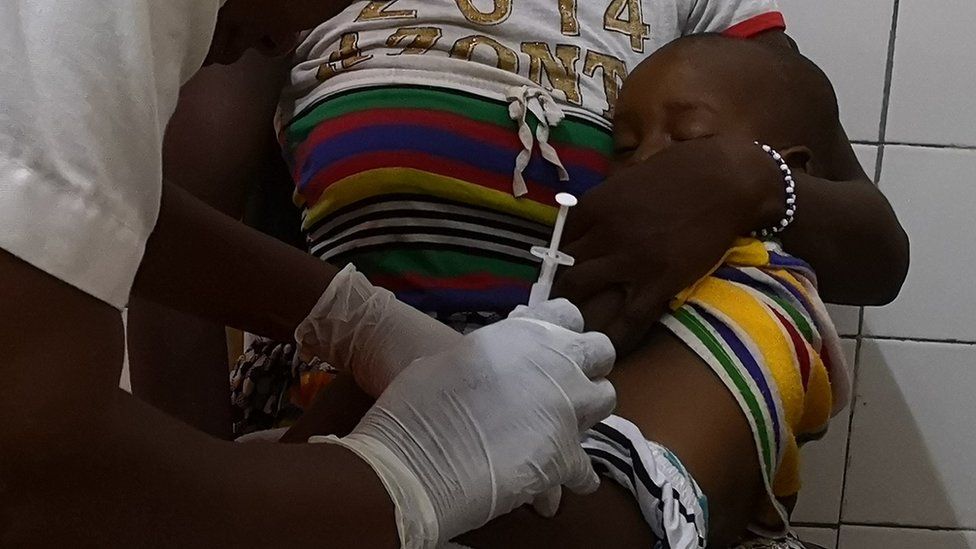
Published20 hours agoSharecloseShare pageCopy linkAbout sharingImage source, Katie EwerMalawi has begun vaccinating children as part of a world-first, large-scale campaign against malaria.The RTS,S vaccine – more than three decades in the making – was developed by pharmaceutical company GSK.However, early trials show just more than 30% of the five to 17-month-olds who received it were protected.But Malawi believes it will still play a key role in the fight against the disease, which killed some 2,500 infants in the country two years ago.”We’re quite aware of its low efficacy… [but] in malaria control there is no single intervention that does it all,” Dr Michael Kayange, the country’s national malaria control programme manager, told the BBC’s Focus on Africa.”We’re not saying that the malaria vaccine has come to eliminate malaria but it’s one tool towards malaria elimination.”Malaria has been one of the biggest scourges on humanity for millennia and mostly kills babies and infants.But the disease, caused by parasites transmitted through the bites of infected female Anopheles mosquitoes, disproportionately affects Africa: in 2020, the continent recorded 95% of malaria cases and 96% of malaria deaths. Children under five accounted for about 80% of all malaria deaths in Africa, according to the World Health Organization.Dr Kayange said the new immunisation campaign will ensure that all children under five, even in the remotest parts of the country, will be covered.The first part will see 11 of the country’s 28 districts will be covered, with 330,000 children expected to be vaccinated in this phase alone.How does it work?The vaccine, which has been through rounds of testing in Malawi, Ghana and Kenya over the past few years, needs to be given four times – once a month for three months and then a fourth dose 18 months later.RTS,S shares similarities with another malaria vaccine developed by University of Oxford. Both target the first stage of the malaria-causing parasite’s lifecycle by intercepting it before it gets to the liver and establishes a foothold in the body.The vaccines are built using a combination of proteins from the malaria parasite and the hepatitis B virus, but Oxford’s version has a higher proportion of malaria proteins. The team think this helps the immune system to focus on malaria rather than the hepatitis. Trial results of the Oxford vaccine from 409 children in Nanoro, Burkina Faso, have been published in the Lancet Infectious Diseases. It shows three initial doses followed by a booster a year later gives up to 80% protection. More on this storyHistoric go-ahead for malaria vaccine in Africa6 October 2021

Read more →

Immensa lab errors may have led to 23 Covid-19 deaths

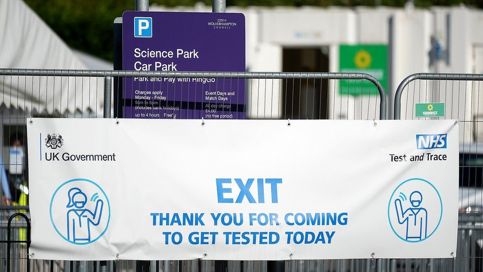
Published33 minutes agoSharecloseShare pageCopy linkAbout sharingImage source, Getty ImagesBy Matthew HillBBC West health correspondentStaff mistakes in a private laboratory may have caused 23 extra deaths from Covid-19.The UK Health Security Agency (UKHSA) makes the claim in a report into errors at the Immensa lab in Wolverhampton.It says as many as 39,000 positive results were wrongly reported as negative in September and October 2021.The mistakes led to “increased numbers of [hospital] admissions and deaths”, the report, published on Tuesday, concluded.Thousands of people, many in the South West, were wrongly told to stop testing after their results were processed by Immensa.The Wolverhampton laboratory was used for additional testing capacity for NHS Test and Trace from early September 2021, but testing was suspended on 12 October following reports of inaccurate results.Immensa lab: What went wrong?Lab ‘not accredited’Experts said high case rates in some areas were down to people unwittingly infecting others when they should have been isolating.UKHSA experts said the mistakes could have led to as many as 55,000 additional infections in areas where the false negatives were reported.”Each incorrect negative test likely led to just over two additional infections,” the report said.”In those same geographical areas, our results also suggest an increased number of admissions and deaths.”Immensa was paid more than £100m to carry out Covid testing for the NHS during the pandemic.The UKHSA said a total of about 400,000 samples had been processed at the Wolverhampton lab.’Staff errors’ to blame”The cause [of the mistakes] was the incorrect setting of the threshold levels for reporting positive and negative results of PCR samples for COVID-19,” said the UKHSA.”Based on background infection rates in different population groups at the time, UKHSA estimated that this error could have led to around 39,000 results being incorrectly reported as negative when they should have been positive.”Richard Gleave, UKHSA director and lead investigator, said: “Through this investigation we have looked carefully at the arrangements in place for overseeing contracts of private labs providing surge testing during this time.”We have concluded that staff errors within Immensa’s Wolverhampton laboratory were the immediate cause of the incorrect reporting of COVID-19 PCR test results in September and October 2021.”It is our view that there was no single action that NHS Test and Trace could have taken differently to prevent this error arising in the private laboratory.”However, our report sets out clear recommendations to both reduce the risk of incidents like this happening again and ensure that concerns are addressed and investigated rapidly.”Jenny Harries, UKHSA chief executive, said: “I fully accept the findings and recommendations made in this report, many of which were implemented as soon as UKHSA discovered the incident.”These ongoing improvements will enhance our ability to spot problems sooner where they do arise.”Dante Labs, the owners of Immensa, have been contacted for comment.Follow BBC West on Facebook, Twitter and Instagram. Send your story ideas to: bristol@bbc.co.uk More on this storyReport into Covid-lab errors must be published- MP7 AprilMonth delay before incorrect Covid tests halted21 December 2021Legal action considered over Covid test lab errors14 December 2021Testing lab ‘chaotic and dangerous’, scientist claims16 October 2020Related Internet LinksUK Health Security AgencyThe BBC is not responsible for the content of external sites.

Read more →