What is known about new Covid variant XBB.1.5?
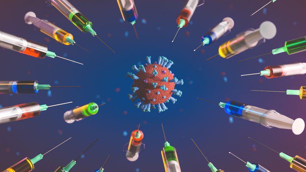
Published14 hours agoShareclose panelShare pageCopy linkAbout sharingImage source, Getty ImagesBy Fergus WalshMedical editor A new Covid sub-variant is causing some concern in the US, where it is spreading rapidly. Some cases have been recorded in the UK, so what do you need to know about XBB.1.5?What is XBB.1.5?It is yet another offshoot of the globally dominant Omicron Covid variant, which itself followed the earlier alpha, beta, gamma and delta variants. Omicron has outperformed all previous versions of coronavirus since it emerged in late 2021, and has given rise to many sub-variants which are even more contagious than the original. Symptoms of XBB.1.5 are thought to be similar to those of previous Omicron strains, but it’s still too early to confirm this. Most people experience cold-like symptoms. Is XBB.1.5 more infectious or dangerous than earlier variants?XBB.1.5 itself evolved from XBB, which began circulating in the UK in September 2022, but which has not been classified as a so-called “variant of concern” by health authorities. XBB had a mutation that helped it beat the body’s immune defences, but this same quality also reduced its ability to infect human cells. Prof Wendy Barclay from Imperial College London said XBB.1.5 had a mutation known as F486P, which restores this ability to bind to cells while continuing to evade immunity. That makes it spread more easily. She said these evolutionary changes were like “stepping stones”, as the virus evolves to find new ways of bypassing the body’s self-defence mechanisms.Scientists from the World Health Organization (WHO) confirmed on Wednesday that XBB.1.5 has a “growth advantage” above all other sub-variants seen so far.But they said there was no indication it was more serious or harmful than previous Omicron variants. The WHO said it would keep a close watch on lab studies, hospital data and infection rates to find out more about its impact on patients.Who can get a Covid booster this winter? Where is XBB.1.5 spreading?Over 40% of Covid cases in the United States are estimated to be caused by XBB.1.5, making it the dominant strain in the country.At the beginning of December, it accounted for only 4% of cases so it has quickly overtaken other versions of Omicron. Covid hospital admissions have been rising in recent weeks across the US. US brings back free at-home Covid testsThe UK Health Security Agency is due to release a report on variants spreading in the UK next week, and may refer to XBB.1.5. Could the XBB.1.5 variant take off in the UK?Nothing is certain, but it does look likely. The UK had five Omicron waves in 2022, and further spikes in cases are inevitable. Figures for the week to Saturday 17 December from the Sanger Institute in Cambridge suggested that one in 25 Covid cases in the UK were XBB.1.5. But that was based on just nine samples, so we’ll need to wait for a week or two to get a better picture of how it is spreading. Prof Barclay said she expected more hospitalisations in the UK if the variant takes off here, “as we expect it to do”. Prof Paul Hunter from the University of East Anglia, said: “The balance of probabilities is that XBB.1.5 will trigger a wave here later this month, but we can’t be sure.” NHS England has said the fears of a “twindemic” of Covid and flu have been realised, with both viruses putting strain on an already stretched NHS. Covid putting massive pressure on NHS – BarclayImage source, Getty ImagesAre scientists worried about XBB.1.5?Prof Barclay said she was not especially concerned about the general UK population because there was “no indication” that XBB.1.5 would “breakthrough” the protection against severe illness provided by vaccines. But she is worried about its effect on the vulnerable, including the immunocompromised, who get less benefit from Covid jabs. Prof Hunter said he’d seen no evidence that XBB.1.5 was more virulent, meaning it was no more likely to “put you in hospital or kill you” than existing Omicron variants. He added: “It’s ironic that everyone is focussing on possible variants emerging from China, but XBB.1.5 came out of the US.” Prof David Heymann from the London School of Hygiene and Tropical Medicine acknowledged that there was still a fair amount to learn about this latest variant. But he said it was unlikely to cause major problems in countries like the UK which have high levels of vaccination and previous infections. His concern was for countries like China, where there was both low take-up of vaccines and little natural immunity because of prolonged lockdowns. “China needs to share clinical information on people infected in order to see how the variant behaves in a non-immune population,” Prof Heymann said. How is China trying to beat its latest Covid surge?Related Internet LinksTracking SARS-CoV-2 variantsLineages (raw) – COVID-19 Genomic Surveillance – Wellcome Sanger InstituteThe BBC is not responsible for the content of external sites.

Read more →
Does COVID change the body's response to other threats? Depends on your sex, study finds

The long-term effects of infection on the immune system have long intrigued John Tsang, a Yale immunobiologist. After the body has faced down a pathogen, does the immune system return to the previous baseline? Or does a single infection change it in ways that alter how it will respond not only to a familiar virus but also to the next new viral or bacterial threat it faces?
Tsang, a professor of immunobiology and biomedical engineering at Yale, has long believed that the immune system reverts to the previous stable baseline after viral infection.
The emergence of the COVID-19 pandemic in 2020 allowed him and colleagues to test that theory. The answer, they found, depends on the individual’s sex, according to a study published Jan. 4 in the journal Nature.
For the study, a team led by Tsang, who at the time was at the National Institute of Allergy and Infectious Diseases (NIAID), and colleagues, including lead author Rachel Sparks, also from NIAID, systematically analyzed immune responses of healthy people who had received the flu vaccine. From that data, they then compared the responses between those who had never been infected by SARS-CoV-2, the virus that causes COVID-19, and those who experienced mild cases but recovered.
To their surprise, they found that immune systems of men who had recovered from mild cases of COVID-19 responded more robustly to flu vaccines than women who had had mild cases or men and women who had never been infected.
In essence, the baseline immune statuses in men previously infected with SARS-CoV-2 was altered in ways that changed the response to an exposure different from SARS-CoV-2, the authors said.

“This was a total surprise,” Tsang said. “Women usually mount a stronger overall immune response to pathogens and vaccines, but are also more likely to suffer from autoimmune diseases.”
The findings may also be linked to an observation made early in the pandemic: Men were much more likely to die from a runaway immune response than women after contracting the COVID-19 virus. Even mild cases of COVID-19, the new findings suggest, might trigger stronger inflammatory responses in males than females, resulting in more pronounced functional changes to the male immune system, even long after recovery.
Their unbiased analysis of immune system status down to the individual cell level revealed several differences between COVID-recovered males and healthy controls and COVID-recovered females, both before and after receiving flu vaccinations. For instance, previously infected males produced more antibodies to influenza and produced increased levels of interferons, which are produced by cells in response to infections or vaccines. Generally, healthy females have stronger interferon responses than their male counterparts.
Understanding the lingering effects of COVID-19 on the immune system is crucial, the authors say, since more than 600 million people worldwide have been infected so far, and the emergence of “long-COVID” symptoms in some people continues to be a major health concern.
“Our findings point to the possibility that any infection or immune challenge may change the immune status to establish new set points,” said Sparks. “The immune status of an individual is likely shaped by a multitude of prior exposures and perturbations.”
Tsang thinks these findings may also help scientists create better vaccines against diverse threats by, for instance, mimicking how mild COVID-19 changes the male immune baseline.
Other researchers included William Lau, a computational biologist at the National Institutes of Health, and Can Liu, a systems immunology graduate student at the University of Maryland who is also affiliated with NIAID.
Story Source:
Materials provided by Yale University. Original written by Bill Hathaway. Note: Content may be edited for style and length.

Read more →

Simple nasal swab can provide early warning of emerging viruses

As the COVID-19 pandemic showed, potentially dangerous new viruses can begin to spread in the population well before the global public health surveillance system can detect them.
However, Yale researchers have found that testing for the presence of a single immune system molecule on nasal swabs can help detect stealthy viruses not identified in standard tests, they report Jan. 3 in the journal Lancet Microbe.
“Finding a dangerous new virus is like searching for a needle in a haystack,” said Ellen Foxman, associate professor of laboratory medicine and immunobiology and senior author of the study. “We found a way to significantly reduce the size of the haystack.”
Public health officials typically look to a few sources for warning signs of emerging disease. They study emerging viruses in animals that may transmit the infection to humans. But determining which of the hundreds, or thousands, of new viral variants represent a true danger is difficult. And they look for outbreaks of unexplained respiratory ailments, which was how SARS-Cov-2, the virus that causes COVID-19, was discovered in China late in 2019.
By the time an outbreak of a novel virus occurs, however, it may be too late to contain its spread.
For the new study, Foxman and her team revisited an observation made in her lab in 2017, which they thought may provide a new way to monitor for unexpected pathogens. Nasal swabs are commonly taken from patients with suspected respiratory infections and are tested to detect specific signatures of 10 to 15 known viruses. Most tests come back negative. But as Foxman’s team observed in 2017, in a few cases the swabs of those who tested negative for the “usual suspect” viruses still exhibited signs that antiviral defenses were activated, indicating the presence of a virus. The telltale sign was a high level of a single antiviral protein made by the cells that line the nasal passages.
Based on that finding, the researchers applied comprehensive genetic sequencing methods to old samples containing the protein and, in one sample, found an unexpected influenza virus, called influenza C.
The researchers also used this same strategy of retesting old samples to search for missed cases of COVID-19 during the first two weeks of March 2020. While cases of the virus had surfaced in New York State around that same time, testing was not readily available until weeks later. Hundreds of nasal swab samples collected from patients at Yale-New Haven Hospital during that time had tested negative for standard signature viruses. When tested for the immune system biomarker, the vast majority of those samples showed no trace of activity of the antiviral defense system. But a few did; among those, the team found four cases of COVID-19 that had gone undiagnosed at the time.
The findings reveal that testing for an antiviral protein made by the body, even if the tests for known respiratory viruses are negative, can help pinpoint which nasal swabs are more likely to contain unexpected viruses.
Specifically, screening for the biomarker can allow researchers to narrow down the search for unexpected pathogens, making it feasible to do surveillance for unexpected viruses using swabs collected during routine patient care. Samples found to possess the biomarker can be analyzed using more complex genetic testing methods to identify unexpected or emerging pathogens circulating in the patient population and jumpstart a response from the health care community.
Yale’s Nagarjuna R. Cheemarla and Jason Bishai are co-lead authors of the paper, as are former Yale researchers Amelia Hanron and Joseph R.Fauver.
Story Source:
Materials provided by Yale University. Original written by Bill Hathaway. Note: Content may be edited for style and length.

Read more →

Gut bacteria may play a role in diabetes

One type of bacteria found in the gut may contribute to the development of Type 2 diabetes, while another may protect from the disease, according to early results from an ongoing, prospective study led by investigators at Cedars-Sinai.
The study, published in the peer-reviewed journal Diabetes, found people with higher levels of a bacterium called Coprococcus tended to have higher insulin sensitivity, while those whose microbiomes had higher levels of the bacterium Flavonifractor tended to have lower insulin sensitivity.
For years, investigators have sought to understand why people develop diabetes by studying the composition of the microbiome, which is a collection of microorganisms that include fungi, bacteria and viruses that live in the digestive tract. The microbiome is thought to be affected by medications and diet. Studies have also found that people who don’t process insulin properly have lower levels of a certain type of bacteria that produce a type of fatty acid called butyrate.
Mark Goodarzi, MD, PhD, the director of the Endocrine Genetics Laboratory at Cedars-Sinai, is leading an ongoing study that is following and observing people at risk for diabetes to learn whether those with lower levels of these bacteria develop the disease.
“The big question we’re hoping to address is: Did the microbiome differences cause the diabetes, or did the diabetes cause the microbiome differences?” said Goodarzi, who is the senior author of the study and principal investigator of the multicenter study called Microbiome and Insulin Longitudinal Evaluation Study (MILES).
Investigators involved in MILES have been collecting information from participating Black and non-Hispanic white adults between 40 and 80 years of age since 2018. An earlier cohort study from the MILES trial found that birth by cesarean section is associated with a higher risk for developing prediabetes and diabetes.

For the most recent study to come out of this ongoing trial, investigators analyzed data from 352 people without known diabetes who were recruited from the Wake Forest Baptist Health System in Winston-Salem, North Carolina.
Study participants were asked to attend three clinic visits and collect stool samples prior to the visits. Investigators analyzed data collected at the first visit. They conducted genetic sequencing on the stool samples, for example, to study the participants’ microbiomes, and specifically look for bacteria that earlier studies have found to be associated with insulin resistance. Each participant also filled out a diet questionnaire and took an oral glucose tolerance test, which was used to determine ability to process glucose.
Investigators found 28 people had oral glucose tolerance results that met the criteria for diabetes. They also found that 135 people had prediabetes, a condition in which a person’s blood-sugar levels are higher than normal but not high enough to meet the definition of diabetes.
The research team analyzed associations between 36 butyrate-producing bacteria found in the stool samples and a person’s ability to maintain normal levels of insulin. They controlled for factors that could also contribute to a person’s diabetes risk, such as age, sex, body mass index and race. Coprococcus and related bacteria formed a network of bacteria with beneficial effects on insulin sensitivity. Despite being a producer of butyrate, Flavonifractor was associated with insulin resistance; prior work by others have found higher levels of Flavonifractor in the stool of people with diabetes.
Investigators are continuing to study samples from patients who participated in this study to learn how insulin production and the composition of the microbiome change over time. They also plan to study how diet may affect the bacterial balance of the microbiome.
Goodarzi emphasized, however, that it is too early to know how people can change their microbiome to reduce their diabetes risk.
“As far as the idea of taking probiotics, that would really be somewhat experimental,” said Goodarzi, who is also the Eris M. Field Chair in Diabetes Research at Cedars-Sinai. “We need more research to identify the specific bacteria that we need to be modulating to prevent or treat diabetes, but it’s coming, probably in the next five to 10 years.”
Jinrui Cui, a biostatistician in the Goodarzi Laboratory at Cedars-Sinai, was the first author of the study.

Read more →

The interior design of our cells: Database of 200,000 cell images yields new mathematical framework to understand our cellular building blocks

Working with hundreds of thousands of high-resolution images, the team at the Allen Institute for Cell Science, a division of the Allen Institute, put numbers on the internal organization of human cells — a biological concept that has to date proven exceptionally difficult to quantify.
Through that work, the scientists also captured details about the rich variation in cell shape even among genetically identical cells grown under identical conditions. The team described their work in a paper published in the journal Nature today.
“The way cells are organized tells us something about their behavior and identity,” said Susanne Rafelski, Ph.D., Deputy Director of the Allen Institute for Cell Science, who led the study along with Senior Scientist Matheus Viana, Ph.D. “What’s been missing from the field, as we all try to understand how cells change in health and disease, is a rigorous way to deal with this kind of organization. We haven’t yet tapped into that information.”
This study provides a roadmap for biologists to understand organization of different kinds of cells in a measurable, quantitative way, Rafelski said. It also reveals some key organizational principles of the cells the Allen Institute team studies, which are known as human induced pluripotent stem cells.
Understanding how cells organize themselves under healthy conditions — and the full range of variability contained within “normal” — can help scientists better understand what goes wrong in disease. The image dataset, genetically engineered stem cells, and code that went into this study are all publicly available for other scientists in the community to use.
“Part of what makes cell biology seem intractable is the fact that every cell looks different, even when they are the same type of cell. This study from the Allen Institute shows that this same variability that has long plagued the field is, in fact, an opportunity to study the rules by which a cell is put together,” said Wallace Marshall, Ph.D., Professor of Biochemistry and Biophysics at the University of California, San Francisco, and a member of the Allen Institute for Cell Science’s Scientific Advisory Board. “This approach is generalizable to virtually any cell, and I expect that many others will adopt the same methodology.”
Computing the pear-ness of our cells

In a body of work launched more than seven years ago, the Allen Institute team first built a collection of stem cells genetically engineered to light up different internal structures under a fluorescent microscope. With cell lines in hand that label 25 individual structures, the scientists then captured high-resolution, 3D images of more than 200,000 different cells.
All this to ask one seemingly straightforward question: How do our cells organize their interiors?
Getting to the answer, it turned out, is really complex. Imagine setting up your office with hundreds of different pieces of furniture, all of which need to be readily accessed, and many of which need to move freely or interact depending on their task. Now imagine your office is a sac of liquid surrounded by a thin membrane, and many of those hundreds of pieces of furniture are even smaller bags of liquid. Talk about an interior design nightmare.
The scientists wanted to know: How do all those tiny cellular structures arrange themselves compared to each other? Is “structure A” always in the same place, or is it random?
The team ran into a challenge comparing the same structure between two different cells. Even though the cells under study were genetically identical and reared in the same laboratory environment, their shapes varied substantially. The scientists realized that it would be impossible to compare the position of structure A in two different cells if one cell was short and blobby and the other was long and pear-shaped. So they put numbers on those stubby blobs and elongated pears.

Using computational analyses, the team developed what they call a “shape space” that objectively describes each stem cell’s external shape. That shape space includes eight different dimensions of shape variation, things like height, volume, elongation, and the aptly described “pear-ness” and “bean-ness.” The scientists could then compare apples to apples (or beans to beans), looking at organization of cellular structures inside all similarly shaped cells.
“We know that in biology, shape and function are interrelated, and understanding cell shape is important to understand how the cells function,” Viana said. “We’ve come up with a framework that allows us to measure a cell’s shape, and the moment you do that you can find cells that are similar shapes, and for those cells you can then look inside and see how everything is arranged.”
Strict organization
When they looked at the position of the 25 highlighted structures, comparing those structures in groups of cells with similar shapes, they found that all the cells set up shop in remarkably similar ways. Despite the massive variations in cell shape, their internal organization was strikingly consistent.
If you’re looking at how thousands of white-collar workers arrange their furniture in a high-rise office building, it’s as if every worker put their desk smack in the middle of their office and their filing cabinet precisely in the far-left corner, no matter the size or shape of the office. Now say you found one office with a filing cabinet thrown on the floor and papers strewn everywhere — that might tell you something about the state of that particular office and its occupant.
The same goes for cells. Finding deviations from the normal state of affairs could give scientists important information about how cells change when they transition from stationary to mobile, are getting ready to divide, or about what goes wrong at the microscopic level in disease. The researchers looked at two variations in their dataset — cells at the edges of colonies of cells, and cells that were undergoing division to create new daughter cells, a process known as mitosis. In these two states, the scientists were able to find changes in internal organization correlating to the cells’ different environments or activities.
“This study brings together everything we’ve been doing at the Allen Institute for Cell Science since the institute was launched,” said Ru Gunawardane, Ph.D., Executive Director of the Allen Institute for Cell Science. “We built all of this from scratch, including the metrics to measure and compare different aspects of how cells are organized. What I’m truly excited about is how we and others in the community can now build on this and ask questions about cell biology that we could never ask before.”

Read more →

How a CRISPR protein might yield new tests for many viruses

In a first for the genetic toolset known as CRISPR, a recently discovered protein has been found to act as a kind of multipurpose self-destruct system for bacteria, capable of degrading single-stranded RNA, single-stranded DNA and double-stranded DNA. With its abilities to target so many types of genetic material, the discovery holds potential for the development of new inexpensive and highly sensitive at-home diagnostic tests for a wide range of infectious diseases, including COVID-19, influenza, Ebola and Zika, according to the authors of a new study in the journal Nature.
Using a high-resolution imaging technique called cryo-EM, the team discovered that when this protein, named Cas12a2, binds to a specific sequence of genetic material from a potentially dangerous virus, called a target RNA, a side portion of Cas12a2 swings out to reveal an active site, similar to a sprung-open switchblade knife. Then, the active site starts to indiscriminately cut any genetic material it comes into contact with. The researchers discovered that, with a single mutation to the Cas12a2 protein, the active site degrades only single-stranded DNA — a feature especially useful in developing new diagnostics tailored for any of a wide range of viruses.
A test based on this technology could theoretically combine the best features of PCR-based tests that detect genetic material from a virus (high sensitivity, high accuracy and the ability to detect an active infection) with the best features of rapid at-home diagnostic tests (inexpensive to produce without requiring specialized lab equipment). It also would be easily adaptable to any new RNA virus.
“If some new virus comes out tomorrow, all you have to do is figure out its genome and then change the guide RNA in your test, and you’d have a test against it,” said David Taylor, an associate professor of molecular biosciences at The University of Texas at Austin and co-corresponding author of the new study.
Such a diagnostic would still require separate work and probably involve collecting saliva or a nasal sample from a patient to be mixed with the team’s modified Cas12a2 protein, the piece of guide RNA that acts like a mugshot to identify a specific virus, and a fluorescent probe designed to light up when its single-stranded DNA gets cut.
CRISPR is the name for a set of tools that occur naturally in bacteria, but which scientists have adapted for use in gene editing. This is the first CRISPR protein that has been found to degrade such a wide range of genetic material.
“Cas12a2 basically grabs the two ends of the DNA double helix and bends it really tightly,” said Jack Bravo, a postdoctoral fellow at UT Austin and co-first author on the paper. “And so, the helix in the middle pops open, and then this allows this active site to destroy the bits of DNA that become single-stranded. This is what makes Cas12a2 different from all the other DNA-targeting systems.”
The paper’s co-corresponding author is Ryan Jackson and co-first author Thomson Hallmark, both of Utah State University. The other co-authors are Bronson Naegle of Utah State and Chase Beisel of the Helmholtz Centre for Infection Research and the University of Würzburg in Germany.
Structural data were collected using the cryo-EM facilities at the Sauer Structural Biology Laboratory at The University of Texas at Austin.
Taylor, Bravo, Hallmark and Jackson are inventors on a patent application covering modifications to the Cas12a2 protein that enable it to cut only single-stranded DNA and for its use in diagnostics. The UT Austin Office of Technology Commercialization is managing the intellectual property and working to find industry partners that can help realize the potential of the technology.
This work was supported in part by the National Institute of General Medical Sciences of the National Institutes of Health, the German Federal Agency for Disruptive Innovation, The Welch Foundation, and the Robert J. Kleberg, Jr. and Helen C. Kleberg Foundation. David Taylor is a CPRIT scholar supported by the Cancer Prevention and Research Institute of Texas.
A companion paper in the same issue of Nature describes the biological functions of Cas12a2, while the paper described in this news release describes the mechanisms by which the protein accomplishes them.
Story Source:
Materials provided by University of Texas at Austin. Note: Content may be edited for style and length.

Read more →

The adverse health effects of disaster-related trauma

Major weather events such as last month’s 6.4 magnitude California earthquake, tornados in Louisiana, and a “once-in-a-generation” multi-state winter storm caused major damage to homes across the US and disrupted daily lives. As climate experts predict these natural disasters will continue to increase in severity and frequency, a new study led by a School of Public Health researcher is shedding new insight on the adverse health impacts that certain vulnerable populations experience following the loss of a home due to severe weather damage.
Published in the American Journal of Epidemiology, the study found that individuals from disadvantaged backgrounds disproportionately experience disaster-related home loss — and that they are more likely to be severely affected by home loss, namely by developing physical and mental functional limitations in the years after they lose their home.
The first-of-its-kind study builds upon previous research that has focused only on population-average data of home loss and adverse health effects; the new study identifies subgroups of vulnerable populations who are more likely to be severely affected by this traumatic experience. These include people who are older, not married, living alone, less educated, and unemployed, as well as those who had health problems prior to the losing their home.
The researchers say that identifying populations at exceptionally high risk of post-disaster functional impairment may better inform resource allocation during disaster mitigation, preparedness, response, and recovery efforts at the local and federal level.
“Our study moved beyond the traditional finding on population-average effects and identified complex effect heterogeneity,” says study lead and corresponding author Dr. Koichiro Shiba, assistant professor of epidemiology at Boston University School of Public Health. “These results help policymakers by providing insights on the impacts that disaster damages may have on health disparities, which analysis of population-average effects ignores. The results can also be used to identify which subpopulations need to be prioritized in post-disaster public health supports.”
For the study, Dr. Shiba and colleagues used machine learning methods to identify variations in the association between disaster-related trauma and functional limitations among a group of older survivors of the 2011 Great East Japan Earthquake and Tsunami. The 9.1-magnitude earthquake and 40-meter tsunami killed nearly 16,000 people and rendered more than 450,000 people homeless. The team used pre- and post-disaster survey data from the Iwanuma Study, part of a larger nationwide study of the living conditions of Japanese older adults, to measure individuals’ functional limitations in 2013 and 2016 based on three indicators: standardized assessments of physical disability (such as whether someone can turn over in bed independently); the ability to accomplish daily activities independently (such as walking, bathing, going to the bathroom); and higher-level functional capacities (such as using public transportation).
The researchers found that individuals experiencing more severe functional limitations after home loss tended to have less education and more pre-disaster health problems, such as depression. But notably, they also found that these individuals had higher income prior to losing their home.
One speculation for this finding could be explained by status inconsistency — “where different aspects of social status contradict with each other and potentially magnify the adverse impacts of disaster-related home loss,” Shiba explains. Previous studies have shown that higher-income people are also more likely to engage in excessive drinking, which could lead to limited functioning. Another explanation, he says, is that “instead of moving to a temporary housing village with other survivors, richer people might have been able to afford and relocate to private housing after home loss, which could result in loss of pre-existing social capital and support.”
Understanding these underlying mechanisms resulting from home loss and other traumatic experiences will help inform more targeted public health interventions following the inevitable occurrence of future severe weather events, he says.
Story Source:
Materials provided by Boston University School of Public Health. Original written by Jillian McKoy. Note: Content may be edited for style and length.

Read more →

'Hospital staff are completely overwhelmed'

The Northern Ireland Ambulance Service is investigating whether a delayed response contributed to the deaths of eight people in recent weeks.All eight deaths occurred between the 12 December and the start of January.At this stage four of the deaths are being treated as serious adverse incidents.The remaining cases are being investigated to determine whether they meet the criteria to be treated as an serious adverse incidents.It comes as an emergency department nurse told BBC News NI he would be worried if any of his loved ones had to seek treatment as staff continued to be overwhelmed.Stephen McKenna, who spent the Christmas period working on the frontline, described conditions as “absolutely horrific”.Read more here.

Read more →

Clinical trial leads to atezolizumab approval for advanced alveolar soft part sarcoma

A clinical trial led by the National Cancer Institute (NCI), part of the National Institutes of Health, has resulted in the first approval of a treatment for advanced alveolar soft part sarcoma (ASPS). The immunotherapy drug atezolizumab (Tecentriq) was recently approved by the U.S. Food and Drug Administration (FDA) for the treatment of adults and children 2 years and older with ASPS that has spread to other parts of the body or cannot be removed by surgery.
ASPS is an extremely rare cancer that affects mostly adolescents and young adults. The approval was based on data from a non-randomized phase 2 trial (NCT03141684) funded by NCI and led by Dr. Alice Chen, M.D., of the Developmental Therapeutics Clinic in NCI’s Division of Cancer Treatment and Diagnosis (DCTD). Genentech, a member of the Roche Group and the manufacturer of atezolizumab, provided the drug to NCI through a cooperative research and development agreement. The results of the study are being prepared for publication.
“Forty percent of the patients were treated at the NIH Clinical Center in Bethesda,” said James H. Doroshow, M.D., director of DCTD. “Our ability to bring patients in from all over the world was a key factor in the ability to do the study.”
“This approval will make a huge impact in terms of a rare disease that has been particularly challenging to treat,” Dr. Chen noted.
This is the largest study on ASPS. It is also the first study conducted in the NCI-funded Experimental Therapeutics Clinical Trials Network that has resulted in a drug approval. The network enabled sarcoma specialists at academic medical centers from across North America to enroll patients in the trial.
“This is a major milestone for investigators in the Experimental Therapeutics Clinical Trials Network, as well as for the ASPS patient community, and for research on rare cancers,” said Elad Sharon, M.D., of DCTD, who is one of the study leaders.

This is also the first time atezolizumab has been approved for children. Dr. Chen noted that this was enabled by the participation of the Pediatric Oncology Branch in NCI’s Center for Cancer Research, which helped enroll children in the trial.
“This study is an important example of collaboration between pediatric and medical oncology, allowing children with very rare cancers access to effective new therapies,” said John W. Glod, M.D., Ph.D., of the Pediatric Oncology Branch. “The entire study team is grateful to the patients who participated in the study and made this work possible.”
About 80 people in the United States are diagnosed with ASPS every year. The disease typically begins in the soft tissue that connects and surrounds the organs and other tissues. Although the disease grows slowly, once it spreads it is often deadly, and chemotherapy is ineffective. About 50% of patients with metastatic disease are still alive after five years. New targeted treatments, including drugs called tyrosine kinase inhibitors, do not have lasting effectiveness. Recently, however, immunotherapy drugs have shown promise as possible therapies for ASPS.
Atezolizumab is an anti-PD-L1 immune checkpoint inhibitor that works by helping the immune system respond more strongly to cancer. FDA has approved atezolizumab for the treatment of patients with several cancer types, including liver cancer, melanoma, and lung cancer.
In 2020, FDA granted breakthrough therapy designation for atezolizumab to treat patients with unresectable or metastatic ASPS. This designation means that atezolizumab, which is intended to treat a serious condition, had met FDA’s criteria for expedited development and review of the drug. Later that year, FDA granted orphan drug designation to atezolizumab for soft tissue sarcoma in general. This status provides incentives for companies to develop a drug for rare diseases.

The phase 2 trial enrolled 49 ethnically diverse patients ages 2 and older with metastatic ASPS, who were given an infusion of atezolizumab every 21 days. About a third of the patients responded to the treatment with some degree of tumor shrinkage, according to their doctor’s assessment. Most of the other patients experienced stable disease.
After two years of treatment, patients were given the opportunity to stop treatment and go on a treatment break for up to two years with close monitoring. None of the patients who took a treatment break had disease progression during that time.
Serious side effects occurred in 41% of patients receiving atezolizumab; these included anemia, diarrhea, rash, dizziness, hyperglycemia, and pain in the extremities. However, no patients came off the study because of side effects.
“This approval represents a victory for rare diseases, which are understudied in clinical trials,” said Dr. Chen. “For this approval to go through in a rare disease, and to be able to make an impact on these young people’s lives, is very significant.”
Research teams are now conducting additional trials with atezolizumab for patients with ASPS, including giving the drug in combination with other therapies.

Read more →

Common fatty acid contributes to temperature and pain sensitivity in psoriasis plaques

A common fatty acid found in the Western diet breaks down into compounds that contribute to increased temperature and pain — but not itch — sensitivity in psoriatic lesions. The finding could lead to better understanding of how lipids communicate with sensory neurons, and potentially to improved pain and sensitivity treatments for psoriasis patients.
Linoleic acid is a fatty acid found in vegetable oils, nuts and seeds, and is one of the predominant fatty acids found in the Western diet. Metabolites from linoleic acid — the products formed when the body breaks it down through digestion — play a role in skin barrier function.
“We noticed high levels of two types of lipids derived from linoleic acid in psoriatic lesions,” says Santosh Mishra, associate professor of neuroscience at North Carolina State University and corresponding author of the research. “That led us to wonder whether the lipids might affect how sensory neurons in these lesions communicate. We decided to investigate whether their presence could be related to the temperature or pain hypersensitivity that many psoriasis patients report.”
The research team used mass spectrometry to create lipid profiles of skin from psoriatic lesions. They focused on two types of linoleic acid-derived lipids, or oxylipids: 13-hydroxy-9,10-epoxy octadecenoate (9,13-EHL) and 9,10,13-trihydroxy-octadecenoate (9,10,13-THL). The first form, 9,13-EHL, can convert into the more stable 9,10,13-THL form via interaction with certain enzymes.
The researchers found that while both forms bind to receptors on sensory neurons within the skin, the more stable form — 9,10,13-THL — had a longer lasting effect than 9,13-EHL.
They also found that once the lipids bind to the neuronal receptor, they activate the neurons expressing TRPA1 and TRPV1 receptors that are involved in temperature and pain hypersensitivity, opening communications channels to the central nervous system.
Interestingly, the lipids did not have any effect on itch.
“It was surprising that these lipids could create hypersensitivity but not impact itch sensation, which is usually the most troublesome symptom associated with psoriasis,” Mishra says. “This most likely has to do with how the neuron is activated — a mechanism we still haven’t uncovered.”
Now that an association between linoleic acid and hypersensitivity to temperature and pain has been established, the researchers want to further explore exactly how this response is being created. They hope that the answers may lead to solutions that can relieve these symptoms in psoriasis patients.
“We know that this lipid moves from one form to another, but don’t yet know what causes that,” Mishra says. “We also know what protein the lipids are binding to, but not where the bond occurs. Answering these questions may hopefully lead to new therapies — or dietary solutions — for some psoriasis sufferers.”
The work appears in JID Innovations, and is supported by the National Institute on Aging and the National Institutes of Health. Josh Wheeler, a postdoctoral researcher at NC State, is first author.
Story Source:
Materials provided by North Carolina State University. Original written by Tracey Peake. Note: Content may be edited for style and length.

Read more →