Scientists make progress in decoding genetics of insomnia

A National Institutes of Health-funded effort involving researchers from Texas A&M University, the Perelman School of Medicine at the University of Pennsylvania and Children’s Hospital of Philadelphia (CHOP) has used human genomics to identify a new genetic pathway involved in regulating sleep from fruit flies to humans — a novel insight that could pave the way for new treatments for insomnia and other sleep-related disorders.
Texas A&M geneticist and evolutionary biologist Alex Keene collaborated with Penn’s Allan Pack and Philip Gehrman and CHOP’s Struan Grant on the groundbreaking research, which is published in Science Advances.
“There have been enormous amounts of effort to use human genomic studies to find sleep genes,” Keene said. “Some studies have hundreds of thousands of individuals. But validation and testing in animal models is critical to understanding function. We have achieved this here, largely because we each bring a different area of expertise that allowed for this collaboration’s ultimate effectiveness.”
Keene says the most exciting thing about the team’s work is that they developed a pipeline starting not with a model organism, but with actual human genomics data.
“There is an abundance of human genome-wide association studies (GWAS) that identify genetic variants associated with sleep in humans,” Keene said. “However, validating them has been an enormous challenge. Our team used a genomics approach called variant-to-gene mapping to predict the genes impacted by each genetic variant. Then we screened the effect of these genes in fruit flies.
“Our studies found that mutations in the gene Pig-Q, which is required for the biosynthesis of a modifier of protein function, increased sleep. We then tested this in a vertebrate model, zebrafish, and found a similar effect. Therefore, in humans, flies and zebrafish, Pig-Q is associated with sleep regulation.”
Keene says the team’s next step is to study the role of a common protein modification, GPI-anchor biosynthesis, on sleep regulation. In addition, he notes that the human-to-fruit flies-to-zebrafish pipeline the team developed will allow them to functionally assess not only sleep genes but also other traits commonly studied using human GWAS, including neurodegeneration, aging and memory.
“Understanding how genes regulate sleep and the role of this pathway in sleep regulation can help unlock future findings on sleep and sleep disorders, such as insomnia,” said Gehrman, an associate professor of clinical psychology in psychiatry at Penn and a clinical psychologist with the Penn Chronobiology and Sleep Institute. “Moving forward, we will continue to use and study this system to identify more genes regulating sleep, which could point in the direction of new treatments for sleep disorders.”
Keene’s research within his Center for Biological Clocks Research-affiliated laboratory lies at the intersection of evolution and neuroscience, with primary focus on understanding the neural mechanisms and evolutionary underpinnings of sleep, memory formation and other behavioral functions in fly and fish models. Specifically, he studies fruit flies (Drosophila melanogaster) and Mexican cavefish that have lost both their eyesight and ability to sleep with the goal of identifying the genetic basis of behavioral choices which factor into human disease, including obesity, diabetes and heart disease.
Story Source:
Materials provided by Texas A&M University. Original written by Shana K. Hutchins. Note: Content may be edited for style and length.
Read more →

Blood-based markers may reveal Alzheimer disease ten years before symptoms show

Alzheimer disease begins decades before any symptoms, such as memory loss, start to show. Consequently, early diagnosis increases the chances of slowing the disease down with drugs. A new study on an inherited form of the disease shows that a protein called GFAP is a possible biomarker for very early stages of the disease. The study, conducted by researchers at Karolinska Institutet and published in the journal Brain, could one day lead to an earlier detection of this serious and common disease.
“Our results suggest that GFAP, a presumed biomarker for activated immune cells in the brain, reflects changes in the brain due to Alzheimer disease that occur before the accumulation of tau protein and measurable neuronal damage,” says the study’s first author Charlotte Johansson, doctoral student at the Department of Neurobiology, Care Sciences and Society, Karolinska Institutet, Sweden. “In the future it could be used as a non-invasive biomarker for the early activation of immune cells such as astrocytes in the central nervous system, which can be valuable to the development of new drugs and to the diagnostics of cognitive diseases.”
Alzheimer disease causes 60 to 70 percent of all dementia cases, according to the Swedish Brain Foundation. In Alzheimer disease, nerve cells in the brain degenerate as a result of the abnormal accumulation of the proteins beta-amyloid and tau. As more brain neurons become damaged, this manifests in dysfunction of cognitive functions such as memory and speech.
The disease progresses insidiously and biological changes in the brain begin already 20 to 25 years before memory loss and other cognitive symptoms become evident. The earlier a patient is correctly diagnosed, the sooner he or she can be offered the right treatment. This is one of many reasons why more research is needed on precise, easy-to-use methods of early diagnosis.
Researchers at Karolinska Institutet and their colleagues at Landspitali University Hospital in Iceland, Gothenburg University and University College London in the UK have been studying biomarkers in blood for very early pathological changes in a rare and inherited form of Alzheimer disease that accounts for less than one percent of all cases. Individuals with a parent with Alzheimer disease caused by a mutation have a 50 percent risk of developing the disease themselves.
For their study, the researchers analysed 164 blood plasma samples from 33 mutation carriers and 42 relatives without the inherited pathogenic predisposition. The data were collected between 1994 and 2018.
Their results reveal clear changes of several blood protein concentrations in the mutation-carriers.
“The first change we observed was an increase in GFAP (glial fibrillary acidic protein) approximately ten years before the first disease symptoms,” says the study’s last author Caroline Graff, professor at the Department of Neurobiology, Care Sciences and Society, Karolinska Institutet. “This was followed by increased concentrations of P-tau181 and, later, NfL (neurofilament light protein), which we already know is directly associated with the extent of neuronal damage in the Alzheimer brain. This finding about GFAP improves the chances of early diagnosis.”
The study was financed by grants from several bodies, including the Swedish Brain Foundation, the Swedish Alzheimer’s Foundation and with ALF project grants (see the study for a full list). Authors Kaj Blennow and Henrik Zetterberg are involved in several collaborations with private pharmaceutical companies. There are no other reported conflicts of interest.
Story Source:
Materials provided by Karolinska Institutet. Note: Content may be edited for style and length.

Read more →

Study identifies neuronal basis of impaired consciousness in 'absence' epilepsy

Imagine slipping in and out of consciousness hundreds of times per day, staying awake the whole time but having no sense of awareness during these lapses.
In children with absence epilepsy, these highly disruptive episodes are known as absence seizures. Children experience brief staring spells, during which they temporarily lose consciousness. Absence seizures can be captured by abnormal rhythms on EEG recordings, but their neuronal cause has never before been identified.
Using a genetic model known as Genetic Absence Epilepsy Rats of Strasbourg (GAERS), Yale researchers have identified the neuronal basis for this condition. Their findings were published Jan. 10 in Nature Communications.
“First, we studied behavior during seizures using an auditory response task, and a spontaneous motivated licking liquid reward task,” said senior author Dr. Hal Blumenfeld, the Mark Loughridge and Michele Williams Professor of Neurology and professor of neuroscience and neurosurgery at Yale School of Medicine. “Next, we imaged the rats using functional magnetic resonance imaging [fMRI] to map brain activity during seizures. Finally, we recorded electrical signals from the brain using EEG and electrical signal from single neurons using multi-contact silicon probes.”
The experiments were led by Cian McCafferty, who at the time was a postdoctoral fellow at Yale and is now a lecturer and principal investigator at University College Cork. The team observed that not only do the rats’ response to external stimuli mimic those of children with absence epilepsy, but the rats also revealed four different types of neuronal activity during seizures.
“Most neurons showed sustained decreases in activity during seizures, explaining the decreased brain function and the impaired consciousness seen during absence seizures in both rats and children,” Blumenfeld said. “However, some neurons showed sustained increases during seizures, some showed transient increases at seizure onset only, and others showed no change.”
Defining four types of neuronal activity could result in more customized treatment for children with absence epilepsy, selectively targeting a certain type of neuron and causing fewer side effects.
Perhaps most importantly, Blumenfeld said, the recordings of electrical brain signals from this study could help epilepsy specialists prevent seizures in the first place and treat patients before their onset.
Having completed this first-of-its-kind study with a rat model, Blumenfeld and his team hope that children whose everyday lives are disrupted by losses of consciousness during absence epilepsy seizures will be able to regain a sense of normalcy and return to the activities they enjoy.

Read more →

Paving the way toward a cure? Study reports new insights into role of proteins in HIV latency

Understanding HIV latency at the molecular level is crucial for efforts to eliminate the viral scourge that causes AIDS. Latent infected cell reservoirs — where the human immunodeficiency virus (HIV) hides and persists in the bodies of infected patients in a kind of silent standby mode — are the reason why antiretroviral treatments never wipe out the virus.
In a nutshell, these latent reservoirs of HIV act as the biggest obstacle to curing the disease.
Now, in a rigorous new study led by uOttawa Faculty of Medicine virologist Dr. Marc-André Langlois, researchers are describing an against-the-grain discovery that is a potential game changer in the field. It has the potential to show the way forward for HIV cure research.
Published today in Nature Communications, the findings demonstrate that a family of host proteins long thought of as purely antiviral are sometimes also helping latent HIV find safe harbor in patients’ bodies.
Using cutting-edge technology and methodical assays in this project started in 2016, Dr. Langlois and his collaborators describe the impact of host-encoded proteins called APOBEC3 (A3). These proteins possess the ability to potently mutate viral DNA and restrict retroviruses like HIV as well as other types of viruses. But his team’s latest findings suggest that these proteins can also play another role outside of their traditional evolutionary one — and it’s not always in a patient’s favor.
“We’re showcasing a new mechanism by which HIV can become latent — and it can become latent through the action of our host proteins that are there to protect us. But in fact, these proteins can end up helping the virus maintain its stealthiness in the body,” says Dr. Langlois, a full professor at the uOttawa Faculty of Medicine and Chair in Pandemic Viruses and Preparedness Research.

“This is an important finding because these proteins were always perceived of as protectors that were on our side. But our work shows there are instances where they appear to have unintended consequences, and one of these unintended consequences is helping HIV become latent. And HIV latency is the biggest hurdle to a cure,” he says.
This raises major questions: Is the action of these proteins ultimately more beneficial or more counterproductive in the case of HIV, a virus that favors a latency phenotype? Can a drug be developed down the line to prevent the action of A3 proteins so the cellular and anatomical reservoir of latently infected cells is reduced?
These are the kind of explorations that Dr. Langlois and his team will be examining moving forward.
“Yes, we can keep HIV under tight control with antiretroviral drugs — and those drugs work wonderfully. But they’re not a cure. We are striving for a cure, and we think part of the countermeasures following an exposure will be to block the activity of A3 proteins to inhibit HIV latency,” says Dr. Langlois, who is also executive director of CoVaRR-Net, a network of interdisciplinary researchersnorth_eastexternal link created to assist the Canadian government’s strategy to address the threat of emerging SARS-CoV-2 variants.
“We’ve done the first demonstration that this mechanism — something that wasn’t on the radar and goes against mainstream thought — is really happening. So this is the first layer of evidence, and we’ll be building on it with follow-up studies.”
For this study, Dr. Langlois and his uOttawa Faculty of Medicine team focused on infection experiments. They provided samples to collaborators at the University of Western Ontario, who provided the “viral deep sequencing” expertise mapping where the virus inserts itself in the human genome after infection. The research was supported by a Canadian Institutes of Health Research (CIHR) grant.
Now demonstrated in vitro in the lab, and to some extent in patient samples, Dr. Langlois wants to take it to the next level with animal models. And while the overarching impact of the A3 proteins’ influence on HIV integration site profiles is unclear at this stage, his research team is committed to exploring potential answers.
The stakes of this research are high. Since its emergence as a new immunodeficiency syndrome in the early 1980s, HIV-AIDs has been one of the globe’s most serious health challenges. There’s been remarkable progress battling the virus, yet there are over 38 million people living with HIV worldwide, and tens of millions of people have died of HIV-related illnesses since the epidemic began.
Story Source:
Materials provided by University of Ottawa. Original written by David McFadden. Note: Content may be edited for style and length.

Read more →

Hydrogel injections treat antibiotic-resistant infections after hip, knee replacements

Total hip and knee replacements are challenging enough for patients. When an infection occurs in the aftermath, the results are often disastrous, requiring potent antibiotics and revisionary surgery.
In APL Bioengineering, by AIP Publishing, researchers from Shanghai Jiao Tong University School of Medicine developed an injectable hydrogel that treats infections around prosthetics without the problems caused by current treatments. Testing showed that the gel inhibits common bacteria and promotes tissue regrowth.
After hip and knee replacement surgeries, pathogenic bacteria can adhere to the surface of the joint prosthesis and form a dangerous biofilm. Gold standard clinical methods use potent antibiotics and further surgery, including removal of infected tissue and transplantation of new tissue, to treat these infections. However, these strategies run into problems with hyper-resistant bacteria caused by the abuse of antibiotics, persistent damage caused by tissue removal, difficulties in obtaining tissue donors, and toxicity and immune system complications.
“It is important to explore a new strategy for treatment of infected soft tissue wounds because it is directly related to prognosis,” said author Ruixin Lin. “We aspire to develop a simpler, safer method to help more patients avoid suffering and help more doctors make the right choices.”
The team created the black phosphorus-enhanced antibacterial injectable hydrogel to reestablish biological barriers in soft tissue and suppress persistent infections. The gel has a porous structure, excellent injectability, and rapid self-healing properties.
In vitro tests showed the hydrogel had good stability and low toxicity to tissue cells. Irradiating the gel with near infrared light causes it to release silver ions. This process was highly efficient at inhibiting S. aureus, common bacteria that cause disease in humans.
“Furthermore, an in vivo infected wound model showed that the hydrogel could not only inhibit the persistent infection of the wound, but also accelerate the deposition of collagen fibers and angiogenesis, thereby realizing the repair of the natural barrier of soft tissue,” said Lin.
The novel hydrogel provides a safe and feasible synergistic antibacterial strategy for infected soft tissue healing. The team believes that it solves current clinical problems, such as stubborn infections caused by antibiotic resistance, and provides new ideas for minimally invasive treatment. They hope to see it used in the clinic after conducting sufficient studies on its underlying mechanisms.
Story Source:
Materials provided by American Institute of Physics. Note: Content may be edited for style and length.

Read more →

School garden-based interventions can improve blood sugar, reduce 'bad' cholesterol in children

School garden-based interventions can improve metabolic parameters such as blood sugar and cholesterol in children, according to a new study from UTHealth Houston.
A cluster randomized controlled trial conducted by researchers with UTHealth Houston School of Public Health and The University of Texas at Austin found that Texas Sprouts — a gardening, nutrition, and cooking intervention implemented in elementary schools in Austin — improved glucose control and reduced bad cholesterol in high-risk minority youth. The results were published today in JAMA Network Open.
“The Dietary Guidelines for Americans recommends 2.5 cups of vegetables per day for children 9 to 13 years old,” said Adriana Pérez, PhD, senior author of the study and professor of biostatistics and data science with the Michael & Susan Dell Center for Healthy Living at UTHealth Houston School of Public Health. “Texas Sprouts incorporates nutrition, gardening, and cooking components that improved glucose control and reduced bad cholesterol in children.”
From 2016 to 2019, researchers analyzed 16 low-income elementary schools in the greater Austin area with majority Hispanic student populations. The schools were randomly assigned to either Texas Sprouts intervention or delayed intervention.
Texas Sprouts spanned the nine-month school year and involved the formation of a Garden Leadership Committee; a quarter-acre outdoor teaching garden; a series of 18 student gardening, nutrition, and cooking lessons taught by trained educators throughout the school year; and nine monthly parent lessons. The delayed intervention was implemented the following academic year and received an identical intervention.
The team measured students’ height, weight, and body mass index (BMI) parameters, as well as their glucose, insulin, insulin resistance, and lipid panel — a blood test that measures the amount of certain fat molecules known as lipids in the blood — via an optional fasting blood draw.
Compared to schools in the control group, Texas Sprouts schools saw a 0.02% reduction in HbA1c, or mean blood sugar levels over the past three months, and a 6.4 mg/dL reduction in bad cholesterol, indicating a reduced risk of diabetes and prediabetes among this population. There were no intervention effects on glucose, insulin, insulin resistance, or other lipid parameters.
Based on the study results, Perez said more elementary schools should incorporate garden-based interventions.
“Small increases in dietary fiber and vegetable intake, as well as reductions in added sugar intake, may have combined effects on lowering bad cholesterol and improving glucose control,” said Pérez, who is based in Austin.
Other co-authors from UTHealth Houston School of Public Health Austin Campus included Deanna M. Hoelscher, PhD, RDN, campus dean and director of the Michael & Susan Dell Center for Healthy Living; and Alexandra E. van den Berg, PhD, MPH, professor in the Department of Health Promotion and Behavioral Sciences and associate director of the center.
Additional co-authors, all with The University of Texas at Austin, were Jaimie N. Davis, PhD, RD; Matthew J. Landry, PhD, RDN; Sarvenaz Vandyousefi, PhD, MS, RD; Matthew R. Jeans, MS, MM; and Erin A. Hudson. Landry is also with Stanford University, while Vandyousefi is also with New York University School of Medicine and Jeans is affiliated with The Health Management Academy in Virginia.

Read more →

New study links hearing loss with dementia in older adults

A new study led by researchers at the Johns Hopkins Bloomberg School of Public Health found that older adults with greater severity of hearing loss were more likely to have dementia, but the likelihood of dementia was lower among hearing aid users compared to non-users.
The findings, from a nationally representative sample of more than 2,400 older adults, are consistent with prior studies showing that hearing loss might be a contributing factor to dementia risk over time, and that treating hearing loss may lower dementia risk.
The findings are highlighted in a research letter published online January 10 in the Journal of the American Medical Association.
“This study refines what we’ve observed about the link between hearing loss and dementia, and builds support for public health action to improve hearing care access,” says lead author Alison Huang, PhD, MPH, a senior research associate in the Bloomberg School’s Department of Epidemiology and at the Cochlear Center for Hearing and Public Health, also at the Bloomberg School.
Hearing loss is a critical public health issue affecting two-thirds of Americans over 70. The growing understanding that hearing loss might be linked to the risk of dementia, which impacts millions, and other adverse outcomes has called attention to implementing possible strategies to treat hearing loss.
For the new study, Huang and colleagues analyzed a nationally representative dataset from the National Health and Aging Trends Study (NHATS). Funded by the National Institute on Aging, the NHATS has been ongoing since 2011, and uses a nationwide sample of Medicare beneficiaries over age 65, with a focus on the 90-and-over group as well as Black individuals.
The analysis covered 2,413 individuals, about half of whom were over 80 and showed a clear association between severity of hearing loss and dementia. Prevalence of dementia among the participants with moderate/severe hearing loss was 61 percent higher than prevalence among participants who had normal hearing. Hearing aid use was associated with a 32 percent lower prevalence of dementia in the 853 participants who had moderate/severe hearing loss.
The authors note that many past studies were limited in that they relied on in-clinic data collection, leaving out vulnerable populations that did not have the means or capacity to get to a clinic. For their study, the researchers collected data from participants through in-home testing and interviews.
How hearing loss is linked to dementia isn’t yet clear, and studies point to several possible mechanisms. Huang’s research adds to a body of work by the Cochlear Center for Hearing and Public Health examining the relationship between hearing loss and dementia.
The study authors expect to have a fuller picture of the effect of hearing loss treatment on cognition and dementia from their Aging and Cognitive Health Evaluation in Elders (ACHIEVE) Study. Results from the three-year randomized trial are expected this year.
“Hearing loss and dementia prevalence in older adults in the United States” was co-authored by Alison Huang, Kening Jiang, Frank Lin, Jennifer Deal, and Nicholas Reed.
Support for the research was provided by the National Institute on Aging (K23AG065443, K01AG054693).

Read more →

Covid: Leftover swabs to be tested for other viruses in the UK

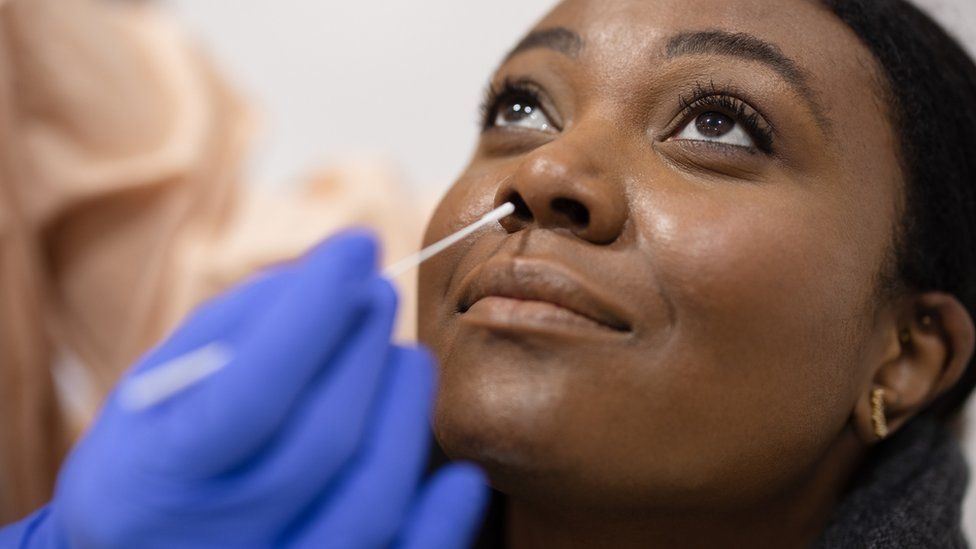
Published14 minutes agoShareclose panelShare pageCopy linkAbout sharingImage source, Getty ImagesBy Michelle RobertsDigital health editorMillions of UK Covid test samples will be analysed for other serious respiratory viruses, including flu, in a new disease-tracking project. The Wellcome Sanger Institute team will run detailed genetic reads on the anonymised nose and throat swabs leftover from the pandemic.The initiative could be an early warning system, quickly spotting future disease threats, say scientists. It could also help identify new treatments and ways to stop outbreaks.If the programme can be scaled up, in a few years the NHS could do these types checks routinely on patients for large-scale surveillance. In the future, it could scan for certain bacteria and fungal infections too. The scientists will work with government and public health teams, and all the data will be made freely and publicly available.Image source, Getty ImagesProf Susan Hopkins, chief medical adviser at the UK Health and Security Agency, said: “Genomic sequencing has been crucial in the response to the Covid pandemic and will continue to be pivotal in global efforts to address all kinds of threats to health in the future.”To begin with, the tests will look for eight or nine viral causes of respiratory tract infections – adenovirus, human metapneumovirus (hMPV), influenza A, parainfluenza, RSV, rhinovirus and several common cold coronaviruses (HCoV-229E. HCoV -NL63, HCoV-OC43).Dr Ewan Harrison, part of the team at the Wellcome Sanger Institute, explained: “We’ll use leftover diagnostic samples from Covid testing. We have millions of them. We can use them rather than waste them.”The nose and throat swabs are from the NHS Covid Test and Trace Lighthouse laboratories that were set up in the pandemic.He said: “We aim to help answer some of the most pressing public health questions, while at the same time addressing some of the gaps in our basic knowledge about respiratory infection and health.”Ultimately, we hope to contribute to global efforts to further establish pathogen genomics for routine public health and research, and as part of pandemic preparedness.”Respiratory infections, carried in coughs and sneezes, can spread very easily between people.Germs can live for hours outside the body on surfaces.If you get a virus on your hands by touching something that is contaminated, it can get into your body when you touch your eyes, nose or mouth. That is why it is good to wash your hands, and use a disposable tissue if you are coughing and sneezing. While most respiratory infections will get better without any treatment in a week or two, they can be dangerous and even deadly for some people.More on this storyInside the lab finding new Covid variants23 March 2021What is known about new Covid variant XBB.1.5?6 hours agoAntibody jab approved for common winter virus RSV10 November 2022Around the BBCThe childhood diseases making a post-lockdown comeback – BBC FutureRelated Internet LinksWellcome Sanger InstituteThe BBC is not responsible for the content of external sites.

Read more →

Aware or not aware: you are affected by food cues either way

Controlling your food intake can be even more difficult than you think. Osaka Metropolitan University scientists show that visual food cues can affect your eating behavior even when you are not aware of them. Their findings were published in PLOS ONE.
Obesity is one of the major pathological conditions that constitute lifestyle-related diseases and is known to be associated with myocardial infarction, stroke, and carcinogenesis. Approaches to regulate eating behavior are widely used in an effort to control obesity, but it has been reported that about half of those who receive dietary guidance return to their original weight within five years.
To explain the limited effectiveness of such guidance, one hypothesis suggests that not only conscious neural processes, which the dietary guidance targets, but also unconscious neural processes play an important role in controlling eating behavior. However, there were no studies directly examining the validity of this hypothesis at the level of neural activity.
The research team led by Professor Takahiro Yoshikawa from the Graduate School of Medicine at Osaka Metropolitan University has revealed that in the inferior frontal gyrus, a region of the brain’s frontal lobe that controls eating behavior, neural activity differs in response to visual food stimuli, or food images, depending on whether those images are presented consciously or unconsciously. Using a questionnaire to assess the study participants, the team found that this difference was associated with their scores on eating behaviors, including emotional eating and cognitive restraint of food intake. These results indicate that eating behavior cannot be understood without taking into account both unconscious and conscious neural processes.
“If we can learn more in future research about how eating behavior is controlled by unconscious neural processes, we can combine that understanding with our current knowledge of conscious neural processes to potentially develop more effective methods for regulating eating behavior,” stated Professor Yoshikawa.
Story Source:
Materials provided by Osaka Metropolitan University. Note: Content may be edited for style and length.

Read more →

Consumption of fast food linked to liver disease

The new year has begun, and with it, resolutions for change.
A study from Keck Medicine of USC published today in Clinical Gastroenterology and Hepatology gives people extra motivation to reduce fast-food consumption.
The study found that eating fast food is associated with nonalcoholic fatty liver disease, a potentially life-threatening condition in which fat builds up in the liver.
Researchers discovered that people with obesity or diabetes who consume 20% or more of their daily calories from fast food have severely elevated levels of fat in their liver compared to those who consume less or no fast food. And the general population has moderate increases of liver fat when one-fifth or more of their diet is fast food.
“Healthy livers contain a small amount of fat, usually less than 5%, and even a moderate increase in fat can lead to nonalcoholic fatty liver disease,” said Ani Kardashian, MD, a hepatologist with Keck Medicine and lead author of the study. “The severe rise in liver fat in those with obesity or diabetes is especially striking, and probably due to the fact that these conditions cause a greater susceptibility for fat to build up in the liver.”
While previous research has shown a link between fast food and obesity and diabetes, this is one of the first studies to demonstrate the negative impact of fast food on liver health, according to Kardashian.

The findings also reveal that a relatively modest amount of fast food, which is high in carbohydrates and fat, can hurt the liver. “If people eat one meal a day at a fast-food restaurant, they may think they aren’t doing harm,” said Kardashian. “However, if that one meal equals at least one-fifth of their daily calories, they are putting their livers at risk.”
Nonalcoholic fatty liver disease, also known as liver steatosis, can lead to cirrhosis, or scarring of the liver, which can cause liver cancer or failure. Liver steatosis affects over 30% of the U.S. population.
Kardashian and colleagues analyzed the most recent data from the nation’s largest annual nutritional survey, the 2017-2018 National Health and Nutrition Examination Survey, to determine the impact of fast-food consumption on liver steatosis.
The study characterized fast food as meals, including pizza, from either a drive-through restaurant or one without wait staff.
The researchers evaluated the fatty liver measurement of approximately 4,000 adults whose fatty liver measurements were included in the survey and compared these measurements to their fast-food consumption.

Of those surveyed, 52% consumed some fast food. Of these, 29% consumed one-fifth or more daily calories from fast food. Only this 29% of survey subjects experienced a rise in liver fat levels.
The association between liver steatosis and a 20% diet of fast food held steady for both the general population and those with obesity or diabetes even after data was adjusted for multiple other factors such as age, sex, race, ethnicity, alcohol use and physical activity.
“Our findings are particularly alarming as fast-food consumption has gone up in the last 50 years, regardless of socioeconomic status,” said Kardashian. “We’ve also seen a substantial surge in fast-food dining during the COVID-19 pandemic, which is probably related to the decline in full-service restaurant dining and rising rates of food insecurity. We worry that the number of those with fatty livers has gone up even more since the time of the survey.”
She hopes the study will encourage health care providers to offer patients more nutrition education, especially to those with obesity or diabetes who are at higher risk of developing a fatty liver from fast food. Currently, the only way to treat liver steatosis is through an improved diet.
Jennifer Dodge, MPH, assistant professor of research medicine and population and public health sciences at the Keck School of Medicine of USC and Norah Terrault, MD, MPH, a Keck Medicine gastroenterologist and division chief of gastroenterology and liver diseases at the Keck School, were also authors on the study.

Read more →