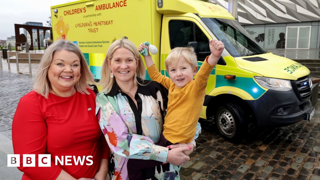
Inside the new children's ambulance for Northern Ireland

A new ambulance which brings children from Northern Ireland to Dublin for life saving treatment has been kitted out with features to make the journey a little easier for families.It’s the first ambulance in Northern Ireland that’s specifically for children and will mainly transport those who need cardiac surgery.It is expected to make about 500 journeys every year.

Read more →

Bill Turnbull's daughter running London Marathon in his memory

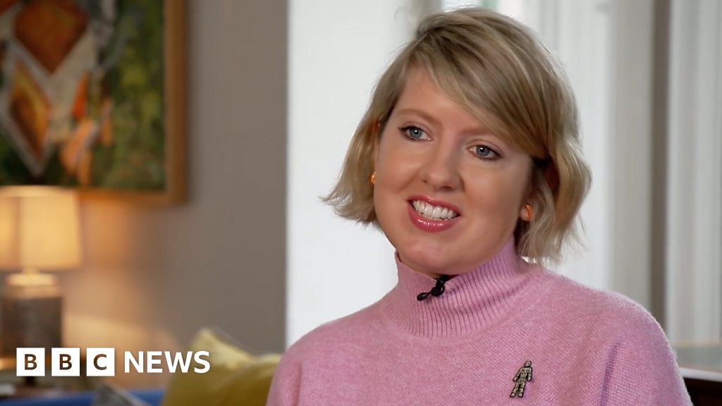
The daughter of former BBC Breakfast presenter Bill Turnbull said she would run this year’s London Marathon in memory of the broadcaster.Flora Turnbull said she wanted to raise awareness of prostate cancer after her father’s death from the disease in August.”I wanted to have a purpose this year and to remember dad in the most purposeful way possible,” she told his former colleagues.Mr Turnbull died at his home in Suffolk after a “challenging and committed fight against prostate cancer”, his family said at the time.Find BBC News: East of England on Facebook, Instagram and Twitter. If you have a story suggestion email eastofenglandnews@bbc.co.uk

Read more →

Body phenotypes say a lot, but not everything, about a person's health

Concordia researchers studying body phenotypes — the observable characteristics like height, behaviour, appearance and more measurables — found that regardless of the muscle they had, high levels of fat mass in an individual were associated with poorer overall health.
The findings, published in the journal Preventive Medicine, used data from a United States longitudinal study. They show that the negative impact of excess adiposity — fat tissue — on a person’s cardiometabolic health was not offset even by high levels of muscle mass.
The researchers based their study on data from NHANES, a cross-sectional representative sample of the US population collected between 1999 and 2006. The data was collected using dual energy X-ray absorptiometry (DEXA), a diagnostic framework that analyzes adiposity and muscle mass. Based on which side of the 50th percentile they ranked, individuals were categorized into one of four proposed phenotypes: low-adiposity/high-muscle, high-adiposity/high-muscle, low-adiposity/high-muscle or low-adiposity/low-muscle.
The researchers looked at how the adiposity/muscle phenotypes related to lipid levels, including cholesterol and triglycerides, as well as blood sugar glucose and blood pressure. Results were also adjusted for age, sex, race and education.
“We wanted to see whether this proposed categorization was better than the traditional body-mass index (BMI) at predicting all these different cardiometabolic outcomes,” says Sylvia Santosa, an associate professor in the Department of Health, Kinesiology and Applied Physiology and one of the authors of the paper.
Surprisingly, they found BMI, though far from perfect, was in some cases a better predictor of cardiometabolic risks like diabetes and hypertension.
Associate professor Lisa Kakinami, Concordia alumna and current Rhodes Scholar Sabine Plummer, BSc 22, PhD student Jessica Murphy and Tamara Cohen of the University of British Columbia co-authored the paper.
Benefits of BMI
Nevertheless, the data did reveal several striking findings. In comparison to the low-adiposity/high-muscle group, which was the healthiest of the four, the researchers noted the following results: The two high-adiposity groups were less likely to be physically active and more likely to have abnormal lipids and less healthy diets. The high-adiposity/low-muscle group had higher total cholesterol levels, lower levels of high-density lipoprotein (“good” cholesterol) and lower nutrient intake. This group was also 56 to 66 per cent less likely to meet weekly physical activity recommendations. The high-adiposity/high-muscle group had unfavourable values for all cardiometabolic and adiposity measures. Nutrient intake was also lower. This group was also 49 to 67 per cent less likely to meet physical activity recommendations, roughly 80 per cent more likely to have hypertension and 23 to 35 per cent more likely to exceed recommended saturated fat intake. Overall, the high-adiposity/high-muscle phenotype was the least likely to meet physical activity and nutrient recommendations and was at the greatest risk of poor cardiometabolic health. The low-adiposity/low-muscle group had significantly lower BMI and waist circumferences. This group also had the lowest grip strength across the four phenotypes.”If we are looking at cardiometabolic risk at the population level, BMI can give you cheap and quick idea about what is happening,” Santosa says.

Read more →

Supplementation with amino acid serine eases neuropathy in diabetic mice

Approximately half of people with type 1 or type 2 diabetes experience peripheral neuropathy — weakness, numbness, and pain, primarily in the hands and feet. The condition occurs when high levels of sugar circulating in the blood damage peripheral nerves. Now, working with mice, Salk Institute researchers have identified another factor contributing to diabetes-associated peripheral neuropathy: altered amino acid metabolism.
The team found that diabetic mice with low levels of two related amino acids, serine and glycine, are at higher risk for peripheral neuropathy. What’s more, the researchers were able to alleviate neuropathy symptoms in diabetic mice by supplementing their diets with serine.
The study, published January 25, 2023 in Nature, adds to growing evidence that some often-underappreciated, “non-essential” amino acids play important roles in the nervous system. The findings may provide a new way to identify people at high risk for peripheral neuropathy, as well as a potential treatment option.
“We were surprised that dialing up and down a non-essential amino acid had such a profound effect on metabolism and diabetic complications,” says senior author Christian Metallo, a professor in Salk’s Molecular and Cell Biology Laboratory. “It just goes to show that what we think of as dogma can change under different circumstances, such as in disease conditions.” Metallo led the study with first author Michal Handzlik, a postdoctoral researcher in his lab.
Amino acids are the building blocks that make up proteins and specialized fat molecules called sphingolipids, which are abundant in the nervous system. Low levels of the amino acid serine force the body to incorporate a different amino acid in sphingolipids, which changes their structure. These atypical sphingolipids then accumulate, which may contribute to peripheral nerve damage. While the team observed this accumulation in diabetic mice, the same amino acid switch and sphingolipid changes occur in a rare human genetic disease marked by peripheral sensory neuropathy, indicating that the phenomenon is consistent across many species.
To determine whether long-term, chronic serine deficiency drives peripheral neuropathy, Metallo’s team fed mice either control or serine-free diets in combination with either low-fat or high-fat diets for up to 12 months. The researchers were surprised to find that low serine, in combination with a high-fat diet, accelerated the onset of peripheral neuropathy in the mice. In contrast, serine supplementation in diabetic mice slowed the progression of peripheral neuropathy, and the mice fared better.

The researchers also tested the compound myriocin, which inhibits the enzyme that switches out serine for another amino acid as sphingolipids are assembled. Myriocin treatment reduced peripheral neuropathy symptoms in mice fed a high-fat, serine-free diet. These findings underscore the importance of amino acid metabolism and sphingolipid production in the maintenance of a healthy peripheral nervous system.
Serine deficiency has also been associated with various neurodegenerative disorders. For example, Metallo and collaborators previously found a link between altered serine and sphingolipid metabolism in patients with macular telangiectasia type 2, a condition that causes vision loss. In mice, reduced serine led to increased levels of atypical retinal sphingolipids and reduced vision. Serine is currently being tested in clinical trials for its safety and efficacy in treating macular telangiectasia and Alzheimer’s disease.
Peripheral neuropathy is typically managed with dietary changes to reduce blood sugar levels, as well as pain relievers, physical therapy, and mobility aids, such as canes and wheelchairs. Foods naturally rich in serine include soybeans, nuts, eggs, chickpeas, lentils, meat, and fish, and serine supplements are inexpensive and available over the counter.
Yet the researchers say it’s premature to advise people with diabetes to take serine supplements to prevent neuropathy.
“You would likely need to take a lot to make a difference, and not everyone needs extra serine,” Metallo says. “We need more time to understand serine physiology in humans and explore potential downsides to supplementation.”
To this end, Metallo and Handzlik are now developing a serine tolerance test, similar to a glucose tolerance test used to diagnose diabetes.

“We want to identify those at highest risk for peripheral neuropathy so we can treat only those who might benefit most,” says Handzlik.
Other authors included: Jivani M. Gengatharan, Grace H. McGregor, and Courtney R. Green of the Salk Institute and UC San Diego; Katie E. Frizzi, Cameron Martino, Gibraan Rahman, Antonio Gonzalez, Ana M. Moreno, Lucie S. Guernsey, Prashant Mali, Rob Knight, and Nigel A. Calcutt of UC San Diego; Terry Lin, Patrick Tseng, and Satchidananda Panda of the Salk Institute; Yoichiro Ideguchi of Scripps Research; Regis J. Fallon and Marin L. Gantner of the Lowy Medical Research Institute; Amandine Chaix of the University of Utah; and Martina Wallace of University College Dublin in Ireland.
The work was funded by the National Institutes of Health (grants R01CA234245, DK076169, R01AG065993, P30
DK120515), a Camille and Henry Dreyfus Teacher-Scholar Award, the Lowy Medical Research Institute, and the American Heart Association (grant 18CDA34110292).

Read more →

Wearable sensor uses ultrasound to provide cardiac imaging on the go

Engineers and physicians have developed a wearable ultrasound device that can assess both the structure and function of the human heart. The portable device, which is roughly the size of a postage stamp, can be worn for up to 24 hours and works even during strenuous exercise.
The goal is to make ultrasound more accessible to a larger population, said Sheng Xu, a professor of nanoengineering at the University of California San Diego, who is leading the project. Currently, echocardiograms- ultrasound examinations for the heart- require highly trained technicians and bulky devices.
“The technology enables anybody to use ultrasound imaging on the go,” Xu said.
Thanks to custom AI algorithms, the device is capable of measuring how much blood the heart is pumping. This is important because the heart not pumping enough blood is at the root of most cardiovascular diseases. And issues with heart function often manifest only when the body is in motion.
The work is described in the Jan. 25 issue of the journal Nature.
Cardiac imaging is an essential clinical tool to assess long-term heart health, detect problems as they arise and care for critically ill patients. This new wearable, non-invasive heart monitor for humans provides real-time, automated insights on the difficult-to-capture pumping activity of the heart, even when a person is exercising.

The wearable heart monitoring system uses ultrasound to continuously capture images of the four chambers of the heart in different angles, and analyze a clinically relevant subset of the images in real time using a custom-built AI technology. The project builds on the team’s previous advances in wearable imaging technologies for deep tissues.
“The increasing risk of heart diseases calls for more advanced and inclusive monitoring procedures,” Xu said. “By providing patients and doctors with more thorough details, continuous and real-time cardiac image monitoring is poised to fundamentally optimize and reshape the paradigm of cardiac diagnoses.”
In comparison, existing non-invasive methods have limited sampling capabilities and provide limited data. The wearable technology developed by Xu’s team enables safe, non-invasive and high-quality cardiac imaging, resulting in images with high spatial resolution, temporal resolution and contrast. “It also minimizes patient discomfort and overcomes some limitations of noninvasive technologies such as CT and PET, which could expose patients to radiation,” said Hao Huang, a PhD student in the Xu group at UC San Diego.
The unique design of the sensor makes it ideal for bodies in motion. “The device can be attached to the chest with minimal constraint to the subjects’ movement, even providing a continuous recording of cardiac activities before, during and after exercise,” said Xiaoxiang Gao, a postdoctoral researcher in the Xu group at UC San Diego.
The importance of cardiac imaging
Cardiac diseases are the leading cause of death among the elderly, and are also becoming more prevalent among the young due to lifestyle factors. The signs of cardiac diseases are transient and unpredictable, making them hard to spot. This has upped demand for more advanced, inclusive, non-invasive and cost-effective monitoring technologies such as long-term cardiac imaging, which this wearable device facilitates.

Cardiac imaging is one of the most powerful tools for screening and diagnosing cardiac issues before they become problems. “The heart undergoes all kinds of different pathologies,” said Hongjie Hu, a postdoctoral researcher in the Xu lab at UC San Diego. “Cardiac imaging will disclose the true story underneath. Whether it be that a strong but normal contraction of heart chambers leads to the fluctuation of volumes, or that a cardiac morphological problem has occurred as an emergency, real-time image monitoring on the heart tells the whole picture in vivid detail and visual effect.”
How it works in detail
The new system gathers information through a wearable patch as soft as human skin, designed for optimal adherence. The patch measures 1.9 cm (L) x 2.2 cm (W) x 0.09 cm (T) , about the size of a postage stamp. It sends and receives the ultrasound waves which are used to generate a constant stream of images of the structure of the heart in real time. This ultrasound patch is soft and stretchable, and it adheres well to human skin, even during exercise.
The system can examine the left ventricle of the heart in separate bi-plane views using ultrasound, generating more clinically useful images than were previously available. As a use case, the team demonstrated imaging of the heart during exercise, which is not possible with the rigid, cumbersome equipment used in clinical settings.
The performance of the heart is characterized by three factors: stroke volume (the volume of blood the heart pumps out each beat), ejection fraction (the percentage of blood pumped out of the left ventricle of the heart every beat) and cardiac output (the volume of blood the heart pumps out every minute).
Xu’s team developed an algorithm to facilitate continuous, AI-assisted automatic processing.
“A deep learning model automatically segments the shape of the left ventricle from the continuous image recording, extracting its volume frame-by-frame and yielding waveforms to measure stroke volume, cardiac output and ejection fraction,” said Mohan Li, a master’s student in the Xu group at UC San Diego.
“Specifically, the AI component involves a deep learning model for image segmentation, an algorithm for heart volume calculation, and a data imputation algorithm,” said Ruixiang Qi, a master’s student in the Xu group at UC San Diego. “We use this machine learning model to calculate the heart volume based on the shape and area of the left ventricle segmentation. The imaging-segmentation deep learning model is the first to be functionalized in wearable ultrasound devices. It enables the device to provide accurate and continuous waveforms of key cardiac indices in different physical states, including static and after exercise, which has never been achieved before.”
Thus, this technology can generate curves of these three indices continuously and noninvasively, as the AI component processes the continuous stream of images to generate numbers and curves.
To create the platform, the team faced some technical challenges that required careful decision-making. To produce the wearable device itself, the researchers used a piezoelectric 1-3 composite bonded with Ag-epoxy backing as the material for transducers in the ultrasound imager, reducing risk and improving efficiency over previous methods. When choosing the transmission configuration of the transducer array, they achieved superior results through wide-beam compounding transmission. They also selected from nine popular models for machine-learning-based image segmentation, landing on FCN-32, which achieved the highest possible accuracy.
In the current iteration, the patch is connected through cables to a computer, which can download the data automatically while the patch is still on. The team has developed a wireless circuit for the patch, which will be covered in a forthcoming publication.
Next steps
Xu plans to commercialize this technology through Softsonics, a company spun off from UC San Diego that he cofounded with engineer Shu Xiang. He also encourages others in his scientific community to follow his lead and work on areas of this research that warrant further exploration.
To follow up on these results, Xu recommends four immediate next steps: B-mode imaging, which allows more diagnostic capabilities involving different organs The design of the soft imager, which allows researchers to fabricate large transducer probes that cover multiple positions simultaneously Miniaturization of the back-end system that powers the soft imager Working toward a general machine learning model that fits more subjectsThis work was supported by the National Institutes of Health (1R21EB025521-01, 1R21EB027303-01A1, 3R21EB027303-02S1, and 1R01EB033464-01).

Read more →