
Whether born naturally or via Cesarean section, babies receive essential microbes from their mothers


For the first time, a research team has identified and analyzed the steps by which immune cells “see” and respond to cancer cells, providing insights into reasons some treatments may be effective for certain patients but not others.
The UCLA Jonsson Comprehensive Cancer Center scientists leading the research believe their findings will lead to better, more personalized immunotherapies — even for patients whose immune systems currently do not appear to respond to treatment.
“This is an important step forward in our understanding of what the T-cell responses see in the tumor and how they change over time while they are in the tumor and in circulation in the blood, searching for new tumor cells to attack,” said Cristina Puig-Saus, Ph.D., a UCLA Jonsson Comprehensive Cancer Center researcher, adjunct assistant professor of medicine at UCLA, and the first author of a study in Nature.
“The deeper understanding of how the T-cell responses clear metastatic tumor masses will help us design better treatments and engineer T cells in multiple ways to mimic them,” she said.
The researchers adapted advanced gene-editing technology to make unprecedented observations about immune responses in patients with metastatic melanoma receiving anti-PD-1 “checkpoint inhibitor” immunotherapy. Although immune cells called T cells have the ability to detect mutations in cancer cells and eliminate them, leaving normal cells unharmed, cancer cells often evade the immune system. Checkpoint inhibitors are designed to improve the T cells’ ability to recognize and attack cancer cells.
“With this work, we can know exactly what the immune system of a particular patient recognized in their cancer to differentiate it from normal cells and attack it,” said Antoni Ribas, M.D., Ph.D., a UCLA Jonsson Comprehensive Cancer Center researcher, professor of medicine at UCLA, a co-senior author of the study.
The investigators showed that when the immunotherapy is effective, it directs a diverse repertoire of T cells against a small group of selected mutations in a tumor. These T-cell responses expand and evolve during the course of treatment, both within the tumor and in the bloodstream. Patients for whom the therapy fails also present a T-cell response against a similarly reduced number of mutations in the tumor, but those immune responses are less focused, and they do not expand during treatment.
“This study demonstrates that patients without response to therapy still induce a tumor-reactive T-cell response,” Puig-Saus said. “These T cells could potentially be isolated and their immune receptors used to genetically modify a larger number of T cells to redirect them against the patient’s tumor. These T cells could be expanded in culture and reinfused into the patients to treat their tumors.”
In the 11 patients studied, seven had a response to PD-1 blockade; four did not. The number of mutations in the tumors ranged between 3,507 and 31. Despite this wide range, the number of mutations seen by tumor-reactive T cells ranged between 13 and one. In patients with clinical benefit from the therapy, the responses were diverse, with a range between 61 and seven different mutation-specific T cells isolated in the blood and the tumor. In contrast, in the patients lacking a response to therapy, the researchers only identified between 14 and two different T cells.
Also, in patients responding to treatment, the researchers were able to isolate tumor-reactive T cells in blood and tumors throughout treatment, but in patients without a response, the T cells were not recurrently detected. Still, the study showed that immune receptors from the T cells isolated from all patients — regardless of response or not — redirected the specificity of immune cells against the tumor, producing antitumor activity.
The work to characterize T-cell activity in patients with and without a clinical response was made possible through the creation of a new technique using sophisticated technology to isolate mutation-reactive T cells from blood and tumor samples. It builds on technology developed through a collaboration with Ribas, James Heath, PhD, president of the Institute for Systems Biology in Seattle, and David Baltimore, PhD, Nobel laureate, emeritus professor at Caltech and a member of the UCLA Jonsson Comprehensive Cancer Center.
As previously published in Nature and presented at the Society for Immunotherapy of Cancer (SITC) 2022 last November, the technology was further developed by PACT Pharma, using CRISPR gene editing to insert genes into immune cells to efficiently redirect them to recognize mutations in a patient’s own cancer cells.
“With this technique, we generated large numbers of T cells expressing the immune receptors from the mutation-reactive T cells isolated from each patient. We used these cells to characterize the reactivity of the immune receptors against the patient’s own cancer cells,” Ribas said. “The new technologies allow us to study these rare immune cells that are the mediators of immune responses to cancer.”
Nearly two dozen experimental therapies targeting the immune system are in clinical trials for Alzheimer’s disease, a reflection of the growing recognition that immune processes play a key role in driving the brain damage that leads to confusion, memory loss and other debilitating symptoms.
Many of the immunity-focused Alzheimer’s drugs under development are aimed at microglia, the brain’s resident immune cells, which can injure brain tissue if they’re activated at the wrong time or in the wrong way. A new study from researchers at Washington University School of Medicine in St. Louis indicates that microglia partner with another type of immune cell — T cells — to cause neurodegeneration.
Studying mice with Alzheimer’s-like damage in their brains due to the protein tau, the researchers discovered that microglia attract powerful cell-killing T cells into the brain, and that most of the neurodegeneration could be avoided by blocking the T cells’ entry or activation. The findings, published March 8 in the journal Nature, suggest that targeting T cells is an alternative route to preventing neurodegeneration and treating Alzheimer’s disease and related diseases involving tau, collectively known as tauopathies.
“This could really change the way we think about developing treatments for Alzheimer’s disease and related conditions,” said senior author David M. Holtzman, MD, the Barbara Burton and Reuben M. Morriss III Distinguished Professor of Neurology. “Before this study, we knew that T cells were increased in the brains of people with Alzheimer’s disease and other tauopathies, but we didn’t know for sure that they caused neurodegeneration. These findings open up exciting new therapeutic approaches. Some widely used drugs target T cells. Fingolomid, for example, is commonly used to treat multiple sclerosis, which is an autoimmune disease of the brain and spinal cord. It’s likely that some drugs that act on T cells could be moved into clinical trials for Alzheimer’s disease and other tauopathies if these drugs are protective in animal models.”
Alzheimer’s develops in two main phases. First, plaques of the protein amyloid beta start to form. The plaques can build up for decades without obvious effects on brain health. But eventually, tau also begins to aggregate, signaling the start of the second phase. From there, the disease quickly worsens: The brain shrinks, nerve cells die, neurodegeneration spreads, and people start having difficulty thinking and remembering.
Microglia and their role in Alzheimer’s have been intensely studied. The cells become activated and dysfunctional as amyloid plaques build up, and even more so once tau begins to aggregate. Microglial dysfunction worsens neurodegeneration and accelerates the course of the disease.
First author Xiaoying Chen, PhD, an instructor in neurology, wondered about the role of other, less studied immune cells in neurodegeneration. She analyzed immune cells in the brains of mice genetically engineered to mimic different aspects of Alzheimer’s disease in people, looking for changes to the immune cell population that occur over the course of the disease.
Mirroring the early phase of the disease in people, two of the mouse strains build up extensive amyloid deposits but do not develop brain atrophy. A third strain, representative of the later phase, develops tau tangles, brain atrophy, neurodegeneration and behavioral deficits by 9½ months of age. A fourth mouse strain does not develop amyloid plaques, tau tangles or cognitive impairments; it was studied for comparison. Along with Chen and Holtzman, the research team included Maxim N. Artyomov, PhD, the Alumni Endowed Professor of Pathology & Immunology, and Jason D. Ulrich, PhD, an associate professor of neurology, among others.
The researchers found many more T cells in the brains of tau mice than the brains of amyloid or comparison mice. Notably, T cells were most plentiful in the parts of the brain with the most degeneration and the highest concentration of microglia. T cells were similarly abundant at sites of tau aggregation and neurodegeneration in the brains of people who had died with Alzheimer’s disease.
Additional mouse studies indicated that the two kinds of immune cells work together to create an inflammatory environment primed for neuronal damage. Microglia release molecular compounds that draw T cells into the brain from the blood and activate them; T cells release compounds that push microglia toward a more pro-inflammatory mode.
Eliminating either microglia or T cells broke the toxic connection between the two and dramatically reduced damage to the brain. For example, when tau mice were given an antibody to deplete their T cells, they had fewer inflammatory microglia in their brains, less neurodegeneration and atrophy, and an improved ability to perform tasks such as building a nest and remembering recent things.
“What got me very excited was the fact that if you prevent T cells from getting into the brain, it blocks the majority of the neurodegeneration,” Holtzman said. “Scientists have put a lot of effort into finding therapies that prevent neurodegeneration by affecting tau or microglia. As a community, we haven’t looked at what we can do to T cells to prevent neurodegeneration. This highlights a new area to better understand and therapeutically explore.”
A new study shows for the first time a connection between a mitochondrial metabolite and the activation of an inflammatory response. Mitochondria are functional units of our cells that fulfil important tasks, i.e. chemical reactions, for the functioning of the cell. One of these tasks is the production of energy that is necessary for cell growth and reproduction. If certain chemical reactions in the mitochondrion change, diseases occur. For example, deficiencies in fumarate hydratase (FH) in the Krebs cycle, one of the most important metabolic pathways in mitochondria, cause an aggressive form of kidney cancer in humans. FH loss leads to the accumulation of the molecule fumarate, which contributes to the development of cancer. For this reason, fumarate is called an oncogenic metabolite, or “oncometabolite” for short.
The research team led by Alexander von Humboldt Professor Dr Christian Frezza, formerly at the University of Cambridge (United Kingdom) and now at the CECAD Cluster of Excellence for Aging Research at the University of Cologne, has now developed a new mouse and cell model together with the research group led by Professor Prudent of the University of Cambridge to deepen the understanding of aggressive kidney cancer. In the models, the silencing of the fumarate hydratase gene can be temporally controlled by the scientists. Using a combination of high-resolution imaging techniques and precise biochemical experiments, the scientists have shown that fumarate causes mitochondrial damage. This in turn releases the genetic material of the mitochondria in small vesicles called mitochondrial-derived vesicles. These vesicles filled with mitochondrial DNA (mtDNA) and RNA (mtRNA) trigger an immune reaction that eventually leads to inflammation. The study titled “Fumarate induces vesicular release of mtDNA to drive innate immunity” was published in Nature.
“Our study shows for the first time a correlation between a mitochondrial metabolite and the onset of inflammation, which could be the trigger for cancer and autoimmune diseases,” said Professor Frezza. “Based on these findings, we can now work on new approaches to treat patients, which will hopefully lead to the development of new therapeutic strategies to treat cancer patients in the future.”
In addition, a group at Trinity Biomedical Sciences Institute in Dublin led by Professor Luke O’Neill in collaboration with Christian Frezza’s research group has described a similar mechanism in macrophages. Macrophages are cells of the body that are responsible for eliminating harmful microbes. Here, the researchers found that mitochondrial RNA released by the macrophages’ mitochondria, rather than DNA, is the main trigger of inflammation. The study “Macrophage fumarate hydratase restrains mtRNA-mediated interferon production” was also published in the journal Nature.
The research was carried out at the University of Cambridge and the CECAD Cluster of Excellence for Aging Research of the University of Cologne. It was funded by Cancer Research UK, the European Research Council, the German Research Foundation (DFG), the Alexander von Humboldt Foundation and the Medical Research Council. The collaborative research was conducted in the laboratory of Luke O’Neill at Trinity Biomedical Sciences Institute in Dublin, Ireland.
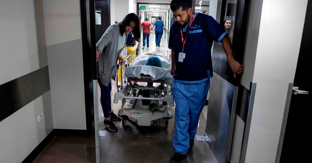
Here’s what experts say about the risks and promises of traveling abroad for cost-efficient care.Last week, four Americans were kidnapped in the Mexican state of Tamaulipas, after crossing the border from Texas. Two were later found dead. A sister of one of the victims said they had gone to Mexico so one of them could get an abdominoplasty, better known as a tummy tuck.Every year, millions of Americans visit Mexico and other countries to obtain health care, a practice often called medical tourism. The National Exterior Commerce Bank in Mexico estimated that the industry was worth $5 billion before it declined during the coronavirus pandemic. For patients, the motivation is often financial.“Some of it is a desperate search for access” to medical care, said Felicia Marie Knaul, director of the Institute for Advanced Study of the Americas at the University of Miami.Many people cross the border for pharmaceuticals at greatly decreased prices from what you pay in the U.S. Others, especially Americans and Canadians in the past two decades, are traveling for surgeries or treatments. The Centers for Disease Control and Prevention says dental care, surgeries, fertility treatments, organ and tissue transplants and cancer treatment are the most common procedures for which people go abroad. Elective procedures are a major component of medical tourism, said Daniel Béland, a professor of political science at McGill University who has studied health policy.In 2016, the C.D.C. surveyed more than 93,000 people; of those who had left the United States for care during the previous year, Mexico was the most common destination.But while crossing national borders might be an affordable way to get high-quality care, medical tourism is largely unregulated, and it’s nearly impossible to track outcomes or the scope of procedures Americans obtain in Mexico.“There are really very few rules,” said David G. Vequist IV, director of the Center for Medical Tourism Research and a professor at the University of the Incarnate Word in San Antonio, Texas. Those that exist are vague, he added, and people are largely “making it up as they go along.”How many people go to Mexico for treatment?It’s hard to find solid data on medical tourism, said Valorie Crooks, a professor of geography at Simon Fraser University in Canada who has studied it for over a decade.She calls the industry a “triple U”: It’s “untracked, untraced and unregulated.”Most of the Mexican hospitals Americans visit are private and do not report their data to the federal government.Josef Woodman, the chief executive of Patients Beyond Borders, which serves as an international health care travel consulting agency and patient guide for people seeking care abroad, estimates that about 1.2 million Americans per year travel to Mexico for medical procedures. After a drop in medical tourism during the pandemic, Mr. Woodman said, he’s seen a spike in people seeking out treatment in Mexico as they got vaccinated.“After the first vax, people just flooded in,” he said.Complex dental treatments like root canals, veneers and full mouth reconstructions are among the most popular procedures, Mr. Woodman said. Los Algodones, near the California-Arizona border, is known as “Molar City” because it caters to this market.Billboards advertising pharmaceutical and medical services in Mexicali, Mexico.Guillermo Arias for The New York TimesThe most common destinations tend to be in Mexican states along the border, like Tamaulipas and Nuevo León, or those with popular beach towns, like Baja California Sur and Quintana Roo, according to Denise Rodriguez, who is studying for a Ph.D. in health geography at the University of Brasília in Brazil and interviewed hundreds of people involved in medical tourism in Los Algodones for her master’s thesis.By counting the numbers of private hospitals and medical professionals available, she found that Baja California Sur was the state with the most medical tourism.Why is the industry booming?Because the overhead costs of running a clinic or health care center in Mexico are much lower, patients typically pay far less than they would for a procedure in the United States, Dr. Crooks said.One study, which surveyed over 400 people near the U.S.–Mexico border about traveling to obtain health care, found that 92 percent cited lower costs in Mexico as guiding their decision. Andrea Miller, a clinical pharmacist in Arizona who led the study, was struck by just how widespread advertising, and infrastructure, for medical services was in a Mexican border town.“You look down the street and it’s like, pharmacy, pharmacy, optical clinic, dental clinic, pharmacy, dental clinic,” she said.Some patients also go abroad to circumvent red tape and restrictions that might stymie them at home, Dr. Crooks said.“You could be too young or too old for an orthopedic surgery; you could be too small or too big for a bariatric surgery — and then you find a surgeon in another country who’s willing to offer you the treatment,” she said.Other patients travel to get procedures that are illegal where they live, including abortions.Money explains only so much, said Ms. Rodriguez, who found that many travelers were simply looking for more personalized care and time with a doctor.“Why do people come back?” she said. “You are treated like a human being.”Medical tourism carries risks.While traveling for health care to certain areas of the world can be dangerous, experts said that for most patients, the risks have more to do with the medical procedure than the journey to obtain it.Patients embarking on medical tourism seek out care on their own and pay out of pocket, Dr. Crooks added. That may mean their primary-care doctors aren’t informed, potentially leading to problems when patients seek follow-up care at home.Foreign patients leaving a hospital in Tijuana. Mexico is a popular destination for Americans seeking cheaper medical care.Guillermo Arias/Agence France-Presse — Getty ImagesThe C.D.C. recommends that patients schedule a consultation with their U.S. health care provider before leaving the country for medical care, said Allison Tayler Walker, lead of the epidemiology and surveillance team in the Travelers’ Health Branch at the agency. The C.D.C. also advises patients to arrange follow-up care ahead of time with the professional who conducts the procedure abroad, as well as with a primary physician in the United States.There are also specific risks that come with certain interventions — for example, doctors caution against flying too soon after some surgeries, Dr. Béland said, because the procedures can make a person more susceptible to blood clots.Mr. Woodman recommended seeking out hospitals accredited by Joint Commission International. It’s important for patients to ensure that anyone giving them medical care has received proper training, said Dr. Patricia Turner, executive director of the American College of Surgeons. That includes not just the doctor performing surgery, for example, but also the person administering anesthesia or interpreting X-rays.Getting any service as complicated as a surgery in another country, with different laws and cultural norms, can be complicated. For instance, someone who receives improper medical care abroad may have little or no legal recourse and may not know what their rights are. And any procedure carries the risk of complications, and in another country, a patient may need to stay longer than expected for follow-up care or to recover, Dr. Crooks cautioned.“It’s not necessarily that those risks are higher when you go abroad,” Dr. Crooks said. “But your ability to remedy or address those risks could become more challenging.”
Read more →
At its first public hearing, the House Select Subcommittee on the Coronavirus Pandemic plunged into the politically charged debate over the origins of the virus.WASHINGTON — The House panel investigating the origins of the coronavirus pandemic opened its first public hearing on Wednesday with Republicans and their witnesses making an aggressive case that the virus may have been the result of a laboratory leak — a notion that has become the subject of intense political and scientific debate.“There is no smoking gun proving a lab origin hypothesis, but the growing body of circumstantial evidence suggests a gun that, at the very least, is warm to the touch,” said Jamie Metzl, a senior fellow at the Atlantic Council and a former State Department official.Dr. Metzl was one of three witnesses invited by Republicans. The others were Dr. Robert R. Redfield, who served as the director of the Centers for Disease Control and Prevention under President Donald J. Trump, and Nicholas Wade, who was the science editor of The New York Times in the 1990s and left the news organization at the end of 2011.The three have previously said the virus may have accidentally escaped from a laboratory. But they all said on Wednesday that the question of how the virus originated remained an open one, and that it was important to settle the question.Dr. Paul G. Auwaerter, the clinical director of the infectious diseases division at the Johns Hopkins University School of Medicine, testified at the invitation of Democrats.Some proponents of the laboratory leak hypothesis have suggested that it was a biological weapon intentionally engineered by China. But Dr. Redfield, a virologist, said he had concluded that the virus was a result of an accident, and that his view was based “primarily on the biology of the virus itself,” including the fact that it was highly infectious, spawning the rapid evolution of new variants.Scientists have said that ability could very well have evolved through a natural spillover from an animal. They have cited, among other things, coronaviruses found in bats in 2020 that carry a molecular hook on their surface that is very similar to a feature on the virus that causes Covid-19. That hook allows the viruses to latch onto human cells.Dr. Redfield also called for a moratorium on “gain of function” research, which involves tinkering with the genes of viruses in a way that could make them more infectious. Many scientists argue that such research is necessary to help develop vaccines and other medical countermeasures that could be used in a pandemic.“I disagree with that assessment,” Dr. Redfield said.
Read more →