Study unravels a cause of resistance to novel drug in patients with acute leukemia

A new targeted drug has not only sparked remissions in patients with a common form of leukemia but also induced the cancer cells to reveal one of their schemes for resisting the drug, investigators at Dana-Farber Cancer Institute and other research centers report in a new pair of studies in the journal Nature.
One of the papers presents results of a clinical trial in which approximately 40% of patients with acute leukemia subtypes had a complete response — a disappearance of all signs of cancer — to treatment with the drug revumenib. The other paper uncovers a molecular countermove by which leukemia cells come to sidestep the drug and reassert their growth.
The papers point to the promise of the targeted approach to acute leukemia treatment exemplified by revumenib and to the potential to extend its benefits with drugs that trip up the resistance mechanism, researchers say.
“The two genetic subtypes of acute leukemia involved in this research account for approximately 40% of all cases of acute myeloid leukemia (AML) in children and adults,” says Scott Armstrong, MD, PhD, president of the Dana-Farber/Boston Children’s Cancer and Blood Disorders Center and co-senior author of the paper on revumenib resistance. “They’re driven by a rearrangement of the MLL1 gene or a mutation in the NPM1 gene. Both types depend on a protein called menin to sustain their growth.”
The first of the new Nature studies reports on a phase I/II clinical trial of the drug revumenib, which targets menin, in 68 patients with acute leukemia that wasn’t responding other therapies. The trial, dubbed the AUGMENT-101 study, found that of 60 patients who could be evaluated, 53% responded to the drug and 30% had a complete response.
“For patients with acute leukemia who have undergone several previous treatments, this is a very encouraging result,” Armstrong says. “However, after the second cycle of treatment, some patients did develop resistance to revumenib.”
In their follow-up study, Armstrong and his associates sought a source of that resistance.

Menin is a critical part of the cell’s “epigenetic” mechanism for switching gene activity on and off. It binds to chromatin — the braid of DNA and protein packaging within the nucleus — and summons large protein complexes to join it. The complexes tighten or loosen the coils of DNA, changing the activity level of the genes at that site. One of the key proteins within these complexes is MLL1, the product of the MLL1 gene.
“Revumenib contains a small molecule that inhibits the interaction of menin and MLL1,” Armstrong says. “The AUGMENT trial shows it can produce remissions in a high percentage of patients.”
When Armstrong and his colleagues analyzed bone marrow samples from some of the patients who became resistant to revumenib, they found that many had developed mutations in MEN1, the gene that gives rise to the menin protein. The mutation leads to a deformed version of menin that doesn’t bind well to revumenib but attaches as firmly as ever to MLL1. As menin and MLL1 renew their interaction, leukemia cell growth restarts.
The finding provides “formal proof in patients that menin itself is a valid target for therapy in both genetic subtypes of AML,” Armstrong relates.
In a sense, revumenib acts as a kind of truth-telling serum on leukemia cells: by placing the cells under pressure, it prompts them to divulge one of their strategies for survival. “The fact that the cell has gone through so much trouble to mutate MEN1 in order to survive is a strong indication that we’re hitting a target the cell truly relies on,” Armstrong states.
The ability of revumenib to stress leukemia cells to the point where they develop a mutation to remain alive is testament to the drug’s effectiveness, he continues. “This is the first time a drug targeting a chromatin-binding protein complex has been shown to put that degree of pressure on cancer in a human patient.”
By tracking the development of mutations in MEN1 and other genes in patients with acute leukemia, doctors may be able to identify patients at high risk of relapsing, Armstrong relates. Such patients might then benefit from further treatment. The discovery of MEN1 mutation as a resistance mechanism suggests that new drugs that specifically target menin or other chromatin-associated proteins could prevent or delay revumenib resistance or treat patients who have become resistant to the drug.
“Seeing Dr. Armstrong’s work translated elegantly into the clinic where patients — including some at Dana-Farber, who had dramatic responses in dire situations — was highly gratifying personally and professionally,” says Dana-Farber’s Richard Stone, MD, a co-author of both Nature studies. “We are now ready to magnify the impact of these results by conducting clinical trials combining revumenib with standard chemotherapy as well as with novel agents, also based on preclinical work done in great part by Dr. Armstrong.”

Read more →

Making sense of scents: Deciphering our sense of smell

Breaking a longstanding impasse in our understanding of olfaction, scientists at UC San Francisco (UCSF) have created the first molecular-level, 3D picture of how an odor molecule activates a human odorant receptor, a crucial step in deciphering the sense of smell.
The findings, appearing online March 15, 2023, in Nature, are poised to reignite interest in the science of smell with implications for fragrances, food science, and beyond. Odorant receptors — proteins that bind odor molecules on the surface of olfactory cells — make up half of the largest, most diverse family of receptors in our bodies; A deeper understanding of them paves the way for new insights about a range of biological processes.
“This has been a huge goal in the field for some time,” said Aashish Manglik, MD, PhD, an associate professor of pharmaceutical chemistry and a senior author of the study. The dream, he said, is to map the interactions of thousands of scent molecules with hundreds of odorant receptors, so that a chemist could design a molecule and predict what it would smell like.
“But we haven’t been able to make this map because, without a picture, we don’t know how odor molecules react with their corresponding odor receptors,” Manglik said.
A Picture Paints the Scent of Cheese
Smell involves about 400 unique receptors. Each of the hundreds of thousands of scents we can detect is made of a mixture of different odor molecules. Each type of molecule may be detected by an array of receptors, creating a puzzle for the brain to solve each time the nose catches a whiff of something new.

“It’s like hitting keys on a piano to produce a chord,” said Hiroaki Matsunami, PhD, professor of molecular genetics and microbiology at Duke University and a close collaborator of Manglik. Matsunami’s work over the past two decades has focused on decoding the sense of smell. “Seeing how an odorant receptor binds an odorant explains how this works at a fundamental level.”
To create that picture, Manglik’s lab used a type of imaging called cryo-electron microscopy (cryo-EM), that allows researchers to see atomic structure and study the molecular shapes of proteins. But before Manglik’s team could visualize the odorant receptor binding a scent molecule, they first needed to purify a sufficient quantity of the receptor protein.
Odorant receptors are notoriously challenging, some say impossible, to make in the lab for such purposes.
The Manglik and Matsunami teams looked for an odorant receptor that was abundant in both the body and the nose, thinking it might be easier to make artificially, and one that also could detect water-soluble odorants. They settled on a receptor called OR51E2, which is known to respond to propionate — a molecule that contributes to the pungent smell of Swiss cheese.
But even OR51E2 proved hard to make in the lab. Typical cryo-EM experiments require a milligram of protein to produce atomic-level images, but co-first author Christian Billesbøelle, PhD, a senior scientist in the Manglik Lab, developed approaches to use only 1/100th of a milligram of OR51E2, putting the snapshot of receptor and odorant within reach.

“We made this happen by overcoming several technical impasses that have stifled the field for a long time,” said Billesbøelle. “Doing that allowed us to catch the first glimpse of an odorant connecting with a human odorant receptor at the very moment a scent is detected.”
This molecular snapshot showed that propionate sticks tightly to OR51E2 thanks to a very specific fit between odorant and receptor. The finding jibes with one of the duties of the olfactory system as a sentinel for danger.
While propionate contributes to the rich, nutty aroma of Swiss cheese, on its own, its scent is much less appetizing.
“This receptor is laser focused on trying to sense propionate and may have evolved to help detect when food has gone bad,” said Manglik. Receptors for pleasing smells like menthol or caraway might instead interact more loosely with odorants, he speculated.
Just a Whiff
Along with employing a large number of receptors at a time, another interesting quality of the sense of smell is our ability to detect tiny amounts of odors that can come and go. To investigate how propionate activates this receptor, the collaboration enlisted quantitative biologist Nagarajan Vaidehi, PhD, at City of Hope, who used physics-based methods to simulate and make movies of how OR51E2 is turned on by propionate.
“We performed computer simulations to understand how propionate causes a shape change in the receptor at an atomic level,” said Vaidehi. “These shape changes play a critical role in how the odorant receptor initiates the cell signaling process leading to our sense of smell.”
The team is now developing more efficient techniques to study other odorant-receptor pairs, and to understand the non-olfactory biology associated with the receptors, which have been implicated in prostate cancer and serotonin release in the gut.
Manglik envisions a future where novel smells can be designed based on an understanding of how a chemical’s shape leads to a perceptual experience, not unlike how pharmaceutical chemists today design drugs based on the atomic shapes of disease-causing proteins.
“We’ve dreamed of tackling this problem for years,” he said. “We now have our first toehold, the first glimpse of how the molecules of smell bind to our odorant receptors. For us, this is just the beginning.”
Funding: This work was supported by NIH grants R01DC020353, K99DC018333 and the UCSF Program for Breakthrough Biomedical Research, funded in part by the Sandler Foundation. Cryo-EM equipment at UCSF is partially supported by NIH grants S10OD020054 and S10OD021741. For other funding, please see the paper.

Read more →

Scientists discover key information about the function of mitochondria in cancer cells

Scientists have long known that mitochondria, the “powerhouses” of cells, play a crucial role in the metabolism and energy production of cancer cells. However, until now, little was known about the relationship between the structural organization of mitochondrial networks and their functional bioenergetic activity at the level of whole tumors.
In a new study, published in Nature, researchers from the UCLA Jonsson Comprehensive Cancer Center used positron emission tomography (PET) in combination with electron microscopy to generate 3-dimensional ultra-resolution maps of mitochondrial networks in lung tumors of genetically engineered mice. They categorized the tumors based on mitochondrial activity and other factors using an artificial intelligence technique called deep learning, quantifying the mitochondrial architecture across hundreds of cells and thousands of mitochondria throughout the tumor.
The authors examined two main subtypes of non-small cell lung cancer (NSCLC) — adenocarcinomas and squamous-cell carcinomas and found distinct subpopulations of mitochondrial networks within these tumors. Importantly, they discovered that the mitochondria frequently organize themselves with organelles such as lipid droplets to create unique subcellular structures that support tumor cell metabolism and mitochondrial activity.
The study was led by Mingqi Han, Ph.D., a post-doctoral researcher in the lab of David Shackelford, Ph.D. Dr. Shackelford is a UCLA Jonsson Comprehensive Cancer Center member and Associate Professor of Pulmonary and Critical Care Medicine at the UCLA David Geffen School of Medicine.
The authors anticipate that mitochondrial populations in human cancer samples will not be mutually exclusive to their respective tumor subtype, but rather there will be a spectrum of activity.
The investigators say these findings provide key information about the function of mitochondria in cancer cells and could lead to new approaches to cancer treatment.
“Our study represents a first step towards generating highly detailed 3-dimensional maps of lung tumors using genetically engineered mouse models,” said Dr. Shackelford. “Using these maps, we have begun to create a structural and functional atlas of lung tumors, which has provided us valuable insight into how tumor cells structurally organize their cellular architecture in response to the high metabolic demands of tumor growth. Our findings hold promise to inform and improve current treatment strategies while illuminating new directions from which to target lung cancer.”
“Our study has uncovered a novel finding in the metabolic flux of lung tumors, revealing that their nutrient preference may be determined by the compartmentalization of their mitochondria with other organelles, either relying on glucose (“sugar”) or free fatty acids (“fat”),” said Dr. Han. “This discovery has important implications for developing effective anti-cancer therapies that target tumor-specific nutrient preferences. Our multi-modality imaging approach has enabled us to uncover this previously unknown aspect of cancer metabolism, and we believe that it can be applied to other types of cancer, paving the way for further research in this area.”

Read more →

Bird flu associated with hundreds of seal deaths in New England in 2022

Researchers at Cummings School of Veterinary Medicine at Tufts University found that an outbreak of highly pathogenic avian influenza (HPAI) was associated with the deaths of more than 330 New England harbor and gray seals along the North Atlantic coast in June and July 2022, and the outbreak was connected to a wave of avian influenza in birds in the region.
The study was published on March 15 in the journal Emerging Infectious Disease.
HPAI is more commonly known as bird flu, and the H5N1 strain has been responsible for about 60 million poultry deaths in the U.S. since October 2020, with similar numbers in Europe. The virus was known to have spilled over from birds into mammals, such as mink, foxes, skunk, and bears, but those were mostly small, localized events. This study is among the first to directly connect HPAI to a larger scale mortality event in wild mammals.
The co-first authors on the paper — virologist and senior scientist Wendy Puryear and post-doctoral researcher Kaitlin Sawatzki, who both work in the Runstadler Lab at Cummings School — have been researching viruses in seals for years. They credit their findings in the new study to a unique and robust data set made possible by a collaboration with wildlife clinics and rehabilitation and response organizations in the region, in particular with Tufts Wildlife Clinic and director Maureen Murray, V03, associate clinical professor at Cummings School, and an author on the paper.
“We have a better resolution and greater depth of detail on this virus than before because we were able to sequence it and detect changes almost in real time,” said Puryear. “And we have pairings of samples, sometimes literally from a bird and a seal on the same beach.”
The clinic has been conducting avian influenza surveillance on birds and some mammals since January 2022, shortly after this strain of avian influenza took a trans-Atlantic journey from Europe into the U.S. Through this testing, the team found a wide range of flu viruses, including at least three strains that crossed the Atlantic, and they witnessed consistent waves of infection in birds.

At the same time, in collaboration with NOAA’s Greater Atlantic Region Marine Mammal Stranding Network, they were able to screen nearly all seals that came through the network, whether or not the animal appeared sick. The stranding network is composed of experts from state and federal wildlife and fisheries agencies, non-profit rehabilitation and response facilities, aquariums, and academic institutions who respond to strandings.
“Because of the genetic data that we gathered, we were the first to see a strain of the virus that’s unique to New England. The data set will allow us to more meaningfully address questions of which animals are passing the virus to which animals and how the virus is changing,” said Sawatzki.
How HPAI Is Transmitted
In addition to poultry, H5N1 also has had a huge impact on wild birds, especially sea birds. Multiple locations around the globe have experienced large die-offs, such as recently in Peru, where the virus killed 60,000 pelicans, penguins, and gulls.
At the time of the seal mortality event in New England, the virus was hitting gulls particularly hard, the researchers found. There are lots of ways gulls and other birds may transmit the virus to seals, they said. Seals and sea birds are coastal animals living in the same areas that have environmental contact, if not direct contact, since they share the same water and shoreline. A seal may contract the virus if it comes in contact with a sick bird’s excrement or water contaminated by that excrement, or if it preys upon an infected bird.

The accepted knowledge is that H5N1 is nearly 100% fatal for domestic and wild birds other than waterfowl, and the same is proving true when it comes to spillover in wild mammals. All the seals that tested positive for HPAI were deceased at the time of sampling or succumbed shortly after. None of the animals that tested positive recovered. However, it’s possible some asymptomatic or recovered cases never came into the stranding networks.
In addition to the seal mortality event in New England, which was the first time H5N1 was detected in marine mammals in the wild, other locations have lost marine mammals to the virus. Peru announced about 3,500 sea lions died from the virus, Canada reported a seal mortality event along the St. Lawrence Estuary, and there was a similar event with seals in the Caspian Sea, according to news reports from Russia.
A hotly debated topic among scientists is whether there has been mammal-to-mammal transmission of HPAI between seals.
“It’s not surprising that you might have transmission between the seals, because it has happened with low pathogenic avian influenza,” said Puryear. “However, we can’t say definitively whether or not there has been mammal-to-mammal transmission of HPAI.”
“To get strong evidence of mammal-to-mammal transmission, you need two things: lots of infected animals and time,” explained Sawatzki. “Time for the virus to mutate, and time for the mutated virus to be transmitted to another seal. As the virus acquires mutations, we can see shared mutations in the sequences that are specific only to mammals and that haven’t been seen in a bird before. We had the numbers, but this outbreak didn’t last long enough to provide evidence for seal-to-seal transmission.”
The research team found evidence that the virus mutated in a small number of seals. But fortunately, they have not seen a case of bird flu in seals along the Atlantic coast since the end of last summer. However, stranding season is about to start for harbor seals and gray seals, so they are bracing themselves for what might happen.
Prevention and Risk to Humans
The risk to the public remains low, according to the Centers for Disease Control and Prevention. Since December 2021, less than 10 human cases of H5N1 have been reported globally, and those cases occurred in people with direct exposure to infected poultry. There are no documented cases of human transmission for this variant.
However, there is the possibility it could become a larger issue for human health. Avian influenza emerged in 1996, and since 2003, 868 cases of human infection with H5N1 have been reported worldwide, according to the World Health Organization. Of those, 457 were fatal, roughly a 50% fatality rate.
“And that’s why people get nervous about it,” Puryear said.
There is a single-dose vaccine available for poultry, but it’s not currently administered on a large scale — in part because of cost and logistics, and in part because there’s some concern it may make future surveillance of the virus more difficult. There’s not much that can be done in terms of responding to the virus for wildlife, particularly given the scale at which infection is occurring.
Biosecurity is important in limiting the ways in which the virus can spread between and within species, the researchers said. For example, wild birds should be kept separate from domestic birds, such as backyard chickens. In addition, thorough and timely surveillance of domestic animals and wildlife is key to understanding how the virus is evolving to prepare the best possible vaccines and treatments.
Citation: Research reported in this article was supported by the National Institutes of Health’s National Institute of Allergy and Infectious Disease under award 75N93021C00014. Complete information on authors, funders, and conflicts of interest is available in the published paper.

Read more →

'Glow-in-the-dark' proteins could help diagnose viral diseases

Despite recent advancements, many highly sensitive diagnostic tests for viral diseases still require complicated techniques to prepare a sample or interpret a result, making them impractical for point-of-care settings or areas with few resources. But now, a team reporting in ACS Central Science has developed a sensitive method that analyzes viral nucleic acids in as little as 20 minutes and can be completed in one step with “glow-in-the-dark” proteins.
The firefly’s flash, the anglerfish’s glowing lure and the ghostly blue of phytoplankton-covered beaches are all powered by the same scientific phenomenon known as bioluminescence. A chemical reaction involving the luciferase protein causes the luminescent, glow-in-the-dark effect. The luciferase protein has been incorporated into sensors that emit an easily observed light when they find their target. This simplicity makes these types of sensors ideal for point-of-care testing, but so far, they’ve lacked the incredibly high sensitivity required of a clinical diagnostic test. The gene-editing technique known as CRISPR could provide this ability, but it requires many steps and additional specialized equipment to detect what can be a low signal in a complex, noisy sample. So, Maarten Merkx and colleagues wanted to use CRISPR-related proteins, but combine them with a bioluminescence technique whose signal could be detected with just a digital camera.
To make sure there was enough sample RNA or DNA to analyze, the researchers performed recombinase polymerase amplification (RPA), a simple method that works at a constant temperature of about 100 F. With the new technique, called LUNAS (luminescent nucleic acid sensor), two CRISPR/Cas9 proteins specific for different neighboring parts of a viral genome each have a distinct fragment of luciferase attached to them. If a specific viral genome that the researchers were testing for was present, the two CRISPR/Cas9 proteins would bind to the targeted nucleic acid sequences and come close to each other, allowing the complete luciferase protein to form and shine blue light in the presence of a chemical substrate. To account for this substrate being used up, the researchers used a control reaction that shined green. A tube that changed from green to blue indicated a positive result.
When tested on clinical samples collected from nasal swabs, RPA-LUNAS successfully detected SARS-CoV-2 RNA within 20 minutes, even at concentrations as low as 200 copies per microliter. The researchers say that the LUNAS assay has great potential for detecting many other viruses effectively and easily.

Read more →

Lawyers to Face Off Before Judge in Closely Watched Abortion Pills Case

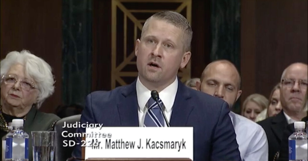
The first hearing in a lawsuit that seeks to overturn F.D.A. approval of the pills takes place Wednesday morning in Texas.AMARILLO, Texas — A hearing Wednesday in a lawsuit that seeks to overturn federal approval of a widely used abortion pill will provide the first opportunity to hear the arguments of the anti-abortion groups that filed the lawsuit and of the Food and Drug Administration, which is fighting to keep the abortion pill legal.The lawsuit, which seeks to end more than 20 years of legal use of medications for abortion, could have widespread implications in states where abortion is legal, not just where it is illegal. Medication abortion is used in more than half of pregnancy terminations in the United States and 40 percent of clinics that provide abortion services offer abortion pills only, not the surgical procedure.The hearing on Wednesday will revolve around the plaintiffs’ request that Judge Matthew J. Kacsmaryk of the Northern District in Texas grant a preliminary injunction ordering the F.D.A. to withdraw its longstanding approval of mifepristone, the first pill in the two-drug medication abortion regimen, while the case proceeds through trial.Judge Kacsmaryk, a Trump appointee who has written critically about Roe v. Wade and previously worked for a Christian conservative legal organization, took some unusual steps leading up to the hearing. In a meeting last Friday with lawyers for the parties in the case, he asked them to keep quiet about the fact that a hearing had been scheduled and told them he planned to delay making the public aware of it and would only enter it into the public court record the evening before.In a transcript of the Friday meeting obtained by The New York Times, Judge Kacsmaryk said that other aspects of the case have “brought a barrage of death threats and protesters and the rest” and that he wanted to avoid an “unnecessary circus-like atmosphere” that might disrupt the lawyers’ presentations in court.Despite the judge’s request, news organizations learned about the hearing and reported it. Members of groups that support abortion rights are planning to demonstrate outside the court building while the hearing is in progress, including by wearing kangaroo and judge costumes to protest what they consider to be a “kangaroo court” and by driving a truck around the city streets with a billboard that will say “a majority of Americans support abortion access.”The lawsuit claims that the F.D.A. did not adequately review the scientific evidence or follow proper protocols when it approved mifepristone in 2000 and that it has since ignored safety risks of the medication. The F.D.A. and the Department of Justice, which is representing the F.D.A., have strongly disputed those claims, saying the F.D.A.’s rigorous reviews of mifepristone over the years had repeatedly reaffirmed its decision to approve mifepristone, which blocks a hormone that allows a pregnancy to develop. It is unclear if the judge will rule at the end of the session Wednesday. Most legal experts expect that he will make his decision at a later date.Here are some of the issues that the judge has asked lawyers to be prepared to discuss at the hearing:Whether the plaintiffs have legal standing to bring the lawsuitThe plaintiffs are led by the Alliance for Hippocratic Medicine, an organization that lists five anti-abortion groups as its members. Shortly after the Supreme Court overturned Roe v. Wade, the alliance was incorporated in August in Amarillo, where Judge Kacsmaryk is the only federal judge.The five groups are not based in Amarillo, but Erik Baptist, senior counsel for the Alliance Defending Freedom, a conservative Christian legal advocacy group that is representing the plaintiffs, said that some of the groups’ members are in the Amarillo area, as is one of the four doctors who are plaintiffs in the case.The plaintiffs contend they have legal standing to sue as parties who experienced injury from the F.D.A. approval because they have treated women who they say have suffered harm from abortion pills. Legal experts, including some conservative legal scholars, said it might be difficult to support a claim that the plaintiffs have standing because the harm they are claiming could be considered several steps removed from the F.D.A.’s approving the drug. The intermediate steps include patients choosing to take the drug and then seeking medical care.If the judge were to decide that the plaintiffs do not have legal standing to sue, then the case could not proceed.Whether this lawsuit is an appropriate legal challenge to the F.D.A.’s authorityThe judge has indicated that he expects the parties to discuss issues related to the F.D.A.’s authority to approve and regulate drugs. Some legal experts have pointed to constitutional provisions and Congressional actions that suggest that the F.D.A. has overarching authority that cannot be second-guessed by a court. Legal experts say that if the judge were to rule for the plaintiffs, it would apparently be the first time a court had acted to order that a drug be removed from the market over the objection of the F.D.A.The F.D.A. has also said in its filing in the case that there is a six-year statute of limitations to challenge the agency’s actions and that the plaintiffs are bringing this lawsuit much too late.The plaintiffs argue that their lawsuit is an appropriate legal action.Did the F.D.A. err in approving mifepristone under the regulation used at the time?Mifepristone was initially approved in 2000 under a set of regulations called “Subpart H,” which was created to expedite the approval of drugs “that have been studied for their safety and effectiveness in treating serious or life-threatening illnesses.”The lawsuit alleges that mifepristone did not qualify for this type of approval because the plaintiffs say that “pregnancy is not an illness.”The approval process for mifepristone was not expedited — it took over four years — but the F.D.A. applied Subpart H, which allowed the agency to impose additional restrictions on use of the drug, including requiring health care providers to have special qualifications to prescribe it and dispense it only in certain medical settings.The F.D.A. argues that “illness” is a term that generally applies to medical conditions, not only to diseases. The agency says that any confusion in semantics was cleared up several years later when Congress created a new regulatory framework that used the term “disease or condition.”Are abortion medications unsafe?The plaintiffs claim that the pills, which they call “chemical abortion,” cause “cramping, heavy bleeding and severe pain” and that the F.D.A. has never adequately evaluated the scientific evidence for safety.The F.D.A. vigorously disputes this claim, as do mainstream medical organizations. They say that bleeding and cramping are normal consequences of the process, a sign that the pregnancy tissue is being expelled, and cite years of scientific studies that show that serious complications are rare. The organizations note that mifepristone has actually been regulated much more strictly and studied more intensively than most other drugs.For a dozen years, the F.D.A. has imposed on mifepristone an additional framework of restrictions and monitoring called a Risk Evaluation and Mitigation Strategy, or REMS. It is a framework that has been used for only about 300 other drugs. In recent years, the F.D.A. has extensively reviewed new data on mifepristone and concluded that the drug was safe enough to lift several of the restrictions, including the requirement that patients obtain the drug in person from a provider.

Read more →

One of the coronaviruses causing common colds boosts immune response to COVID-19 in children, study finds

During the pandemic, medical doctors and researchers noticed that children and adolescents infected with COVID-19 became less ill than adults. A possible explanation for this is that children already had a prior level of immunity to COVID-19 provided by memory T cells generated by common colds.
After studying unique blood samples from children taken before the pandemic, researchers from Karolinska Institutet in Sweden have now identified memory T cells that react to cells infected with SARS-CoV-2, the virus that causes COVID-19.
A possible explanation for this immunity in children is that they already had colds caused by one of the four coronaviruses causing seasonal common cold symptoms. This could stimulate an immune response with T cells able to also react to cells infected with SARS-CoV-2.
This new study reinforces this hypothesis and shows that T cells previously activated by the OC43 virus can cross-react against SARS-CoV-2.
“These reactions are especially strong early in life and grow much weaker as we get older,” says the study’s corresponding author Annika Karlsson, research group leader at the Department of Laboratory Medicine, Karolinska Institutet. “Our findings show how the T-cell response develops and changes over time and can guide the future monitoring and development of vaccines.”
The results indicate that the memory T-cell response to coronaviruses develops as early as the age of two. The study was based on 48 blood samples from two- and six-year-old children, and 94 samples from adults between the ages of 26 and 83. The analysis also included blood samples from 58 people who had recently recovered from COVID-19.
“Next, we’d like to do analogous studies of younger and older children, teenagers and young adults to better track how the immune response to coronaviruses develops from childhood to adulthood,” says Marion Humbert, postdoctoral researcher currently at the Department of Medicine Huddinge, Karolinska Institutet, joint first author with Anna Olofsson, doctoral student at the Department of Laboratory Medicine.
The paper is the result of a collaborative study among researchers at Karolinska Institutet, the universities of Bern (Switzerland), Oslo (Norway) and Linköping University (Sweden).
The study was financed by the Swedish Research Council, Region Stockholm (CIMED), Karolinska Institutet, the Knut and Alice Wallenberg Foundation and the European Research Council. Karl-Johan Malmberg, Ebba Sohlberg and Soo Aleman receive fees from companies and organisations outside this research project (see the paper for more details); all other researchers report no conflicts of interest.

Read more →

Researchers discover way to reverse infertility by reducing HDL cholesterol

Houston Methodist scientists reversed infertility in sterile mice by reducing high-circulating cholesterol with a bacterial protein, showing further evidence that links high cholesterol to female infertility. This is a promising development, with one in every five women of childbearing age in the U.S. unable to get pregnant after trying for a year.
“We are working with a protein, called serum opacity factor, with unique characteristics,” said Corina Rosales, Ph.D., assistant research professor of molecular biology in medicine with the Houston Methodist Research Institute and lead author on the study. “In our experiments, serum opacity factor lowered cholesterol levels by over 40% in three hours. So, this protein is quite potent.”
The results are published in the American Society for Biochemistry and Molecular Biology’s Journal of Lipid Research.
While this protein’s primary function is to increase bacterial colonization, it also alters the structure of cholesterol-carrying high-density lipoproteins, or HDLs, making it easier for the liver to dispose of the excess cholesterol that’s preventing conception. The researchers also noted that serum opacity factor’s dramatic action on HDL could be leveraged as a potential alternative to statins, which are the current gold standard for lowering cholesterol in people with atherosclerosis.
HDL, known as the “good cholesterol,” carries excess cholesterol from different tissues to the liver for breakdown, thereby bringing down cholesterol levels. However, if there is HDL dysfunction, lipid metabolism gets altered, which could then be harmful, like its counterpart LDL, or low-density lipoprotein. Often called “bad cholesterol,” LDL carries cholesterol from the liver to other tissues, with high levels of it causing accumulation and diseases.
“Both HDLs and LDLs contain a mixture of free and esterified cholesterol, and free cholesterol is known to be toxic to many tissues,” said Henry J. Pownall, Ph.D., professor of biochemistry in medicine at the Houston Methodist Research Institute and corresponding author on the study. “So, any dysfunction in HDL could be a risk factor for several diseases, too.”
To study HDL dysfunction, the researchers worked with preclinical mouse models that had unnaturally high levels of HDL cholesterol circulating in their bloodstream. While this made them ideal for studying atherosclerosis, Rosales observed that these mice were also completely sterile.

“Cholesterol is the backbone of all steroidal hormones, and an orchestra of hormones is needed to have a fertile animal,” Rosales said. “We know that the ovaries are studded with receptors for HDL, so the metabolism of HDL had to play a very important role in fertility for that reason.”
As predicted, when the researchers fed the sterile mice with a lipid-lowering drug, both LDL and HDL cholesterol levels reduced, and the animals were temporarily rescued from infertility. Motivated by these results, they turned to the bacterial protein serum opacity factor, known to be highly selective for HDL.
“Serum opacity factor is known mainly in the context of bacterial strep infections where it serves as a virulence factor. But it was also discovered that this protein only reacts to HDL and not to LDL or other lipoproteins,” Rosales said. “We hypothesized that perhaps administering serum opacity factor to these mice might help restore their fertility, as well.”
For their next set of experiments, the team engineered an adeno-associated virus to deliver the gene for serum opacity factor to the mice lacking HDL receptors that had high blood cholesterol. When the gene was expressed and the bacterial protein was produced, the animals’ HDL cholesterol significantly lowered, and their fertility was restored.
Based on these promising preclinical results, the researchers next plan to conduct a clinical study to investigate lipid levels in women undergoing treatments for idiopathic infertility, where the underlying causes are not fully known. If these patients have high HDL levels, then the researchers say serum opacity factor may be a line of future treatment.
“Even if we were to help 1% of women who are struggling to conceive, it would be life-changing for them, and I think that’s where we can make the most impact with our research,” Rosales said.
Rosales and Pownall’s collaborators on this study were Dedipya Yelamanchili, Baiba K. Gillard and Jing Liu with the Center for Bioenergetics and Department of Medicine at the Houston Methodist Research Institute; and Antonio M. Gotto Jr. with Weill Cornell Medicine’s Department of Medicine.
This research is funded by the National Institutes of Health (HL149804) and the Bass Endowment.

Read more →

How neuroimaging can be better utilized to yield diagnostic information about individuals

Since the development of functional magnetic resonance imaging in the 1990s, the reliance on neuroimaging has skyrocketed as researchers investigate how fMRI data from the brain at rest, and anatomical brain structure itself, can be used to predict individual traits, such as depression, cognitive decline, and brain disorders.
Brain imaging has the potential to reveal the neural underpinnings of many traits, from disorders like depression and chronic widespread pain to why one person has a better memory than another, and why some people’s memories are resilient as they age. But how reliable brain imaging is for detecting traits has been a subject of wide debate.
Prior research on brain-wide associated studies (termed ‘BWAS’) has shown that links between brain function and structure and traits are so weak that thousands of participants are needed to detect replicable effects. Research of this scale requires millions of dollars in investment in each study, limiting which traits and brain disorders can be studied.
However, according to a new commentary published in Nature, stronger links between brain measures and traits can be obtained when state-of-the-art pattern recognition (or ‘machine learning’) algorithms are utilized, which can garner high-powered results from moderate sample sizes.
In their article, researchers from Dartmouth and University Medicine Essen provide a response to an earlier analysis of brain-wide association studies led by Scott Marek at Washington University School of Medicine in St. Louis, Brenden Tervo-Clemmens at Massachusetts General Hospital/Harvard Medical School, and colleagues. The earlier study found very weak associations across a range of traits in several large brain imaging studies, concluding that thousands of participants would be needed to detect these associations.
The new article explains that the very weak effects found in the earlier paper do not apply to all brain images and all traits, but rather are limited to specific cases. It outlines how fMRI data from hundreds of participants, as opposed to thousands, can be better leveraged to yield important diagnostic information about individuals.

One key to stronger associations between brain images and traits such as memory and intelligence is the use of state-of-the-art pattern recognition algorithms. “Given that there’s virtually no mental function performed entirely by one area of the brain, we recommend using pattern recognition to develop models of how multiple brain areas contribute to predicting traits, rather than testing brain areas individually,” says senior author Tor Wager, the Diana L. Taylor Distinguished Professor of Psychological and Brain Sciences and director of the Brain Imaging Center at Dartmouth.
“If models of multiple brain areas working together rather than in isolation are applied, this provides for a much more powerful approach in neuroimaging studies, yielding predictive effects that are four times larger than when testing brain areas in isolation,” says lead author Tamas Spisak, head of the Predictive Neuroimaging Lab at the Institute of Diagnostic and Interventional Radiology and Neuroradiology at University Medicine Essen.
However, not all pattern recognition algorithms are equal and finding the algorithms that work best for specific types of brain imaging data is an active area of research. The earlier paper by Marek, Tervo-Clemmens et al. also tested whether pattern recognition can be used to predict traits from brain images, but Spisak and colleagues found that the algorithm they used is suboptimal.
When the researchers applied a more powerful algorithm, the effects got even larger and reliable associations could be detected in much smaller samples. “When you do the power calculations on how many participants are needed to detect replicable effects, the number drops to below 500 people,” Spisak says.
“This opens the field to studies of many traits and clinical conditions for which obtaining thousands of patients is not possible, including rare brain disorders,” says co-author Ulrike Bingel at University Medicine Essen, who is the head of the University Centre for Pain Medicine. “Identifying markers, including those involving the central nervous system, are urgently needed, as they are critical to improve diagnostics and individually tailored treatment approaches. We need to move towards a personalized medicine approach grounded in neuroscience. The potential for multivariate BWAS to move us towards this goal should not be underestimated.”
The team explains that the weak associations found in the earlier analysis, particularly through brain images, were collected while people were simply resting in the scanner, rather than performing tasks. But fMRI can also capture brain activity linked to specific moment-by-moment thoughts and experiences.
Wager believes that linking brain patterns to these experiences may be a key to understanding and predicting differences among individuals. “One of the challenges associated with using brain imaging to predict traits is that many traits aren’t stable or reliable. If we use brain imaging to focus on studying mental states and experiences, such as pain, empathy, and drug craving, the effects can be much larger and more reliable,” says Wager. “The key is finding the right task to capture the state.”
“For example, showing images of drugs to people with substance use disorders can elicit drug cravings, according to an earlier study revealing a neuromarker for cravings,” says Wager.
“Identifying which approaches to understanding the brain and mind are most likely to succeed is important, as this affects how stakeholders view and ultimately fund translational research in neuroimaging,” says Bingel. “Finding the limitations and working together to overcome them is key to developing new ways of diagnosing and caring for patients with brain and mental health disorders.”

Read more →