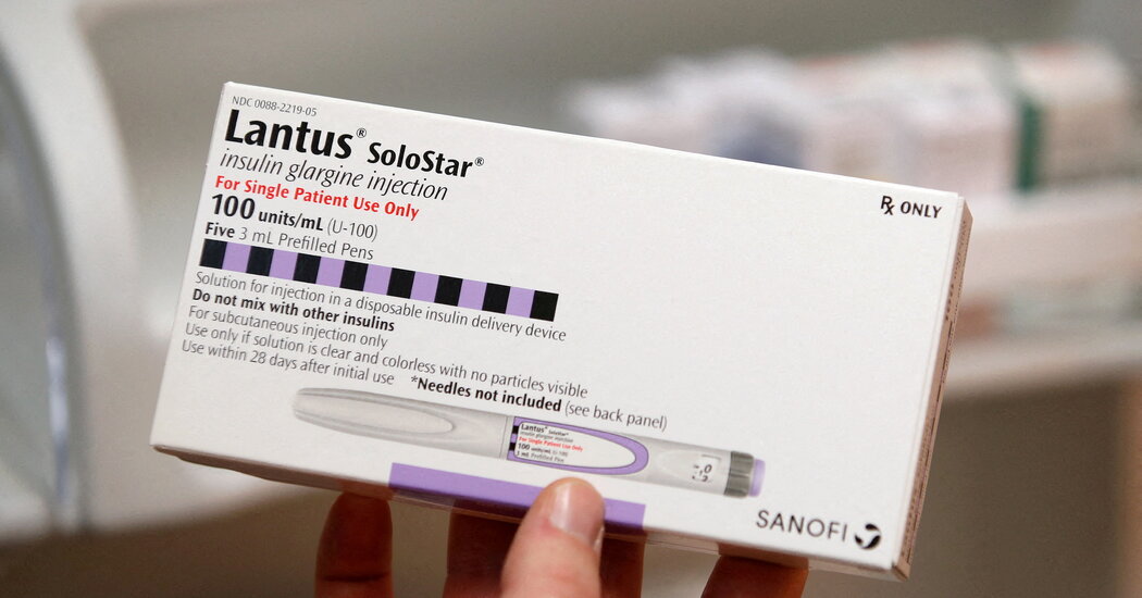
Sanofi Plans to Cut the Price of Insulin

The company is the third of the three major insulin manufacturers that dominate the U.S. market to announce such a move this month.Facing pressure to follow a wave of industry price cuts, the drug maker Sanofi said on Thursday that it would reduce the sticker price of its most commonly used insulin by 78 percent.The company said it would also cap, at $35 per month, that product’s out-of-pocket costs for diabetes patients with private health plans.Sanofi’s moves, which will go into effect at the start of next year, follow similar announcements this month by the two other large insulin manufacturers, Eli Lilly and Novo Nordisk. Together, the three companies control about 90 percent of the insulin market in the United States.The price cuts are likely to reduce how often Americans with diabetes struggle to pay for insulin, which millions depend on to stay alive. A federal law that went into effect at the start of this year had already capped out-of-pocket costs for insulin at $35 per month for people covered by Medicare.President Biden and Democratic lawmakers have taken credit for the drug makers’ moves, but the companies were facing fewer financial incentives to keep prices high on their older insulin products. Their businesses have grown more reliant on newer drugs for diabetes and obesity. They were also facing looming penalties that would have forced them to pay Medicaid back for raising their prices faster than inflation.For years, Sanofi repeatedly increased the list price of its most frequently prescribed insulin, Lantus, which the Food and Drug Administration first approved in 2000. The company said it was bringing in less from its insulin products after discounts and rebates were accounted for, compared with a decade ago, and it has blamed insurers for not passing savings down to patients.Sanofi already had a program capping monthly insulin costs at $35 per patient for the uninsured. Previously, all commercially insured patients were eligible for a Sanofi co-pay assistance program that significantly limited costs for most of them, but a cap was not in place. Under Sanofi’s new policy, the cap will automatically go into effect at the pharmacy counter, making it easier for patients to take advantage of.

Read more →

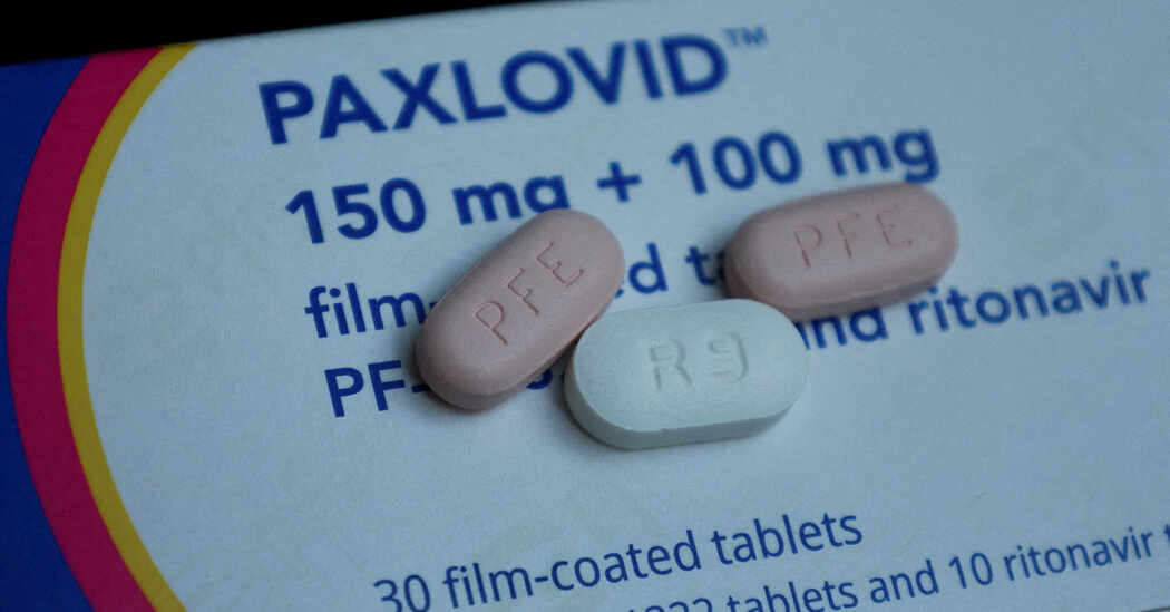
F.D.A. Advisers Endorse Paxlovid’s Benefits as a Covid Treatment

While an agency analysis did find signs of Covid “rebound,” the drug significantly reduces hospitalizations and deaths, researchers said.A panel of expert advisers to the Food and Drug Administration on Thursday endorsed Paxlovid as a treatment for adults with Covid who are at high risk for progression to severe illness. The move is likely to lead to full approval of the drug, which has been available under emergency use authorization.The 16-1 vote came after the agency released a new analysis showing that Paxlovid reduced hospitalizations and deaths among both unvaccinated and vaccinated people. Agency researchers estimated, based on Covid rates in January, that Paxlovid could “lead to 1,500 lives saved and 13,000 hospitalizations averted each week in the United States.”“I’d say, besides oxygen, Paxlovid has probably been the single most important treatment tool in this epidemic and continues to be,” Dr. Richard A. Murphy, chief of infectious diseases at Veterans Affairs White River Junction Medical Center in Vermont, said explaining his vote in favor of the treatment.Consumers have been particularly concerned about reports that people who take the drug experience a “rebound” of Covid, in which symptoms return a few days after disappearing. The F.D.A.’s analysis did find evidence of rebound among patients receiving the treatment, but the data also showed that some Covid patients who did not receive Paxlovid experienced rebound as well.The agency concluded that there was not a significant difference in rebound rates between the two groups and that rebound had no impact on the risk of developing severe illness. At Thursday’s meeting of experts, Dr. Stephanie Troy, an F.D.A. researcher, suggested that Covid rebound “may be a natural part of Covid-19 clinical course in a small subset of patients,” whether or not they take Paxlovid.Currently, only one antiviral drug, remdesivir, has full F.D.A. approval as a Covid treatment. Its use is limited, because patients have to visit a clinic for infusions three days in a row. Paxlovid, a pill, can be taken at home over the course of five days.Representatives of Pfizer, the manufacturer of Paxlovid, said on Thursday that the company was continuing to study the drug in patients who are immunocompromised or pregnant, as well as for the prevention of long Covid.Another pill, molnupiravir, also has emergency use authorization as a Covid treatment. But concerns have been raised about its safety, prompting regulators in Europe to recommend against its approval there.In Japan another antiviral pill, called Xocova, has emergency approval. The drug is in a clinical trial in the United States.The F.D.A. gave emergency use authorization for Paxlovid in December 2021 based on preliminary data from a clinical trial. In that trial, unvaccinated people who were at high risk of severe Covid — people over age 60 or with conditions such as diabetes — saw an 88 percent reduction in their risk of hospitalization.Pfizer has continued that trial and others. In one study, participants were vaccinated and at high risk, or unvaccinated and at low risk. In the latest analysis, Paxlovid reduced hospitalization and death by 86 percent in unvaccinated, high-risk subjects.Among high-risk people who were vaccinated, the reduction was 58 percent. People who gained immunity from a previous infection also saw a reduced risk after taking Paxlovid.The rebound phenomenon gained attention after such famous patients as President Biden and Dr. Anthony Fauci took the drug, tested negative for Covid and then, days later, tested positive again.The F.D.A. found that a small fraction of people who took Paxlovid experienced a rebound, as did those who took a placebo. In one trial, they estimated that 8.3 percent of people who took Paxlovid tested positive after an initial negative test, compared with 5.7 percent of patients given a placebo.Yet the rebound seen among those who took Paxlovid was not associated with a longer illness or a worsening of symptoms.“It’s good to know that this phenomenon exists, but data shows us that in a real-life, clinical sense, it’s not making much of on-the-ground impact in patients,” said Dr. Adi Shah, an infectious disease specialist at the Mayo Clinic who was not on the review panel.Since December 2021, when Paxlovid received emergency use authorization, federal officials have delivered more than 12.5 million doses nationwide. At the advisers meeting, Pfizer officials said 10 million doses had been taken in the United States and 14 million worldwide.Yet uptake has been more sluggish than expected. One concern is related to interactions between drugs commonly taken by older adults. The F.D.A. analysis examined that problem, too.Agency researchers found that a “sizable” proportion of patients who are eligible for Paxlovid also take drugs — whether to lower bad cholesterol or to treat high blood pressure — that can lead to interactions. All told, the F.D.A. has received 147 reports of hospitalizations and six deaths following drug-drug interactions with Paxlovid.“The drug-drug interactions is a significant concern” that requires ongoing attention, said Dr. Lindsey Baden of Harvard Medical School and chair of the expert committee.Apoorva Mandavilli

Read more →

Protein engineers navigate toward more targeted therapeutics

More than a third of FDA-approved drugs work by targeting a G protein-coupled receptor, or GPCR. The human body has more than 800 types of GPCRs that provide cells with information about the external environment to calibrate responses. Drugs that either block or activate GPCRs are used to treat a wide range of diseases including hypertension, pain and inflammation. Most drugs bind to the outside of the receptor, but this can result in adverse side effects since receptors often resemble one another.
In a new study published in Nature, Sivaraj Sivaramakrishnan, a professor in the College of Biological Sciences, along with graduate student Fred Sadler and co-authors Michael Ritt and Yatharth Sharma, uncovered the role of the third intracellular loop in the GPCR’s signaling mechanism, suggesting the possibility of a more targeted approach to drug discovery and a paradigm shift for new therapeutics.
“Typical GPCR drugs act as on or off switches for cellular signaling outcomes,” said Sivaramakrishnan. “Drugs that leverage the loop effectively can act as signaling dimmer switches to more precisely control drug responses.”
The authors developed new biochemical and biophysical tools, combined with computational measurements by collaborators Ning Ma and Nagarajan Vaidehi at the City of Hope Cancer Center. They tracked how the third intracellular loop changes in shape, or conformation, through the receptor signaling process. In a breakthrough for the field, their data show that the loop acts as a kind of gate to ensure that receptors activate the correct type of G protein signaling at the right intensity.
“A key advantage of this loop is that it is highly unique, even among closely related receptors, making it an outstanding drug target,” said Sadler. “Developing drugs through this newly discovered mechanism would allow for far more targeted therapeutics.”
Funding was provided by the National Institutes of Health and the University of Minnesota Graduate School.

Read more →

DNA treatment could delay paralysis that strikes nearly all patients with ALS

In virtually all persons with amyotrophic lateral sclerosis (ALS) and in up to half of all cases of Alzheimer’s disease (AD) and frontotemporal dementia, a protein called TDP-43 is lost from its normal location in the nucleus of the cell. In turn, this triggers the loss of stathmin-2, a protein crucial to regeneration of neurons and the maintenance of their connections to muscle fibers, essential to contraction and movement.
Writing in the March 16, 2023 issue of Science, a team of scientists, led by senior study author Don Cleveland, PhD, Distinguished Professor of Medicine, Neurosciences and Cellular and Molecular Medicine at University of California San Diego School of Medicine, with colleagues and elsewhere, demonstrate that stathmin-2 loss can be rescued using designer DNA drugs that restore normal processing of protein-encoding RNA.
“With mouse models we engineered to misprocess their stathmin-2 encoding RNAs, like in these human diseases, we show that administration of one of these designer DNA drugs into the fluid that surrounds the brain and spinal cord restores normal stathmin-2 levels throughout the nervous system,” Cleveland said.
Cleveland is broadly credited with developing the concept of designer DNA drugs, which act to either turn on or turn off genes associated with many degenerative diseases of the aging human nervous system, including ALS, AD, Huntington’s disease and cancer.
Several designer DNA drugs are currently in clinical trials for multiple diseases. One such drug has been approved to treat a childhood neurodegenerative disease called spinal muscular atrophy.
The new study builds upon ongoing research by Cleveland and others regarding the role and loss of TDP-43, a protein associated with ALS, AD and other neurodegenerative disorders. In ALS, TDP-43 loss impacts the motor neurons that innervate and trigger contraction of skeletal muscles, causing them to degenerate, eventually resulting in paralysis.
“In almost all of instances of ALS, there is aggregation of TDP-43, a protein that functions in maturation of the RNA intermediates that encode many proteins. Reduced TDP-43 activity causes misassembly of the RNA-encoding stathmin-2, a protein required for maintenance of the connection of motor neurons to muscle,” said Cleveland.
“Without stathmin-2, motor neurons disconnect from muscle, driving paralysis that is characteristic of ALS. What we have now found is that we can mimic TDP-43 function with a designer DNA drug, thereby restoring correct stathmin-2 RNA and protein level in the mammalian nervous system.”
Specifically, the researchers edited genes in mice to contain human STMN2 gene sequences and then injected antisense oligonucleotides — small bits of DNA or RNA that can bind to specific RNA molecules, blocking their ability to make a protein or changing how their final RNAs are assembled — into cerebral spinal fluid. The injections corrected STMN2 pre-mRNA misprocessing and restored stathmin-2 protein expression fully independent of TDP-43 function.
“Our findings lay the foundation for a clinical trial to delay paralysis in ALS by maintaining stathmin-2 protein levels in patients using our designer DNA drug,” Cleveland said.
Co-authors include: Michael W. Baughn, Jone López-Erauskin, Melinda S. Beccari, Roy Maimon, Sonia Vazquez-Sanchez, Jonathan W. Artates and Eitan Acks, all at Ludwig Institute for Cancer Research-UC San Diego and UC San Diego; Ze’ev Melamed, Ludwig Institute for Cancer Research-UC San Diego, UC San Diego, and The Hebrew University of Jerusalem; Karen Ling, Paayman Jafar-nejad, Frank Rigo and C. Frank Bennett, all at Ionis Pharmaceuticals; Aamir Zuberi, Maximilliano Presa, Elena Gonzalo-Gil and Cathleen Lutz, all at The Jackson Laboratory; Som Chaturvedi, Mariana Bravo-Hernández, Vanessa Taupin and Stephen Moore, all at UC San Diego; L. Sandra Ndayambaje and Ana R. Agra de Almeida Quadros, Harvard Medical School; Clotilde Lagier-Tourenne, Harvard University and Broad Institute of Harvard University and Massachusetts Institute of Technology.

Read more →

Breast cancer gene linked to Orkney islands

Published12 hours agoShareclose panelShare pageCopy linkAbout sharingImage source, FrancisXTA gene variant which is known to increase the risk of breast and ovarian cancer has been identified in people with Orkney heritage.A new study suggests one in 100 people with grandparents from Orkney has a specific mutation of the BRCA1 gene.It found that most of them could trace their family ancestry back to the island of Westray.It is believed to be the first time a geographic ancestral link of this kind has been made within the UK. The researchers also discovered the specific Orkney gene variant in smaller numbers in genetic testing across the UK and even in the US. Previous research has found that women from certain ethnic backgrounds, such as Ashkenazi Jews, also have a high rate of a specific BRCA gene variant.Across the UK about 1 in 1,000 people have a BRCA1 mutation, which can leaves women at a higher risk of ovarian and breast cancer.The BRCA genes are present in every person, both men and women, but when a fault occurs in one of them it can result in DNA damage and lead to cells becoming cancerous.People with a genetic variant have a 50% chance of passing it on to their children.Awareness of the faulty gene was raised a decade ago when Hollywood actress Angelina Jolie underwent a double mastectomy after discovering she had a BRCA1 variant.The operation was said to reduce her chances of getting breast cancer from 87% to 5%.However the NHS advises that risk-reducing surgery is not the only option.It also advises awareness of changes to breasts, annual breast screenings and MRI scans can help detect breast cancer, while lifestyle changes like healthy eating and exercise can “sometimes reduce risk”.There is currently no reliable screening test for ovarian cancer, it adds.Analysis by Laura Goodwin, BBC Scotland Science and Innovation CorrespondentIdentifying this variant – BRCA1 V1736A – is the result of 25 years of clinical research by Prof Zosia Miedzybrodzka, the director of the NHS Grampian Clinical Genetics service.Back then the breast cancer screening centre started to detect an increase in the number of families they were seeing and wanted to know why. Image source, University of AberdeenAs genetic testing grew, the team saw the same gene variant appearing time after time and became suspicious of its significance. Speaking to patients about their ancestry helped make the link to Westray, an outer Orkney island with a population of just 600.BRCA1 V1736A is likely to have arisen in a founder individual from Westray at least 250 years ago. So far 37 women of Orcadian heritage with the variant have been identified. Some won’t ever go on to develop cancer but others have chosen to have risk-reducing surgery. There are 20 people found to have the gene variant who don’t yet know they carry it. They were among more than 2,000 volunteers who gave genetic data to the Orcades (Orkney Complex Disease Study) study. The design of the study at the time meant information would not be disclosed. The team behind it have now asked the Research Ethics Committee for permission to contact the women identified to tell them they have the BRCA1 gene variant.Prof Jim Wilson of the University of Edinburgh, who ran the study, said: “In a person here on this island hundreds of years ago this variation happened in a BRCA 1 gene and now we are finding many descendants both in Westray and further afield in Scotland and beyond. “I think this is the most significant thing that I have ever really discovered. Its going to have immediate benefit to society at large.”‘It is important that we know’Former nurse Linda Hagan was born on Westray and has lived there most of her life.Her parents and grandparents and most of her ancestors were also from the island.Linda, 69, told the BBC that her younger sister died of breast cancer four years ago. She should get the results of her genetic screening shortly.Linda has three daughters and said it would be tough to think she might have passed the gene variant to them.”But it is important that we know and hopefully something can be done about it,” she said.Grandparents from WestrayThe latest research from the Universities of Aberdeen and Edinburgh was published in the European Journal of Human Genetics.The study looked back at the 80 grandparents of the BRCA1 variant carriers identified in the Orcades genetic study and found 60% were from Westray.Further ancestral links to the island went back to the early 18th century.The Orkney population is just 22,000 but there are people with a shared ancestry around the world and the researchers said they should be offered a targeted test for the variant.Currently in Scotland the test is available to those who know of a direct family connection to the gene or have a history of ovarian or breast cancer in their family.Planning is under way for a small pilot trial that will offer testing to anyone living in Westray with a Westray-born grandparent, regardless of a family history.If the pilot is successful, the long-term aim is to offer the test to everyone in Scotland with a Westray-born grandparent who wants it. NHS Grampian genetics clinic is running a helpline for queries about the gene variant linked to breast and ovarian cancer for those who have grandparents from Orkney. The number to call is 01224 553940. Email inquiries can be directed to gram.orkBRCAgene@nhs.scot GPs will not be able to assist with gene testing and any questions about this research and next steps should be directed to the helpline.More on this storyCancer survival ‘unaffected by faulty gene’12 January 2018Beating breast cancer with gene testing14 March 2018Angelina Jolie has double mastectomy14 May 2013’Screen more’ for cancer risk genes1 December 2014’I spotted a lump when preparing for my mikveh’3 July 2019

Read more →

Researchers chart a course for understanding, preventing, and treating young-onset colorectal cancer

Colorectal cancer among young people is increasing globally and rapidly. Experts expect it to become the leading cause of cancer death in individuals aged 20-49 in the U.S. by the year 2030.
Yet no one is certain why this disease is suddenly affecting so many young people. In a new paper published in Science, Dana-Farber Cancer Institute researchers outline the complexities of the disease and the research needed to map out a path toward understanding it.
“The rising incidence of young-onset colorectal cancer is extremely concerning, and it is urgent that the scientific community comes together to better understand the underlying causes and biology,” said co-author Kimmie Ng, MD, MPH, associate chief of gastrointestinal oncology and director of the Young-Onset Colorectal Cancer Center at Dana-Farber. The Center provides expert care for patients and conducts the multidisciplinary research required to understand colorectal cancer in young adults and develop new ways to prevent, detect and treat it.
Young-onset Colorectal Cancer: a unique challenge
Young-onset colorectal cancer (CRC), also called early-onset CRC, differs from later-onset CRC in several ways, according to the authors. Young-onset disease is often more aggressive, presents on the left side of the colon rather than the right, and often presents with rectal bleeding and abdominal pain.
At a molecular level, however, studies have shown conflicting results that suggest both similarities and differences in the genetic mutations that drive the diseases. This is likely due to the complexity of the disease, according to the authors, and future research should account for this variability.

More study is also needed to determine if CRC risk factors for young people are similar to those for older adults. Obesity and environmental exposures, for instance, have been associated with young-onset disease, but other factors could also play a role, such as increased antibiotic use or the frequency of Cesarean sections, both of which could influence the microbiome. To begin to understand the risk factors, the authors suggest that investigations should include a combination of genetics, environmental exposures, diet and lifestyle measures, as well as immune system interactions and the microbiome composition.
One clear difference is that young-onset CRC is typically discovered after the disease has advanced. This is due in part to the fact that screening for colorectal cancer starts at age 45 in the U.S., so the disease often goes undetected in younger people.
“It’s important not to dismiss the idea that a young person could have colorectal cancer even though the disease is still more common in older adults,” said co-author Marios Giannakis, MD, PhD, a gastrointestinal oncologist at Dana-Farber.
Responding with multidisciplinary research involving diverse populations
To account for the complexity of young-onset CRC, Ng and Giannakis said that research should be multidisciplinary and include many areas of investigation simultaneously. For instance, genome-wide association studies, which aim to find risk genes for the disease, should also include data about environmental exposures that could also increase risk.
These types of studies could point to new ways to identify young people who are at high risk of young-onset disease and should be screened for CRC. “Risk stratification is going to be very important as we think about screening for young-onset disease,” said Giannakis.
Clinical studies should also include the collection of blood, tissue, and stool samples from patients over time to shed light on the role of immune cells, environmental exposures and the microbiome in disease onset, progression, and treatment response. Ng and Giannakis encourage global collaborations aimed at facilitating the collection of these specimens, such as the Count Me In Colorectal Cancer Project, which directly partners with patients in the U.S. and Canada and makes all data available for research.
Ng and Giannakis also call for more effort in ensuring diverse populations are included in studies of young-onset CRC. Studies show underrepresented minorities are disproportionally burdened by young-onset CRC and non-Hispanic Black patients have a higher mortality rate when compared to non-Hispanic whites.
“Although each of these steps require commitment and perseverance,” said the authors, “it is the growing numbers of young patients bravely battling this disease that will be the compass that keeps us on the path towards better understanding, preventing, and treating young-onset colorectal cancer.”

Read more →

Artificial pancreas improves blood sugar control for kids ages 2-6, study finds

An artificial pancreas originally developed at the University of Virginia Center for Diabetes Technology improves blood sugar control in children ages 2 to 6 with type 1 diabetes, according to a new study. Details of the clinical study and its findings were just published in the New England Journal of Medicine.
Trial participants using the artificial pancreas spent approximately three more hours per day in their target blood sugar range compared with participants in a control group who continued relying on the methods they were already using to manage their blood sugar.
The Control-IQ system, manufactured by Tandem Diabetes Care, is a diabetes management device thatautomatically monitors and regulates blood glucose. The artificial pancreas has an insulin pump that uses advanced control algorithms based on the person’s glucose-monitoring information to adjust the insulin dose as needed.
Based on findings from two earlier studies, the system has previously been approved by the U.S. Food and Drug Administration for people ages 6 and older with type 1 diabetes.
“After the resounding success of Control-IQ technology in people ages 6 and up, it is very rewarding to see our youngest patients, and often the most challenging patients to help, benefit as well,” said Marc D. Breton, PhD, a UVA School of Medicine researcher who served as the trial’s principal investigator and was recently honored as UVA’s 2022 Innovator of the Year. “With these results, we have now accumulated years of clinical validation of this system across all age groups and look forward to seeing this life-changing technology made available to the broadest possible population.”
Used During Everyday Life
The study enrolled 102 children between ages 2 and 6 at three U.S. sites (UVA, Stanford University and the University of Colorado) and randomly assigned 68 of them to use the artificial pancreas system for 13 weeks, while the remaining 34 children were assigned to the control group. All participants maintained their regular daily routines during the study.

On average, the time participants using the artificial pancreas spent within their target blood glucose range was about 12 percentage points higher than participants in the control group overall and 18 percentage points higher during the overnight hours of 10 p.m. to 6 a.m. Nighttime blood glucose control is particularly important, as severe, unchecked hypoglycemia (very low blood glucose levels) can lead to seizures, coma or even death.
Overall, the researchers found, participants were able to use the artificial pancreas safely. There were two cases of severe hypoglycemia in the artificial pancreas group, compared with one in the control group. There was also one case of diabetic ketoacidosis in the artificial pancreas group, caused by a failure of the thin plastic tube that connects the insulin pump to the patient’s body.
Of note, most of the study-related visits — including 80% of the training sessions on the artificial pancreas and more than 90% of the overall visits — were conducted virtually. Achieving the reported results under these conditions highlights the ease of use of the technology and its potential for areas without easy access to endocrinologists.
“At the end of the day, this technology significantly improved glycemia and ensured safety of our youngest patients, but perhaps just as importantly it lessened these families’ constant anxiety about glucose levels, especially during the night.” Breton said. “It is incredibly rewarding for us to hear about these families’ experiences and how they manage to integrate these new tools in their life, offering some reprieve to the challenges they face.”
Findings Published
The study results have been published in the New England Journal of Medicine. The study’s authors are R. Paul Wadwa, Zachariah W. Reed, Bruce A. Buckingham, Mark D. DeBoer, Laya Ekhlaspour, Gregory P. Forlenza, Melissa Schoelwer, John Lum, Craig Kollman, Roy W. Beck and Breton.
This study was funded by the National Institutes of Health’s National Institute of Diabetes and Digestive and Kidney Diseases, grant U01DK127551. Tandem Diabetes Care provided the investigational closed-loop systems used in the trial, while Dexcom, Inc. provided the continuous glucose monitor supplies used in the trial.

Read more →

Maintaining heart function in donors declared 'dead by circulatory criteria' could improve access to heart transplantation

More donated hearts could be suitable for transplantation if they are kept functioning within the body for a short time following the death of the donor, new research has concluded.
The organs are kept functioning by restarting local circulation to the heart, lungs and abdominal organs — but, crucially, not to the brain — of patients whose hearts have stopped beating for five minutes or longer and have been declared dead by circulatory criteria (donation after circulatory death, or DCD).
It is hoped that this technique could increase the number of usable donated hearts by as much as 30% in the future, helping address the shortage of transplant organs. In 2021, 8,409 heart transplants were reported to the Global Observatory on Donation and Transplantation (GODT) by 54 countries. This activity is in contrast with the 21,935 patients who were on a heart waiting list during the year 2021, of whom 1,511 died while waiting and many others became too sick to receive a transplant.
John Louca, a final year medical student at Gonville & Caius College, University of Cambridge, and the study’s first author, said: “Heart transplants are the last bastion for patients with end-stage heart failure. They are successful — patients who receive a transplant live on average a further 13 to 16 years. The biggest problem they face is actually getting access to a donated heart: many patients will die before an organ becomes available. That’s why we urgently need to find ways to increase the suitability of donor organs.”
Though the first heart transplant performed at the Groote Schuur Hospital in Cape Town (South Africa) in 1967 was obtained from a DCD donor, this technique was abandoned and replaced by heart transplants obtained from donors confirmed dead using neurological criteria (donation after brain death, or DBD) — in other words, their brain has stopped functioning entirely.
Until recently, heart transplants worldwide were still performed only with organs obtained from DBD donors. However, in recent years, heart transplants from DCD donors have become a clinical reality worldwide thanks to years of research carried out in Cambridge.

DCD is the donation of organs by patients who tragically have a non-survivable illness. These patients are typically unconscious in intensive care in hospital and dependent on ventilation. Detailed discussions between doctors, specialist nurses and the patient’s family take place and if the family agree to organ donation, the process starts.
After treatment is withdrawn, the heart stops beating and it begins to sustain damage to its tissues. After 30 minutes, it is thought that this damage becomes irreversible and the heart unusable. To prevent this damage, at the time of death these non-beating hearts are transferred to a portable machine known as the Organ Care System (OCS) where the organ is perfused with oxygenated blood and assessed to see whether it is suitable for transplantation.
This technique was pioneered by Royal Papworth Hospital NHS Foundation Trust in Cambridge, whose transplant team carried out the first DCD heart transplant in Europe in 2015. Royal Papworth has since become the largest and most experienced DCD heart transplant centre in the world.
DCD heart transplantation started simultaneously in Australia, followed by Belgium, The Netherlands, Spain and USA. According to the GODT, 295 DCD heart transplants were performed in these six countries in 2021.
Organ Care Systems are expensive, costing around US$400,000 per machine plus an additional $75,000 for consumables for each perfused organ. An alternative, and much more cost-effective approach, is known as thoraco-abdominal normothermic reperfusion (taNRP). This involves perfusing the organ in situ in the donor’s body and is estimated to cost around $3,000. Its use was first reported in 2016 by a team at Royal Papworth Hospital.

In a study published in eClinical Medicine, an international team of clinical scientists and heart specialists from 15 major transplant centres worldwide, including the UK, Spain, the USA and Belgium, looked at clinical outcomes of 157 DCD donor hearts recovered and transplanted from donors undergoing taNRP. They compared these with the outcomes from 673 DBD heart transplants, which represents the ‘gold-standard’.
The team found that overall, the use of taNRP increased the donor pool significantly, increasing the number of heart transplantations performed by 23%.
Mr Stephen Large, Consultant Cardiothoracic Surgeon at Royal Papworth Hospital and chief investigator, said: “Withdrawing life support from a patient is a difficult decision for both the families and medical staff involved and we have a duty to honour the wishes of the donor as best we can. At present, one in ten retrieved hearts is turned down, but restoring function of the heart in situ could help us ensure more donor hearts find a recipient.”
Survival rates were comparable between DCD and DBD heart transplantation, with 97% of patients surviving for more than 30 days following taNRP DCD heart transplant, 93% for more than a year and 84% of patients still alive after five years.
Professor Filip Rega, Head of Clinic at the Department of Cardiac Surgery, UZ Leuven, Belgium, said: “This promising new approach will allow us to offer heart transplantation, a last resort treatment, to many more patients in need of a new heart.”
The researchers say that some of the benefits from taNRP are likely thanks to the reduced amount of time the heart was not receiving oxygenated blood, known as its warm ischaemic time, when compared to direct procurement (that is, when the heart is removed immediately for transplant, and perfused outside the body). The median average time was 16.7 minutes, significantly less than the 30 minutes associated with permanent damage to the heart cells.
An added benefit to this approach is that it allows medical teams to simultaneously preserve several organs, such as the liver, pancreas and kidneys, without the need of several organ-specific external machine perfusion devices. This decreases complexity and costs.
Professor Ashish Shah, Head of the Department of Cardiac Surgery at Vanderbilt University Hospitals, Nashville, USA, said: “Heart transplantation has been and always will be a uniquely international effort. The current study is another example of effective international collaboration and opens a new frontier, not just in transplantation, but in our basic understanding of how all hearts can be rescued.”
Dr Beatriz Domínguez-Gil, Director General of the National Organisation of Transplantation in Spain, said: “The results of this collaborative study bring hope to thousands of patients in need for a heart transplant every year throughout the world. Its findings reveal that DCD heart transplantation based on taNRP can lead to results at least similar to the gold standard and increase hearts available for transplantation in a manner that contributes to the sustainability of health-care systems.”

Read more →

Where the sidewalk ends

It’s easier than ever to view maps of any place you’d like to go — by car, that is. By foot is another matter. Most cities and towns in the U.S. do not have sidewalk maps, and pedestrians are usually left to fend for themselves: Can you walk from your hotel to the restaurants on the other side of the highway? Is there a shortcut from downtown to the sports arena? And how do you get to that bus stop, anyway?
Now MIT researchers, along with colleagues from multiple other universities, have developed an open-source tool that uses aerial imagery and image-recognition to create complete maps of sidewalks and crosswalks. The tool can help planners, policymakers, and urbanists who want to expand pedestrian infrastructure.
“In the urban planning and urban policy fields, this is a huge gap,” says Andres Sevtsuk, an associate professor at MIT and a co-author of a new paper detailing the tool’s capabilities. “Most U.S. city governments know very little about their sidewalk networks. There is no data on it. The private sector hasn’t taken on the task of mapping it. It seemed like a really important technology to develop, especially in an open-source way that can be used by other places.”
The tool, called TILE2NET, has been developed using a few U.S. areas as initial sources of data, but it can be refined and adapted for use anywhere.
“We thought we needed a method that can be scalable and used in different cities,” says Maryam Hosseini, a postdoc in MIT’s City Form Lab in the Department of Urban Studies and Planning (DUSP), whose research has focused extensively on the development of the tool.
The paper, “Mapping the Walk: A Scalable Computer Vision Approach for Generating Sidewalk Network Datasets from Aerial Imagery,” appears online in the journal Computers, Environment and Urban Systems. The authors are Hosseini; Sevtsuk, who is the Charles and Ann Spaulding Career Development Associate Professor of Urban Science and Planning in DUSP and head of MIT’s City Form Lab; Fabio Miranda, an assistant professor of computer science at the University of Illinois at Chicago; Roberto M. Cesar, a professor of computer science at the University of Sao Paulo; and Claudio T. Silva, Institute Professor of Computer Science and Engineering at New York University (NYU) Tandon School of Engineering, and professor of data science at the NYU Center for Data Science.

Significant research for the project was conducted at NYU when Hosseini was a student there, working with Silva as a co-advisor.
There are multiple ways to attempt to map sidewalks and other pedestrian pathways in cities and towns. Planners could make maps manually, which is accurate but time-consuming; or they could use roads and make assumptions about the extent of sidewalks, which would reduce accuracy; or they could try tracking pedestrians, which probably would be limited in showing the full reach of walking networks.
Instead, the research team used computerized image-recognition techniques to build a tool that will visually recognize sidewalks, crosswalks, and footpaths. To do that, the researchers first used 20,000 aerial images from Boston, Cambridge, New York City, and Washington — places where comprehensive pedestrian maps already existed. By training the image-recognition model on such clearly defined objects and using portions of those cities as a starting point, they were able to see how well TILE2NET would work elsewhere in those cities.
Ultimately the tool worked well, recognizing 90 percent or more of all sidewalks and crosswalks in Boston and Cambridge, for instance. Having been trained visually on those cities, the tool can be applied to other metro areas; people elsewhere can now plug their aerial imagery into TILE2NET as well.
“We wanted to make it easier for cities in different parts of the world to do such a thing without needing to do the heavy lifting of training [the tool],” says Hosseini. “Collaboratively we will make it better and better, hopefully, as we go along.”
The need for such a tool is vast, emphasizes Sevtsuk, whose research centers on pedestrian and nonmotorized movement in cities, and who has developed multiple kinds of pedestrian-mapping tools in his career. Most cities have wildly incomplete networks of sidewalks and paths for pedestrians, he notes. And yet it is hard to expand those networks efficiently without mapping them.

“Imagine that we had the same gaps in car networks that pedestrians have in their networks,” Sevtsuk says. “You would drive to an intersection and then the road just ends. Or you can’t take a right turn since there is no road. That’s what [pedestrians] are constantly up against, and we don’t realize how important continuity is for [pedestrian] networks.”
In the still larger picture, Sevtsuk observes, the continuation of climate change means that cities will have to expand their infrastructure for pedestrians and cyclists, among other measures; transportation remains a huge source of carbon dioxide emissions.
“When cities talk about cutting carbon emissions, there’s no other way to make a big dent than to address transportation,” Sevtsuk says. “The whole world of urban data for public transit and pedestrians and bicycles is really far behind [vehicle data] in quality. Analyzing how cities can be operational without a car requires this kind of data.”
On the bright side, Sevtsuk suggests, adding pedestrian and bike infrastructure “is being done more aggressively than in many decades in the past. In the 20th century, it was the other way around, we would take away sidewalks to make space for vehicular roads. We’re now seeing the opposite trend. To make best use of pedestrian infrastructure, it’s important that cities have the network data about it. Now you can truly tell how somebody can get to a bus stop.”

Read more →

Study offers a potential strategy to improve T cell therapy in solid tumors

A new approach that delivers a “one-two punch” to help T cells attack solid tumors is the focus of a preclinical study by researchers from the Perelman School of Medicine at the University of Pennsylvania. The findings, published in the Proceedings of the National Academy of Sciences (PNAS), showed that targeting two regulators that control gene functions related to inflammation led to at least 10 times greater T cell expansion in models, resulting in increased antitumor immune activity and durability.
CAR T cell therapy was pioneered at Penn Medicine by Carl H. June, MD, the Richard W. Vague Professor in Immunotherapy at Penn and director of the Center for Cellular Immunotherapies (CCI) at Abramson Cancer Center, whose work led to the first approved CAR T cell therapy for B-cell acute lymphoblastic leukemia in 2017. Since then, personalized cellular therapies have revolutionized blood cancer treatment, but remained stubbornly ineffective against solid tumors, such as lung cancer and breast cancer.
“We want to unlock CAR T cell therapy for patients with solid tumors, which include the most commonly diagnosed cancer types,” said June, the new study’s senior author. “Our study shows that immune inflammatory regulator targeting is worth additional investigation to enhance T cell potency.”
One of the challenges for CAR T cell therapy in solid tumors is a phenomenon known as T cell exhaustion, where the persistent antigen exposure from the solid mass of tumor cells wears out the T cells to the point that they aren’t able to mount an antitumor response. Engineering already exhausted T cells from patients for CAR T cell therapy results in a less effective product because the T cells don’t multiply enough or remember their task as well.
Previous observational studies hinted at the inflammatory regulator Regnase-1 as a potential target to indirectly overcome the effects of T cell exhaustion because it can cause hyperinflammation when disrupted in T cells — reviving them to produce an antitumor response. The research team, including lead author David Mai, a Bioengineering graduate student in the School of Engineering and Applied Science, and co-corresponding author Neil Sheppard, DPhil, head of the CCI T Cell Engineering Lab, hypothesized that targeting the related, but independent Roquin-1 regulator at the same time could boost responses further.
“Each of these two regulatory genes has been implicated in restricting T cell inflammatory responses, but we found that disrupting them together produced much greater anticancer effects than disrupting them individually,” Mai said. “By building on previous research, we are starting to get closer to strategies that seem to be promising in the solid tumor context.”
The team used CRISPR-Cas9 gene editing to knock out Regnase-1 and Roquin-1 individually and together in healthy donor T cells with two different immune receptors that are currently being investigated in Phase I clinical trials: the mesothelin-targeting M5 CAR (mesoCAR) and the NY-ESO-1-targeting 8F TCR (NYESO TCR). Neither engineered T cell product targets CD19, the antigen targeted by most approved CAR T cell therapies, as this antigen is not present in solid tumors.
After CRISPR editing, the T cells were expanded and infused in solid tumor mice models, where researchers observed the double knockout led to at least 10 times as many engineered T cells compared to disabling Regnase-1 alone, as well as increased antitumor immune activity and longevity of the engineered T cells. In some mice, it also led to overproduction of lymphocytes, causing toxicity.
“CRISPR is a useful tool for completely ablating the expression of target genes like Regnase and Roquin, resulting in a clear phenotype, however there are other strategies to consider for translating this work to the clinical setting, such as forms of conditional gene regulation,” Sheppard said. “We’re certainly impressed by the antitumor potency that was unleased by knocking out these two non-redundant proteins in combination. In solid tumor studies, we often see limited expansion of CAR T cells, but if we’re able to make each T cell more potent, and replicate them to greater quantities, we expect T cell therapies to have a better shot at attacking solid tumors.”
Additional authors include Omar Johnson, Jordan Reff, Ting-Jia Fan, and John Scholler. The research was supported by the National Institutes of Health (1P01CA214278, R01CA226983), the Parker Institute for Cancer Immunotherapy, the Emerson Collective, the Fontaine Fellowship, the Norman and Selma Kron Research Fellowship, and the Robert Wood Johnson Foundation Health Policy Research Scholars.

Read more →