
Kombucha to kimchi: Which fermented foods are best for your brain?

Many countries around the world have their own staple fermented foods which are ingrained into culture and diet. It can’t […]
Read more →
Many countries around the world have their own staple fermented foods which are ingrained into culture and diet. It can’t […]
Read more →
The intestinal or gut barrier is crucial for nutrient absorption and preventing harmful substances from leaking into the blood stream. Under diseased conditions, the disruption of the gut barrier may increase its permeability and result in a “leaky gut.”
The “leaky gut” syndrome often comes with symptoms like chronic diarrhea, constipation, or bloating. It has been associated with many diseases, including inflammatory bowel disease and non-alcoholic fatty liver disease.
Both diseases are highly prevalent in the general population, with the latter affecting around 40% of the Singaporean population, while inflammatory bowel disease affects 1-3 in 10,000 Singaporeans. However, treatment options for these two highly common diseases are limited. Repairing the leaky gut is thus a potential strategy for the treatment of these diseases.
At the same time, milk, as nature’s first functional food, plays essential roles in the development of the intestinal barrier and gut immune system. Both human and bovine milk are rich in extracellular vesicles (mEVs), which are nanosized particles containing beneficial components that can improve gut immunity and quality of gut bacteria.
However, it is unclear whether mEVs protect the gut barrier and treat the leaky gut.
To this end, Assistant Professor Jiong-Wei Wang from the Nanomedicine Translational Research Programme and Centre for NanoMedicine at the Yong Loo Lin School of Medicine, National University of Singapore (NUS Medicine), in collaboration with Professor Huaxi Yi from Ocean University of China, led a research team to investigate the potential treatment effects of mEVs on the leaky gut. This study is published in Science Advances.
The mEVs are obtained by removing milk fat, proteins and lactose with an in-house approach developed by Asst Prof Wang and his team. They discovered that large amounts of proteins and small nucleic acids carried in mEVs are associated with gut barrier function. The mEVs extracted from both human breast milk and cow milk carry similar therapeutic contents. The treatment efficacy of mEVs was demonstrated in laboratory models.
After orally administering mEVs to the models, the researchers observed that their intestinal inflammation was suppressed, and the damaged gut barrier was repaired. More importantly, the mEVs prevented the leakage of gut bacterial toxins into the blood stream, effectively averting toxin-induced liver damage.
This illustrates that oral administration of mEVs can potentially heal the leaky gut, and effectively slow down the progression of inflammatory bowel disease and nonalcoholic fatty liver disease.
“A leaky gut is a common effect of many diseases. However, whether the leaky gut is a symptom, or a cause of those diseases, remains debatable. Our research shows that treating the leaky gut with mEVs can ameliorate both inflammatory bowel disease and nonalcoholic fatty liver disease, two types of diseases that are seemingly unrelated,” said Asst Prof Wang, the lead author of this study.
“Another interesting finding is that mEVs extracted from milk produced at different stages after pregnancy¾colostrum, transient or mature milk, all exert similar gut barrier protection,” Asst Prof Wang added.
Colostrum milk refers to the milk initially produced by the breast during pregnancy, while transitional milk refers to milk produced 2 to 3 days postpartum. The milk then gradually changes from colostrum to mature milk.
According to the research team, a human adult may need to drink 1 litre of milk a day to achieve therapeutic effects on the aforementioned disease conditions. The mEVs are thus more beneficial for individuals with lactose intolerance.
Currently, the researchers are exploring the mechanisms underlying the treatment effects. The team is also working with doctors to explore clinical trials with patients in the near future.
MIT neuroscientists have found a way to reverse neurodegeneration and other symptoms of Alzheimer’s disease by interfering with an enzyme that is typically overactive in the brains of Alzheimer’s patients.
When the researchers treated mice with a peptide that blocks the hyperactive version of an enzyme called CDK5, they found dramatic reductions in neurodegeneration and DNA damage in the brain. These mice also showed improvements in their ability to perform tasks such as learning to navigate a water maze.
“We found that the effect of this peptide is just remarkable,” says Li-Huei Tsai, director of MIT’s Picower Institute for Learning and Memory and the senior author of the study. “We saw wonderful effects in terms of reducing neurodegeneration and neuroinflammatory responses, and even rescuing behavior deficits.”
With further testing, the researchers hope that the peptide could eventually be used as a treatment for patients with Alzheimer’s disease and other forms of dementia that have CDK5 overactivation. The peptide does not interfere with CDK1, an essential enzyme that is structurally similar to CDK5, and it is similar in size to other peptide drugs that are used in clinical applications.
Picower Institute Research Scientist Ping-Chieh Pao is the lead author of the paper, which appears this week in the Proceedings of the National Academy of Sciences.
Targeting CDK5
Tsai has been studying CDK5’s role in Alzheimer’s disease and other neurodegenerative diseases since early in her career. As a postdoc, she identified and cloned the CDK5 gene, which encodes a type of enzyme known as a cyclin-dependent kinase. Most of the other cyclin-dependent kinases are involved in controlling cell division, but CDK5 is not. Instead, it plays important roles in the development of the central nervous system, and also helps to regulate synaptic function.
CDK5 is activated by a smaller protein that it interacts with, known as P35. When P35 binds to CDK5, the enzyme’s structure changes, allowing it to phosphorylate — add a phosphate molecule to — its targets. However, in Alzheimer’s and other neurodegenerative diseases, P35 is cleaved into a smaller protein called P25, which can also bind to CDK5 but has a longer half-life than P35.
When bound to P25, CDK5 becomes more active in cells. P25 also allows CDK5 to phosphorylate molecules other than its usual targets, including the Tau protein. Hyperphosphorylated Tau proteins form the neurofibrillary tangles that are one of the characteristic features of Alzheimer’s disease.
In previous work, Tsai’s lab has shown that transgenic mice engineered to express P25 develop severe neurodegeneration. In humans, P25 has been linked to several diseases, including not only Alzheimer’s but also Parkinson’s disease and frontotemporal dementia.
Pharmaceutical companies have tried to target P25 with small-molecule drugs, but these drugs tend to cause side effects because they also interfere with other cyclin-dependent kinases, so none of them have been tested in patients.
The MIT team decided to take a different approach to targeting P25, by using a peptide instead of a small molecule. They designed their peptide with a sequence identical to that of a segment of CDK5 known as the T loop, which is a structure critical to the binding of CDK5 to P25. The entire peptide is only 12 amino acids long — slightly longer than most existing peptide drugs, which are five to 10 amino acids long.
“From a peptide drug point of view, usually smaller is better,” Tsai says. “Our peptide is almost within that ideal molecular size.”
Dramatic effects
In tests in neurons grown in a lab dish, the researchers found that treatment with the peptide led to a moderate reduction in CDK5 activity. Those tests also showed that the peptide does not inhibit the normal CDK5-P35 complex, nor does it affect other cyclin-dependent kinases.
When the researchers tested the peptide in a mouse model of Alzheimer’s disease that has hyperactive CDK5, they saw a myriad of beneficial effects, including reductions in DNA damage, neural inflammation, and neuron loss. These effects were much more pronounced in the mouse studies than in tests in cultured cells.
The peptide treatment also produced dramatic improvements in a different mouse model of Alzheimer’s, which has a mutant form of the Tau protein that leads to neurofibrillary tangles. After treatment, those mice showed reductions in both Tau pathologies and neuron loss. Along with those effects in the brain, the researchers also observed behavioral improvements. Mice treated with the peptide performed much better in a task that required learning to navigate a water maze, which relies on spatial memory, than mice that were treated with a control peptide (a scrambled version of the peptide used to inhibit CDK5-P25).
In those mouse studies, the researchers injected the peptide and found that it was able to cross the blood-brain barrier and reach neurons of the hippocampus and other parts of the brain.
The researchers also analyzed the changes in gene expression that occur in mouse neurons following treatment with the peptide. Among the changes they observed was an increase in expression of about 20 genes that are typically activated by a family of gene regulators called MEF2. Tsai’s lab has previously shown that MEF2 activation of these genes can confer resilience to cognitive impairment in the brains of people with Tau tangles, and she hypothesizes that the peptide treatment may have similar effects.
Tsai now plans to do further studies in other mouse models of diseases that involve P25-associated neurodegeneration, such as frontotemporal dementia, HIV-induced dementia, and diabetes-linked cognitive impairment.
“It’s very hard to say precisely which disease will most benefit, so I think a lot more work is needed,” she says.
The research was funded by the National Institutes of Health.
Houston Methodist nanomedicine researchers have found a way to tame pancreatic cancer — one of the most aggressive and difficult to treat cancers — by delivering immunotherapy directly into the tumor with a device that is smaller than a grain of rice.
In a paper recently published in Advanced Science, Houston Methodist Research Institute researchers used an implantable nanofluidic device they invented to deliver CD40 monoclonal antibodies (mAb), a promising immunotherapeutic agent, at a sustained low-dose via the nanofluidic drug-eluting seed (NDES). The result, found in murine models, was tumor reduction at a fourfold lower dosage than traditional systemic immunotherapy treatment.
“One of the most exciting findings was that even though the NDES device was only inserted in one of two tumors in the same animal model, we noted shrinkage in the tumor without the device,” said Corrine Ying Xuan Chua, Ph.D., co-corresponding author and assistant professor of nanomedicine at Houston Methodist Academic Institute. “This means that local treatment with immunotherapy was able to activate the immune response to target other tumors. In fact, one animal model remained tumor-free for the 100-days of continued observation.”
Pancreatic ductal adenocarcinoma is frequently diagnosed at advanced stages. In fact, about 85% of patients already have metastatic disease at diagnosis.
The Houston Methodist researchers are studying similar nanofluidic delivery technology on the International Space Station. Grattoni’s nanomedicine lab at Houston Methodist focuses on implantable nanofluidics-based platforms for controlled and long-term drug delivery and cell transplantation to treat chronic diseases.
Immunotherapy holds promise in treating cancers that previously did not have good treatment options. However, because immunotherapy is delivered throughout the entire body, it causes many side effects that are sometimes long-lasting, if not life-long. By focusing the delivery directly into the tumor, the body is protected from being exposed to toxic drugs and fewer side effects, essentially allowing patients undergoing treatment to have a better quality of life.
“Our goal is to transform the way cancer is treated. We see this device as a viable approach to penetrating the pancreatic tumor in a minimally invasive and effective manner, allowing for a more focused therapy using less medication,” said Alessandro Grattoni, Ph.D., co-corresponding author and chair of the Department of Nanomedicine at Houston Methodist Research Institute.
The NDES device consists of a stainless-steel drug reservoir containing nanochannels, thus creating a membrane that allows for sustained diffusion when the drug is released.
Other medical technology companies offer intratumoral drug-eluting implants for cancer therapeutics, but those are intended for shorter duration use. The Houston Methodist nanofluidic device is intended for long-term controlled and sustained release, avoiding repeated systemic treatment that often leads to adverse side effects.
Additional lab research is underway to determine the effectiveness and safety of this delivery technology, but researchers would like to see this become a viable option for cancer patients in the next five years.
Houston Methodist Research Institute collaborators on this study include Hsuan-Chen Liu, Daniel Davila Gonzalez, Dixita Ishani Viswanath, Robin Shae Vander Pol, Shani Zakiya Saunders, Nicola Di Trani, Yitian Xu, Junjun Zheng and Shu-Hsia Chen.
This research received funding support from the Golfers Against Cancer and the National Institutes of Health (NIH-NIGMS R01GM127558).
People with myotonic dystrophy experience progressive muscle weakness and repeated episodes of painless muscle stiffness called myotonia.
Investigators at Massachusetts General Hospital (MGH) recently used a targeted drug to restore muscle strength and correct myotonia in mice with myotonic dystrophy. The research, which is published in Nature Communications, could lead to new treatments for affected patients.
Myotonia in myotonic dystrophy is caused by abnormal processing (or splicing) of the transcript created from the gene that codes for the muscle chloride channel Clcn1, a protein that controls the flow of chloride ions into muscle cells.
The abnormal splicing leads to a truncated and poorly functioning Clcn1.
Also, the degree of weakness in patients with myotonic dystrophy is associated with higher amounts of oxidative, rather than glycolytic, muscle fibers. These fibers differ in how they obtain energy for contraction.
To correct the abnormal splicing in mice with myotonic dystrophy, a team led by Thurman Wheeler, MD, a neuromuscular researcher at MGH and an associate professor of Neurology at Harvard Medical School, used a genetic therapy involving small pieces of DNA called antisense oligonucleotides (ASOs).
The ASOs were based on a code that targets the abnormal splicing of Clcn1, and when injected directly into the animals’ muscles, the ASOs corrected the abnormality, boosted the abundance of functional Clcn1, increased the amount of glycolytic muscle fibers, and restored muscle health.
“Our findings show that muscle fiber type transitions in myotonic dystrophy result from myotonia and are reversible,” says Wheeler. “Our results also support Clcn1-targeting therapies as a way to increase strength and reduce muscle injury in patients.”
Additional co-authors include Ningyan Hu, Eunjoo Kim, and Layal Antoury.
This work was supported by the Elaine and Richard Slye Fund and the Muscular Dystrophy Association.
Researchers have created the largest atlas of post-zygotic genome mutations in healthy human tissue ever assembled — a scientific advancement that could unlock new avenues for diagnosing and treating genetic disease. It is the largest ever in terms of the combined number of tissues and number of donors sampled.
The study, led by researchers at Oregon Health & Science University, published today in the journal Science.
The development points the way toward understanding the genetic underpinnings of disease associated with cancer as well as innumerable conditions caused by cellular malfunction, including aging. The atlas could be useful in diagnosing medical conditions, and potentially useful in reversing genetic mutations that cause disease.
“If you’re talking about genetic changes being the basis of disease, there are a wide variety of technologies now that allow us to make changes to the genome,” said senior author Don Conrad, Ph.D., associate professor in the OHSU School of Medicine who directs the Division of Genetics at the Oregon National Primate Research Center. “It may be possible to change those mutations we’ve acquired due to bad luck or bad habits, and change them back to what they were before.”
Window into cell mutations
Researchers generated the atlas using 54 tissue and cell types compiled after death from 948 individuals who donated their bodies for the National Institute of Health’s Genotype-Tissue Expression Program.
Every person begins as a single cell at the moment of conception, carrying a DNA blueprint within the nucleus of that first fertilized cell. Using those original DNA instructions, the cell divides and replicates into vast groups of cells that form distinct tissues carrying out unique functions in the body. At any one time, the average person is comprised of about 30 trillion cells; in the course of a lifetime, that same person produces quadrillions of cells.
Over time, an individual cell is damaged again and again. In some cases, they repair themselves thousands of times a day.
“Every once in a while, the cell makes a mistake during DNA repair, or misses one — and that’s a change that gets propagated on,” Conrad said. “Our work gives us a window to the extent that those changes occur in different organs and tissues, and during different periods of our lives.”
This situation, known as somatic mosaicism, is a result of cells mutating from the original DNA blueprint.
Building the atlas
Until now, genetic research investigating mutations that occur post-zygotically, or after fertilization, has generally been conducted in biopsies of cancerous tissue such as skin melanomas and lung tumors, or in easily accessible tissues such as blood.
The new atlas opens a field of inquiry into mutations that occur over the course of a lifetime.
“Going from a single cell to a child is a remarkable process,” said lead author Nicole Rockweiler, Ph.D., previously part of Conrad’s lab at OHSU and Washington University in St. Louis, and now a postdoctoral associate at the Broad Institute of Massachusetts Institute of Technology and Harvard University. “When you add on layers of mutations happening at such an important part of our lives, it’s amazing that we can come out pretty well at the end of our gestation.”
To generate the new atlas, researchers developed a computational method using bulk RNA sequencing to characterize the mutations in a massive catalogue of tissue samples throughout the body. They were able to trace the point at which mutations occurred by mapping them to a “developmental tree,” indexing them across tissues and among many people. They found many mutations arose systematically and somewhat predictably as people age, although roughly 10% of mutations appeared to be the result of something intrinsic to an individual, be it genes or environment.
They found most detectable mutations occurred later in life, although many occurred before birth.
“We learned that some tissues, like the esophagus and liver, acquire a lot of mutations whereas other tissues like the brain, acquire fewer mutations,” Rockweiler writes in a post on the Conrad Lab website describing the research. “This made sense to us because the esophagus and liver are exposed to many environmental toxins; here the cells must transmit the message in a noisy environment. A low number of mutations in the brain also makes sense because the brain is primarily composed of cells that don’t replicate.”
Funding for the research was supported by the National Institutes of Health awards R01MH101810, R01HG007178 and R01HD078641; the National Human Genome Research Institute award T32HG000045; and the Medical Research Council awards MR/M004422/1 and MR/R023131/1.
“Don’t poke the bear”, they said. But that’s exactly what a team of scientists have been doing, to discover the secrets of blood clotting.
Hibernating bears, paralysed humans, and pigs kept in small enclosures all avoid dangerous blood clots, despite being immobile for extremely long periods.
Research from the University of Reading, with partners in Denmark, Germany, Norway and Sweden, shows that reduction of a key protein prevents the formation of blood clots in all three mammal species when they are still for days, weeks, months, or even years at a time. The study is published today (13 April 2023), in Science.
If you’ve ever taken a long haul flight, you might have taken advice to prevent a dangerous blood clot – deep vein thrombosis – from forming in one or both of your legs, while you sit still for multiple hours, dreaming of your destination. Perhaps you set a reminder to get up and walk around, and you wore compression socks to keep the blood from pooling in your legs.
Most people won’t experience a clot if they take care on a flight, but there is a serious risk for some people who are pre-disposed to blood clots, due to genetic factors.
The discovery that a protein known as Hsp47 is dramatically reduced, by 55 times, when someone is immobilised for a much longer period than a flight, could lead to new medicines to help those who have inherited blood clotting disorders that put them at risk for pulmonary embolism, heart attack, and stroke.
Professor Jon Gibbins led the work at the University of Reading. He said: “It seems counterintuitive that people who have severe paralysis don’t appear to be at higher risk of blood clots. This tells us that something interesting is happening. And it turns out that reducing levels of Hsp47 plays a key role in preventing clots, not just in humans, but in other mammals, including bears and pigs.
“When we see something like this in multiple species, that reinforces its importance. Having Hsp47 must have been an evolutionary advantage.”
Hsp47 is released by platelets – the sticky blood cells that trigger blood clotting. Usually clotting is an important response to an injury, to prevent blood loss, and Hsp47 is one of the necessary ingredients to enable platelets to do their job. Examining the role of Hsp47 in clotting function the team found that when released into the blood of bears, mice and humans that it promoted conditions that may give rise to deep vein thrombosis.
Professor Gibbins said “We aren’t totally sure how, but it appears that there is something about movement that keeps Hsp47 at an appropriate level. It could be that the mechanical forces involved in moving around actually have an impact on gene expression, dramatically increasing the amount of Hsp47 that circulates in the blood.”
The team took blood samples from bears in winter, while hibernating, and in summer, while awake and moving around. They also compared people who were immobilised with those who can move and walk. And finally, pigs kept in small pens were compared with others that were free to move around in barns. In all three cases, proteomics experiments showed that the absence of movement was associated with having far less Hsp47.
Professor Gibbins said: “Now we know that Hsp47 is so important, we can begin to look for new or existing medicines that might be able to inhibit the function of this protein in blood clotting and protect mobile people who are prone to clots.”
Responsible for over 5 million infections and 100,000 deaths every year, influenza remains one of the most challenging public health issues for populations globally, particularly First Nations communities.
New research from the Peter Doherty Institute for Infection and Immunity (Doherty Institute) has found that First Nations populations around the world are significantly more likely to be hospitalised and die from influenza compared to non-Indigenous populations.
Researchers from the Doherty Institute analysed 36 studies that examined influenza hospitalisations and deaths for First Nations and non-First Nations populations globally, finding that hospitalisation and mortality rates were consistently higher in First Nations communities than corresponding benchmark populations.
In Canada, New Zealand and Australia, First Nations people were over five times more likely to be hospitalised with the flu than the benchmark population.
The study’s authors note that data on the rates of severe flu in First Nations populations from low and middle income countries was scarce.
Senior author of the study and Epidemiologist at the Doherty Institute, Royal Melbourne Hospital’s Dr Katherine Gibney said that more needs to be done to determine the disease burden among First Nations populations in Australia and around the world.
“It is critical that governments ensure that people who have the flu have equitable access to healthcare and that vaccination rates are as high as possible,” Dr Gibney said.
“When we are planning for seasonal flu, but especially pandemic flu, we need to have specific and targeted plans for First Nations people that are generated by First Nations people.
“‘Australia did a fantastic job during COVID of having First Nations-led plans that worked well. And if that can be applied to the flu, it would be incredibly valuable.”
Dr Gibney added that surveillance of respiratory virus information is vital for management of the disease.
“When we get information about flu hospitalisations and deaths, we need to capture that individual’s First Nations status to determine whether the gap we have described is closing over time, and to continue to advocate for resources to reduce the disease burden in First Nations populations.”
Co-author of the study, Monash University’s Dr Juliana Betts said the study also shows the need for systemic and political reform.
“Our research emphasises the widespread and ongoing impacts of colonisation on the health outcomes of First Nations communities,” Dr Betts said.
“Solutions to these health gaps largely sit outside of the health sector, in policies that address the many social determinants of health including poverty, housing, education and racism.”
Need a little spring — or buzz — in your step? A wearable electrical-stimulation and vibration-therapy system designed by Rice University engineering students might be just what the doctor ordered for people experiencing foot pain and balance loss due to diabetic neuropathy.
Rice engineering students in the StimuSock team — Abby Dowse, Yannie Guo, Andrei Mitrofan, Sarah Park and Kelly Xu — designed a sock with a smart insole that can deliver both transcutaneous electrical nerve stimulation (TENS) and vibration therapy that block pain signals to the brain and provide haptic feedback to help with balance issues, respectively.
The team will present its device at the annual Oshman Engineering Design Kitchen showcase and competition April 13 at the Ion.
According to the Centers for Disease Control and Prevention’s 2022 estimates, over 37 million people in the U.S. suffer from diabetes. About half of them will develop some form of diabetic neuropathy, a type of nerve damage that occurs most often in the legs and feet.
The StimuSock team sought to combine the best aspects of existing therapies into a single, user-centered design.
“Existing products or devices used to treat the symptoms of diabetic neuropathy are either pharmaceuticals or large at-home vibration devices users stand on,” Dowse said. “But none of them can both treat pain and improve balance, which our device aims to do by combining the TENS and the vibrational therapy in one wearable, portable, user-controllable and easy-to-use device.”
A lot of the team’s effort went into making the device as low-profile as possible.
“The intent is for the patient to be able to wear the device for the whole day,” Guo said. “Even when everything’s off and they don’t want the electrostimulation or haptics effect, they can still wear their device. … You don’t want it to look like you’re wearing an ankle monitor.”
Patients use a smartphone app to control the type, intensity and duration of the desired therapeutic stimulus. The system also allows users to target a specific area of the foot.
“We have three regions: one in the front of the insole, one in the middle and one at the back,” Park said. “Our aim is to allow patients to be able to control both the amplitude of the vibration and the location where it’s delivered. Some patients might only want vibration at the front of their feet and some only at the back.”
Mitrofan said the team anticipates the device’s final form will have sufficient battery life to provide the recommended maximum of four 30-minute sessions of TENS therapy per day and operate on standby the rest of the day.
The team’s mentor is bioengineering assistant teaching professor Sabia Abidi .
Sign up for CNN’s Wonder Theory science newsletter. Explore the universe with news on fascinating discoveries, scientific advancements and more.
CNN
—
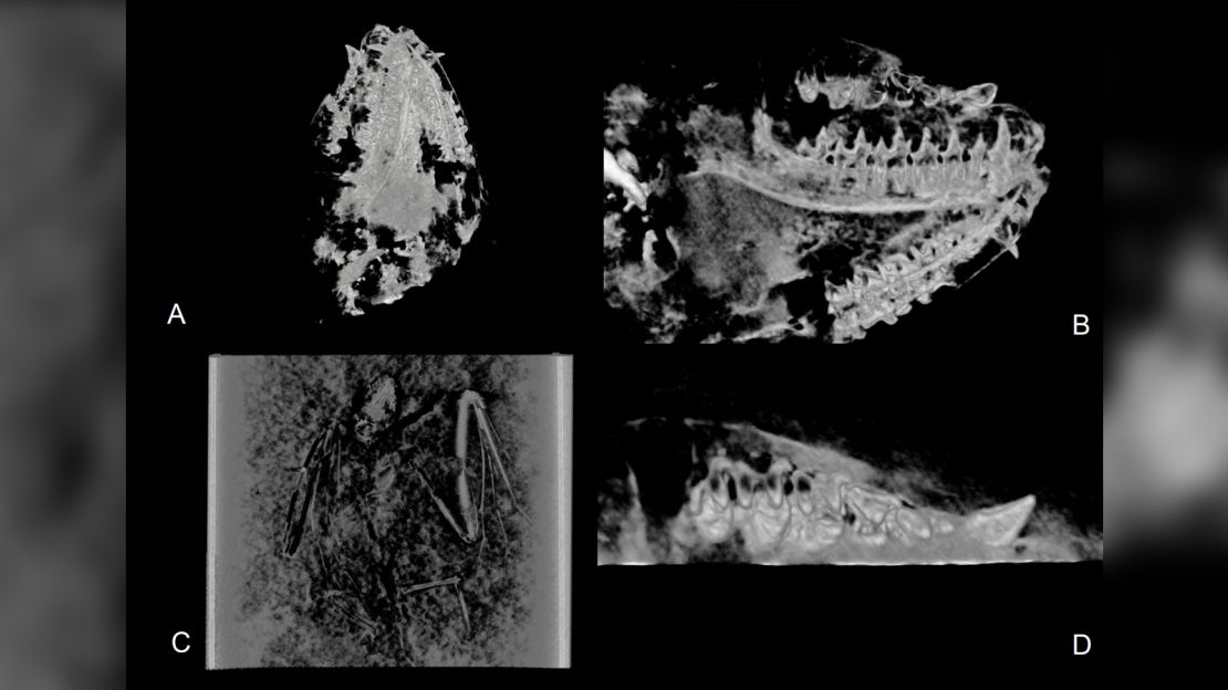
Two 52 million-year-old bat skeletons discovered in an ancient lake bed in Wyoming are the oldest bat fossils ever found — and they reveal a new species.
Tim Rietbergen, an evolutionary biologist at the Naturalis Biodiversity Center in Leiden, the Netherlands, identified the previously unknown bat species when he began collecting measurements and other data from museum specimens.
“This new research is a step forward in understanding what happened in terms of evolution and diversity back in the early days of bat,” he said.
Today, there are more than 1,400 living bat species found all over the world, with the exception of polar regions. But how the creatures evolved to be the only mammal capable of powered flight isn’t well understood.
The bat fossil record is patchy, and the two fossils Rietbergen identified as a new species were lucky finds — exceptionally well-preserved and revealing the animals’ complete skeletons, including teeth.
“Bat skeletons are small, light and fragile, which is very unfavorable for the fossilization process. They simply do not preserve well,” he said.
The newly discovered extinct bat species -— Icaronycteris gunnelli — was not much different from bats that fly around today. Its teeth revealed that it lived on a diet of insects. It was tiny, weighing in at only 25 grams (0.88 ounces).
“If it folds his wings next to its body, it would easily fit inside your hand. Its wings were relatively short and broad, reflecting a more fluttering flight style,” Rietbergen said.
This particular bat lived when Earth’s climate was warm and humid. The two skeletons Rietbergen studied survived the eons likely because the creatures fell into a lake, putting them out of reach of predators and into an environment more conducive to fossilization. The ancient lake bed is part of Wyoming’s Green River Formation and has yielded a number of bat fossils.
One of the two fossils was collected by a private collector in 2017 and purchased by the American Museum of Natural History. The other belonged to the Royal Ontario Museum in Toronto and was found in 1994.
The research was published in the scientific journal PLOS One on Wednesday.
Read more →