Landmark study involving babies in Ireland supports use of Cystic Fibrosis drug in infants from four weeks of age

A Cystic Fibrosis drug targeting the basic defect that causes the condition has been shown to be safe and effective in newborns aged four weeks and above, new research involving RCSI University of Medicine and Health Sciences and Children’s Health Ireland has found.
The finding is described as a ‘huge moment’ for Cystic Fibrosis by one of the lead researchers. The study included the first baby in the world with Cystic Fibrosis to be diagnosed from birth and enrolled directly onto a trial of this sort.
The drug, Ivacaftor (Kalydeko), is the first drug designed to treat the basic defect in Cystic Fibrosis. It was originally approved for adults, then sequentially over several years for older and younger children. Currently, it is approved for babies aged four months and older, however, this new research suggests that it is safe and effective for babies as young as four weeks of age.
Cystic Fibrosis experts predict that the earlier treatments can begin, the more likely that progression of the condition can be slowed down or halted in children, and this is the focus of several international research studies led by RCSI and Children’s Health Ireland. The findings of this study could pave the way for eligible newborns to start treatment on the medicine at the time of diagnosis (typically at newborn screening) rather than having to wait until they are four months old.
“This is a huge moment in Cystic Fibrosis,” said Paul McNally, Associate Professor of Paediatrics at RCSI and Consultant in Respiratory Medicine at CHI. McNally is one of the authors of the new study, which was published in the Journal of Cystic Fibrosis.
“Over the years Ivacaftor, or Kalydeko, has been put through clinical trials in younger and younger children. Now, through this study, it has been shown to be safe and effective all the way down to four weeks of age,” he said. “This is an important development because almost all children are diagnosed through newborn screening at around this time. The availability of a treatment that targets the underlying cause of the disease in newborns and can be started immediately at diagnosis will provide a huge sense of reassurance and hope for families.”
Cystic Fibrosis is an inherited disease that mainly affects the lungs and digestive system. Ireland has the highest incidence of the condition in the world: approximately 1,400 children and adults in Ireland live with the condition and more than 30 new cases of
Cystic Fibrosis are diagnosed here each year, typically around 4 weeks of age through the newborn screening programme.

In recent years, new medicines have emerged that target the basic defect that causes Cystic Fibrosis. Ivacaftor (Kalydeko) is one such treatment. It targets a genetic change seen in around 4% of people with Cystic Fibrosis worldwide, and around 10% in Ireland.
Siblings Kara (aged 5) and Isaac Moss (aged 2) both participated in the study through Children’s Health Ireland. Kara was part of an earlier phase of the study that paved the approval of the drug in older infants and led to the latest trial that Isaac took part in.
Isaac was the first baby with Cystic Fibrosis in the world to be diagnosed from birth and enrolled directly onto a trial of these ground-breaking treatments.
“Both Kara and Isaac are doing really well and remarkably are not experiencing any of the typical symptoms of Cystic Fibrosis at the moment,” said their mother Debbie.
“Research studies like this one are so important to ensuring that children get access to the right treatments as early as possible. With the right medications, they can enjoy a healthy childhood and look forward to a brighter future”
Ivacaftor is manufactured by pharmaceutical company Vertex Pharmaceuticals, who are currently applying to the European Medicines Agency for an extension to the marketing authorisation for Ivacaftor down to one month of age.
Read more →

Parkinson’s Disease: New theory on the disease’s origins and spread

The nose or the gut? For the past two decades, the scientific community has debated the wellspring of the toxic proteins at the source of Parkinson’s disease. In 2003, a German pathologist, Heiko Braak, MD, first proposed that the disease begins outside the brain. More recently, Per Borghammer, MD, with Aarhus University Hospital in Denmark, and his colleagues argue that the disease is the result of processes that start in either the brain’s smell center (brain-first) or the body’s intestinal tract (body-first).
A new hypothesis paper appearing in the Journal of Parkinson’s Disease on World Parkinson’s Day unites the brain- and body-first models with some of the likely causes of the disease-environmental toxicants that are either inhaled or ingested. The authors of the new study, who include Borghammer, argue that inhalation of certain pesticides, common dry cleaning chemicals, and air pollution predispose to a brain-first model of the disease. Other ingested toxicants, such as tainted food and contaminated drinking water, lead to body-first model of the disease.
“In both the brain-first and body-first scenarios the pathology arises in structures in the body closely connected to the outside world,” said Ray Dorsey, MD, a professor of Neurology at the University of Rochester Medical Center and co-author of the piece. “Here we propose that Parkinson’s is a systemic disease and that its initial roots likely begin in the nose and in the gut and are tied to environmental factors increasingly recognized as major contributors, if not causes, of the disease. This further reinforces the idea that Parkinson’s, the world’s fastest growing brain disease, may be fueled by toxicants and is therefore largely preventable.”
Different pathways to the brain, different forms of disease
A misfolded protein called alpha-synuclein has been in scientists’ sights for the last 25 years as one of the driving forces behind Parkinson’s. Over time, the protein accumulates in the brain in clumps, called Lewy bodies, and causes progressive dysfunction and death of many types of nerve cells, including those in the dopamine-producing regions of the brain that control motor function. When first proposed, Braak thought that an unidentified pathogen, such as a virus, may be responsible for the disease.
The new piece argues that toxins encountered in the environment, specifically the dry cleaning and degreasing chemicals trichloroethylene (TCE) and perchloroethylene (PCE), the weed killer paraquat, and air pollution, could be common causes for the formation of toxic alpha-synuclein. TCE and PCE contaminates thousands of former industrial, commercial, and military sites, most notably the Marine Corps base Camp Lejeune, and paraquat is one of the most widely used herbicides in the US, despite being banned for safety concerns in more than 30 countries, including the European Union and China. Air pollution was at toxic levels in nineteenth century London when James Parkinson, whose 269th birthday is celebrated today, first described the condition.
The nose and the gut are lined with a soft permeable tissue, and both have well established connections to the brain. In the brain-first model, the chemicals are inhaled and may enter the brain via the nerve responsible for smell. From the brain’s smell center, alpha-synuclein spreads to other parts of the brain principally on one side, including regions with concentrations of dopamine-producing neurons. The death of these cells is a hallmark of Parkinson’s disease. The disease may cause asymmetric tremor and slowness in movement and, a slower rate of progression after diagnosis, and only much later, significant cognitive impairment or dementia.

When ingested, the chemicals pass through the lining of the gastrointestinal tract. Initial alpha-synuclein pathology may begin in the gut’s own nervous system from where it can spread to both sides of the brain and spinal cord. This body-first pathway is often associated with Lewy body dementia, a disease in the same family as Parkinson’s, which is characterized by early constipation and sleep disturbance, followed by more symmetric slowing in movements and earlier dementia, as the disease spreads through both brain hemispheres.
New models to understand and study brain diseases
“These environmental toxicants are widespread and not everyone has Parkinson’s disease,” said Dorsey. “The timing, dose, and duration of exposure and interactions with genetic and other environmental factors are probably key to determining who ultimately develops Parkinson’s. In most instances, these exposures likely occurred years or decades before symptoms develop.”
Pointing to a growing body of research linking environmental exposure to Parkinson’s disease, the authors believe the new models may enable the scientific community to connect specific exposures to specific forms of the disease. This effort will be aided by increasing public awareness of the adverse health effects of many chemicals in our environment. The authors conclude that their hypothesis “may explain many of the mysteries of Parkinson’s disease and open the door toward the ultimate goal-prevention.”
In addition to Parkinson’s, these models of environmental exposure may advance understanding of how toxicants contribute to other brain disorders, including autism in children, ALS in adults, and Alzheimer’s in seniors. Dorsey and his colleagues at the University of Rochester have organized a symposium on the Brain and the Environment in Washington, DC, on May 20 that will examine the role toxicants in our food, water, and air are playing in all these brain diseases.
Additional authors of the hypothesis paper include Briana De Miranda, PhD, with the University of Alabama at Birmingham, and Jacob Horsager, MD, PhD, with Aarhus University Hospital in Denmark.

Read more →

Synthetic platelets stanch bleeding, promote healing in animal models

Researchers have developed synthetic platelets that can be used to stop bleeding and enhance healing at the site of an injury. The researchers have demonstrated that the synthetic platelets work well in animal models but have not yet begun clinical trials in humans.
A number of medical situations require platelet transfusions — such as cases of severe bleeding, or for patients who are going into surgery or receiving chemotherapy. Currently, patients in any of those situations receive platelets harvested from blood donors, ideally from donors with a compatible blood type. This is challenging, because there is a very limited supply of platelets available, those platelets have a limited shelf life, and the platelets must be stored under controlled conditions.
“We’ve developed synthetic platelets that can be used with patients of any blood type and are engineered to go directly to the site of injury and promote healing,” says Ashley Brown, corresponding author of a paper on the synthetic platelets and an associate professor in the joint biomedical engineering program at North Carolina State University and the University of North Carolina at Chapel Hill. “The synthetic platelets are also easy to store and transport, making it possible to give the synthetic platelets to patients in clinical situations sooner — such as in an ambulance or on the battlefield.”
The synthetic platelets are made of hydrogel nanoparticles that mimic the size, shape and mechanical properties of human platelets. Hydrogels are water-based gels that are composed of water and a small proportion of polymer molecules.
“Our synthetic platelets are deformable — meaning they can change shape — in the same way that normal platelets are,” Brown says.
The researchers engineered the surface of the synthetic platelets to incorporate antibody fragments that bind to a protein called fibrin. When a body is injured, it synthesizes fibrin at the site of the wound. The fibrin then forms a mesh-like substance to promote clotting.
“Because the synthetic platelets are coated with these antibody fragments, the synthetic platelets travel freely through the blood stream until they reach the wound site,” Brown says. “Once there, the antibody fragments bind to the fibrin, and the synthetic platelets expedite the clotting process.”
In addition to forming a clot within the fibrin network, the synthetic platelets act to contract the clot over time — just like normal platelets.

“This expedites the process of healing, allowing the body to move forward with tissue repair and recovery,” Brown says.
The researchers initially demonstrated the efficacy of the antibody fragments via in vitro testing, as well as demonstrating that the antibody fragments and synthetic platelets could be produced at scales that would make them viable for large-scale manufacturing.
The researchers then used a mouse model to determine the optimal dose of synthetic platelets necessary to stop bleeding.
Subsequent research in both mouse and pig models demonstrated that the synthetic platelets traveled to the site of a wound, expedited clotting, did not cause any clotting problems in areas outside of the wound, and accelerated healing.
“In the mouse and pig models, healing rates were comparable in animals that received platelet transfusions and synthetic platelet transfusions,” Brown says. “And both groups fared better than animals that did not receive either transfusion. We also found that the animals in both mouse and pig models were able to safely clear the synthetic platelets over time through normal kidney function. We didn’t see any adverse health effects associated with the use of the synthetic platelets.
“In addition, based on our preliminary estimates, we anticipate that the cost of the synthetic platelets — if they are approved for clinical use — would be comparable to the current cost of platelets,” Brown says.

“We are wrapping up preclinical efficacy testing and are in the process of securing funding for preclinical safety work that should allow us to obtain FDA approval to begin clinical trials within two years.”
The paper, “Ultrasoft Platelet-like Particles Stop Bleeding in Rodent and Porcine Models of Trauma,” is published in the journal Science Translational Medicine. Co-lead authors of the paper are Kimberly Nellenbach, Seema Nandi and Emily Mihalko. Nellenbach and Nandi are former postdoctoral researchers in the joint biomedical engineering program at NC State and UNC; Mihalko is a former Ph.D. student in the program.
The paper was co-authored by Jennifer Sollinger, laboratory manager in Brown’s lab; Nina Moiseiwitsch, a former Ph.D. student in the joint biomedical engineering program at NC State and UNC; Ana Sheridan and Sanika Pandit, current Ph.D. students in the joint program; Drew Koch, a former grad student at NC State; Lauren Schnabel, a professor of clinical sciences in NC State’s College of Veterinary Medicine; Jagathpala Shetty, Leandro Moretti and Thomas Barker, of the University of Virginia; Maureane Hoffman of Duke University; and Andrew Lyon of Chapman University.
Brown, Nandi, Lyon and Barker are all co-founders of a start-up company called SelSym Biotech that is focused on developing and marketing synthetic platelets for clinical use. Nellenbach owns stock in SelSym Biotech.
This research was done with support from the National Heart, Lung, and Blood Institute under grants R01HL130918, R01HL146701, R01HL162809 and F30HL163869; the National Institute of General Medical Sciences, under grant 1T32GM133366; the National Institutes of Health, under grant T32OD011130; the Department of Defense, under grant W81XWH-15-1-0485; North Carolina Biotechnology Translational Research Grant TRG-573547; the National Science Foundation, under grant 1847488; the American Heart Association, under grant AHA18PRE33990338; the U.S. Department of Veterans Affairs; and the North Carolina State University Chancellor’s Innovation Fund.

Read more →

Study lays the basis for new knowledge on gastrointestinal diseases

The meeting point of the stomach and esophagus, the so-called gastro-esophageal junction, is a region of the human body that is not well-suited to the modern lifestyle. Stress, alcohol, nicotine and severe obesity are often triggers for pathological changes to the mucosal membrane in this area, often resulting in esophageal cancer.
An international research team has now gained new insights into the development of the cells, their communication with each other, and their regulation at the junction of the esophagus and stomach. With the help of specially developed mini-organs, so-called organoids, and with techniques that make it possible to track and profile individual cells, they have been able to follow the development of the gastro-esophageal junction from embryonic to adult stage in detail using animal experiments.
New Insights into the Development of the Gastrointestinal Tract
Their results reveal the complex communication at the cellular level and the specific pathways that these cells use to communicate. They provide new insights into the development of the gastro-esophageal junction and thus have significant implications for the understanding, prevention and treatment of gastrointestinal diseases. At the same time, they present new starting points for medical research and the development of new therapies.
Cindrilla Chumduri is responsible for this study, which has now been published in the journal Nature Communications. Until recently, the infection and cancer biologist was a research group leader at the Department of Microbiology at Julius-Maximilians-Universität Würzburg (JMU); she is now an associate professor at Aarhus University (Denmark). Other participants came from Charité — Universitätsmedizin and the Max Planck Institute for Infection Biology in Berlin.
“This collaboration underlines the importance of different expertise to improve our understanding of the biology of the gastrointestinal tract,” says Chumduri. She herself has many years of experience in research with organoids. Among other things, she has used mini-organs she developed to study how cells in the cervix degenerate and turn cancerous — another region where different types of mucosal cells collide.
Where Different Epithelia Meet
“The squamous epithelia of the esophagus and the columnar epithelia of the stomach meet at the gastroesophageal junction,” explains Dr. Naveen Kumar Nirchal, one of the first authors of the study. The area is known as a “hotspot for the development of metaplasia” — the replacement of one type of cell by another.

Barrett’s esophagus, a precursor to esophageal cancer, often develops there, the number of cases of which has increased dramatically in the Western world over the past four decades. “Barrett’s esophagus is characterized by the replacement of the resident squamous epithelium of the esophagus by other cell types that are not normally found in this tissue,” says the scientist.
However, it is still unclear why this region is so susceptible to this process. In order to better understand this transformation, it is therefore first necessary to decipher the normal development process in detail — from embryo to mature adult. “This is the only way to determine the tissue changes that trigger the progression of the disease, explains Dr. Rajendra Kumar Gurumurthy, another researcher of the study.
A Never-Before-Seen Insight into the Development of this Region
This has now been achieved: By using a novel approach that combines organoid and mouse models with advanced single-cell transcriptome analyses over time and space, the research team has shed light on the complex developmental process of the gastroesophageal junction. “We were able to provide unprecedented insight into the development of this region from the embryonic stage to adulthood in mice and identify the intricate composition of the cells involved and how they develop,” explains Pon Ganish Prakash, another scientist involved in the study.
The work shows the sophisticated communication between different cell types within the gastroesophageal junction and the signaling pathways involved. “This understanding opens up new avenues for research into gastrointestinal diseases,” says Cindrilla Chumduri.
Above all, the precision of the single-cell analysis in their study opens new doors to understanding how pathological processes develop and to developing innovative treatments, the team writes in its study. The work will therefore be a “cornerstone for understanding the development of such diseases” and will significantly influence the approach to the early detection and treatment of diseases in this important part of the digestive system.

Read more →

Chemicals stored in home garages linked to ALS risk

Over the last decade, researchers at University of Michigan continue to find that exposure to environmental toxins — from pesticides used in agriculture to volatile organic compounds in the manufacturing industry — is linked to the development of amyotrophic lateral sclerosis, or ALS.
The buildup of exposures, which researchers call the ALS exposome, is possibly associated with recreational activities such as woodworking and gardening.
Now, a Michigan Medicine study finds that storing chemicals in a garage at home may associate with an increased risk of ALS.
The results are published in Amyotrophic Lateral Sclerosis and Frontotemporal Degeneration.
“Identifying disease-provoking exposures can inform and motivate interventions to reduce exposure, risk and, ultimately, the ALS burden,” said first author Stephen Goutman, M.D., M.S., director of the Pranger ALS Clinic and associate director of the ALS Center of Excellence at University of Michigan.
“Exposures in the home setting are an important part of the ALS exposome, as it is one place where behavior modifications could possibly lessen ALS risk.”
Storage containing volatile chemicals in garages is extremely common, whether it’s in a car or motorcycle, equipment like a chainsaw, or solvents, cleaners, paints and other items.

Investigators assessed exposures in the residential setting from a survey of more than 600 participants both with and without ALS. Through statistical analysis, they found that the storage of chemicals — including gasoline and gasoline powered equipment, lawn care products, pesticides, paint and woodworking supplies — were significantly associated with ALS risk.
All of the reported chemicals linked to disease development were volatile with toxic components. Most participants reported storing several of the items in their attached garage.
Storing chemicals in a detached garage, however, did not show as strong of an association with risk.
Researchers say the flow of air and airborne pollutants from attached garages to the living space may explain the finding.
“Especially in colder climates, air in the garage tends to rush into the house when the entry door is opened, and air flows occur more or less continuously through small cracks and openings in walls and floors,” said Stuart Batterman, Ph.D., senior author and professor of environmental health science at the U-M School of Public Health.
“Thus, it makes sense that keeping volatile chemicals in an attached garage shows the stronger effect.”
The latest building codes, Batterman notes, tackle this problem by specifying measures to reduce or eliminate these air flows.

“We are beginning to see risk factors across multiple settings that may associate with a greater ALS risk; we also see some relationships across the studies, for example, woodworking and woodworking supplies and gardening and lawn care supplies,” Goutman said.
“This begs the question: is it the activities that associate with ALS risk or the exposures to related products? This requires further research.”
In 2016, the research team found that people with ALS had higher concentrations of pesticides in their blood compared to people without the condition.
A subsequent study published in 2019 linked organochlorine pesticides and polychlorinated biphenyls, or PCBS, to worsening survival for ALS.
“With each study, we better understand the types of exposures that increase the risk of developing ALS,” said senior author Eva Feldman, M.D., Ph.D., director of the ALS Center of Excellence at U-M and James W. Albers Distinguished University Professor at U-M.
“We now need to build on these discoveries to understand how these exposures increase ALS risk. In parallel, we must continue to advocate to make ALS a reportable disease. Only then we will fully understand the array of exposures that increase disease risk.”
Studies to understand how environmental exposures contribute to the development of ALS and other neurodegenerative diseases, both of people with and without family history of the condition, are underway.

Read more →

In the drive to deprescribe, heartburn drug study teaches key lessons

Whether it’s costs, safety risks or “pill fatigue” they’re trying to reduce, many health systems and clinics have started working on ways to encourage deprescribing of medications that patients may not need.
Now, a new study published in the BMJ shows the potential promise, and pitfalls, of a massive effort to reduce overuse of a common class of heartburn medications known as proton pump inhibitors or PPIs.
The findings also reveal that some of the feared risks from PPIs may be overblown.
The study tracks the impact of an intervention that imposed limits on PPI prescription size and refills for patients without a documented reason to be on the medication, discontinued old prescriptions, and provided education to patients and clinicians on alternatives.
The effort was carried out in one region of the Veterans Health Administration system, called VISN 17, and involved a quarter of a million patients, making it one of the largest ever studies on deprescribing.
Key findings
In all, the intervention led to a massive reduction in PPI use: a nearly 30% reduction in prescriptions of PPIs compared to other VA regions.

But the drive to reduce potentially unnecessary PPI use had one unintended consequence: a drop in prescribing to veterans who actually have an ongoing need to take PPIs because their other medicines carry a high risk of gastrointestinal bleeding. Strong evidence shows that PPIs are effective for preventing gastrointestinal bleeding and they are recommended in clinical guidelines.
Reassuringly, no matter the reason for taking PPIs, the deprescribing effort didn’t lead to increases in health care visits with gastrointestinal diagnoses. Nor did it lead to increases in gastrointestinal bleeding in patients at high risk, which suggests that the deprescribing initiative itself was safe.
Interestingly, the rate of purported negative PPI effects — such as kidney disease, stroke, heart attack or pneumonia — didn’t go down in VISN17 relative to the other regions. Hip fractures, another risk linked with PPIs in past studies, only went down by a small percentage.
This supports evidence from other high-quality studies that suggest PPIs may be a marker of patients at risk for certain adverse outcomes, but that the drugs are unlikely to be the cause.
For this reason, the main benefits to deprescribing PPIs have more to do with cost and hassle of taking more pills than clinical risk reduction.
More about the study
The new VA-funded study uses data from multiple years before and after VISN 17 implemented its PPI deprescribing program for most veterans living in Texas, and parts of New Mexico and Oklahoma.

It was led by a multi-institutional team that includes investigators from University of Michigan and the VA Center for Clinical Management Research (CCMR) in Ann Arbor; the University of Pennsylvania and the VA Center for Health Equity Research and Promotion (CHERP) in Philadelphia; and the Yale School of Medicine and VA Center for Pain Research, Informatics, Multi-morbidities, and Education (PRIME).
“This intervention worked so well because it was involuntary to some degree — refills could no longer be on autopilot for patients without a clear indication for the medication,” says Jacob Kurlander, M.D., M.S., first author of the study and a gastroenterologist at Michigan Medicine, U-M’s academic medical center, and the Lieutenant Colonel Charles S. Kettles VA Ann Arbor Medical Center. “At the same time, what we saw is that is that patients who benefit from PPIs for bleeding prevention — which is sometimes overlooked by doctors — got swept up in this effort, too.”
This signals that deprescribing efforts need to take even more care to ensure providers don’t allow a patient who has a need for the drug to inadvertently go off it, Kurlander said.
“Our findings also suggest that PPIs may not be as harmful as some have feared,” he adds.
Before the VISN 17 program started, about 26% of veterans across the country who got their primary care from a VA provider were prescribed a PPI in a six-month period.
By the end of the study period in 2019, only about 15% of veterans in VISN 17 had a PPI prescription, compared with about 22% of those in the other regions.
This means PPI prescribing dropped by 30% within VISN 17, and that there was more than a 7% absolute reduction in PPI use between VISN 17 and other regions by the end of the study period.
The researchers even connected veterans’ VA records with their Medicare data in case they received care outside the VA, and also used information from death certificates to look for causes of cardiovascular-related death. There were no differences between VISN 17 and the other regions.
Kurlander is a member of the VA Center for Clinical Management Research, which is directed by co-senior author Sameer Saini, M.D., M.S. Both are members of the U-M Institute for Healthcare Policy and Innovation, and faculty members in the Division of Gastroenterology at the U-M Medical School’s Department of Internal Medicine.
In addition to Kurlander and Saini, the study’s authors are co-senior author Yu-Xiao Yang, M.D., M.S.C.E., of the University of Pennsylvania and the VA CHERP in Philadelphia; VA CCMR researchers Hyungjin Myra Kim, Sc.D., Darcy Saffar, Aimee Myers, Robert Holleman, Yuqing Gao, Jane Forman and Sarah L Krein, Ph.D. as well as Loren Laine, M.D., of the Yale School of Medicine and VA Connecticut; Christopher B Roberts, of CHERP, Michelle Shank, who was the pharmacy executive of VISN 17 during the time covered by the study, Richard Nelson, Ph.D. of the University of Utah and Salt Lake City VA Center for Informatics, Decision Enhancement, & Analytics Sciences (IDEAS); and Christian Helfrich, Ph.D. of the Seattle-Denver Center of Innovation for Veteran-Centered and Value-Driven Care.
The study was funded by the Department of Veterans Affairs Health Services Research and Development Service (HX002693-01). Kurlander’s work is also funded by the National Institute of Diabetes and Digestive and Kidney Diseases, part of the National Institutes of Health (DK118179).

Read more →

This outdated diabetes drug still has something to offer

Thiazolidinediones (TZDs) are a class of drug that can be used to treat type 2 diabetes by reversing insulin resistance, one of the main hallmarks of the disease. While TZDs were extremely popular in the 1990’s and early 2000’s, they have fallen out of use among physicians in recent decades because they were discovered to cause unwanted side effects, including weight gain and excess fluid accumulation in body tissues.
Now, researchers at University of California San Diego School of Medicine are exploring how to isolate the positive effects of these drugs, which could help yield new treatments that don’t come with the old side effects. In a new study published in Nature Metabolism, the researchers discovered how one of the most well-known TZD drugs works at the molecular level and were able to replicate its positive effects in mice without giving them the drug itself.
“For decades, TZDs have been the only drugs we have that can reverse insulin resistance, but we seldom use them anymore because of their side effects profile,” said Jerrold Olefsky, M.D., a professor of medicine and assistant vice chancellor for integrative research at UC San Diego Health Sciences. “Impaired insulin sensitivity is the root cause of type 2 diabetes, so any treatment we can develop to safely restore this would be a major step forward for patients.”
The main driver of insulin resistance in type 2 diabetes is obesity, which currently affects more than 40 percent of Americans and in 2021 bore an annual medical cost of nearly $173 billion. In addition to causing adipose tissue (fat) to expand, obesity also causes low levels of inflammation. This inflammation causes immune cells, called macrophages, to accumulate in adipose tissue, where they can comprise up to 40 percent of the total number of cells in the tissue.
When adipose tissue is inflamed, these macrophages release tiny nanoparticles containing instructions for surrounding cells in the form of microRNAs, small fragments of genetic material that help regulate gene expression. These microRNA-containing capsules, called exosomes, are released into the circulation and can travel through the bloodstream to be absorbed by other tissues, such as the liver and muscles. This can then lead to the varied metabolic changes associated with obesity, including insulin resistance. For the current study, the researchers wanted to understand how TZD drugs, which restore insulin resistance, affect this exosome system.
The researchers treated a group of obese mice with rosiglitazone, a type of TZD drug. Those mice became more sensitive to insulin, but they also gained weight and retained excess fluid, known side effects of rosiglitazone. However, by isolating exosomes from the adipose tissue macrophages of the mice who had received the drug and injecting them into another group of obese mice that had not received it, the researchers were able to deliver the positive effects of rosiglitazone without transferring the negative effects.
“The exosomes were just as effective in reversing insulin resistance as the drug itself but without the same side effects,” said Olefsky. “This indicates that exosomes can ultimately link obesity-related inflammation and insulin resistance to diabetes. It also tells us that we may be able to leverage this system to boost insulin sensitivity.”
The researchers were also able to identify the specific microRNA within the exosomes that was responsible for the beneficial metabolic effects of rosiglitazone. This molecule, called miR-690, could eventually be leveraged into new therapies for type 2 diabetes.
“It’s likely not practical to develop exosomes themselves as a treatment because it would be difficult to produce and administer them, but learning what drives the beneficial effects of exosomes at the molecular level makes it possible to develop drugs that can mimic these effects,” said Olefsky. “There’s also plenty of precedent for using microRNAs themselves as drugs, so that’s the possibility we’re most excited about exploring for miR-690 going forward.”

Read more →

Safety of a potential new treatment to manage complications from sickle cell disease

A drug approved to treat pulmonary arterial hypertension may be effective at managing hypertension and end-organ damage in patients with sickle cell disease, according to a new study published in Lancet Haematology. An early phase randomized clinical trial involving 130 patients with sickle cell disease found that the drug, called riociguat, was found to be safe to use and well tolerated in these patients and significantly improved their blood pressure. Preliminary efficacy data suggested the medication might improve heart function.
An estimated 100,000 Americans have sickle cell disease, and the disease occurs in about 1 out of every 365 Black or African-American births, according to the Centers for Disease Control and Prevention. People with sickle cell disease are at high risk for vascular complications that can lead to pulmonary hypertension, stroke, and kidney failure as well as severe pain when red blood cells block blood flow through tiny blood vessels in the chest, abdomen, and joints. These complications can be worsened by hypertension.
Unfortunately, previous research found that sildenafil, an effective treatment for pulmonary hypertension, caused unacceptable side effects in patients with sickle cell disease. It found that those who took this drug experienced high levels of pain that caused increased admissions to the hospital compared to those who took a placebo treatment.
This new study was designed to test the safety of riociguat and how well it works in preventing or reducing the clinical complications for patients with sickle cell disease.
In the study, patients with sickle cell disease and mild hypertension or protein in their urine (an early sign of kidney disease) were randomly assigned to receive either riociguat or a placebo in a double-blind clinical trial. Both groups received the study drug at a starting dose of 1 milligram, which was gradually increased up to 2.5 milligram, taken three times a day for 12 weeks. The researchers found that among the participants who took riociguat, 22.7 percent experienced at least one serious adverse event related to the treatment. In comparison, in the group that received the placebo, 31.3 percent of participants had at least one serious adverse event during the study.
The differences were not statistically significant. There were no differences between the two groups in the rates of pain severity, pain interference in their daily lives, and in vascular events related to their sickle cell disease. When it comes to the effectiveness of the drug treatment, participants who took riociguat had their blood pressure drop by 8.20 mmHg, while those who took a placebo only saw a decrease of about 1.24 mmHg. The result was highly statistically significant, meaning riociguat was much more effective at lowering blood pressure compared to the placebo, with a difference of approximately 6.96 mmHg. In summary, riociguat was found to be safe and led to a significant improvement of blood pressure over the duration of the study.
“Our results are encouraging and open the door to larger clinical trials involving this class of drugs in patients with sickle cell disease who have pulmonary hypertension or kidney disease. Having a drug that’s easy to tolerate can help them better manage their blood pressure and help prevent serious complications down the road,” said study leader Mark T. Gladwin, MD, who is the John Z. and Akiko K. Bowers Distinguished Professor and Dean of UMSOM, and Vice President for Medical Affairs at University of Maryland, Baltimore.
Bayer Pharmaceuticals, manufacturer of riociguat, provided funding (as well as the drug and placebo) for the study.
The study was led by the clinical and data coordinating centers at the University of Pittsburgh. Study co-authors included faculty from the University of Illinois at Chicago, Albert Einstein College of Medicine, University of Pittsburgh, Emory University, Duke University, Johns Hopkins School of Medicine, and other institutions.

Read more →

Hypertensive disorders of pregnancy increase risk of cardiovascular death after giving birth

Rutgers Health researchers have found that hypertensive disorders in pregnancy are strongly associated with fatal cardiovascular disease for up to a year after birth.
Among the hypertensive disorders that cause dangerously high blood pressure during pregnancy — chronic hypertension, gestational hypertension, preeclampsia without severe features, preeclampsia with severe features, superimposed preeclampsia and eclampsia — all but gestational diabetes were associated with a doubling in the risk of fatal cardiovascular disease compared to women with normal blood pressure.
Eclampsia, a condition whereby hypertensive disorders cause seizures, was associated with a nearly 58-fold increase in fatal cardiovascular disease, according to a study published in Paediatric and Perinatal Epidemiology.
“Maternal and postpartum mortality rates in the U.S. are higher than in other high-income countries and rising, but more than half of cardiovascular disease-related deaths are preventable,” said lead author Rachel Lee, a data analyst at Rutgers Robert Wood Johnson Medical School. “This study provides new information about how each hypertensive disorder is related to fatal cardiovascular disease, so healthcare providers can monitor patients with such complications more closely and develop strategies for keeping them healthy postpartum.”
The researchers used the Nationwide Readmissions Database to examine pregnancy-related mortality rates for females 15 to 54 years old from 2010 to 2018. Data from more than 33 million delivery hospitalizations identified hypertensive disorders in 11 percent of patients, but that number increased with time. In 2010, 9.4 percent of patients in the study had hypertensive disorders of pregnancy. By 2018, that figure had risen by more than half to 14.4 percent.
“We’ve gotten better at predicting, diagnosing, and treating preeclampsia in this country, so the risk of death is falling for any individual patient with that condition,” said Cande Ananth, chief of the Division of Epidemiology and Biostatistics in the Department of Obstetrics, Gynecology, and Reproductive Sciences at Rutgers Robert Wood Johnson Medical School and senior author of the study.
Unfortunately, Ananth noted, the sharp increase in the number of patients who develop chronic hypertension has more than offset the improved ability to treat it.
“Cases of chronic hypertension are rising sharply among people of childbearing age, but optimal treatment strategies remain uncertain,” he said. “While we’re treating more pregnant people with mild hypertension with antihypertensive medications, there remain many questions about the right definitions of hypertension in pregnant compared to non-pregnant individuals.”
Pregnant people with hypertensive disorders, especially those with pre-existing hypertension, need high-quality care as heart disease and related cardiac symptoms can be confused with common symptoms of normal pregnancy. Delays in diagnosis are associated with an increased incidence of preventable complications, the study authors said. Early identification and optimal treatment of hypertensive disorders, especially preeclampsia-eclampsia, are crucial for the primary prevention of maternal stroke.
Guidelines for ongoing care for up to one year after delivery are needed for each hypertensive disorder, the researchers conclude.

Read more →

PM defends NHS record as targets missed in England

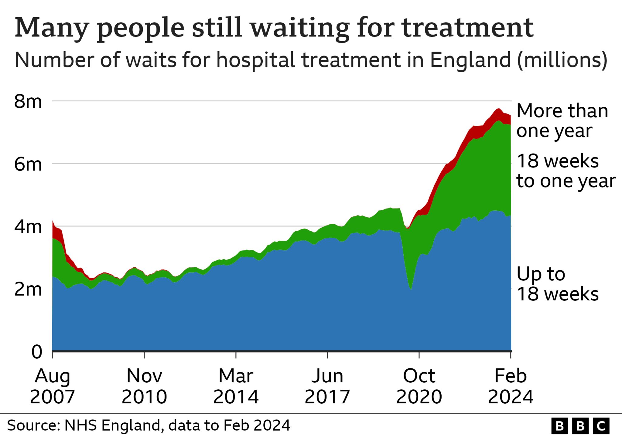
Published10 minutes agoShareclose panelShare pageCopy linkAbout sharingThis video can not be playedTo play this video you need to enable JavaScript in your browser.By Smitha MundasadHealth reporter The government is making progress on cutting NHS waiting lists in England, the prime minister insists, as latest figures show targets are being missed. Rishi Sunak said: “While we haven’t made as much progress as I would like, today’s figures show we are making headway towards that goal.”Waiting lists for routine treatments were around 7.54 million in February, down from 7.58 million in January.Opponents say waiting times are still too long.Labour highlighted that waiting lists were still higher than when Mr Sunak pledged to bring them down at the start of 2023.The Liberal Democrats accused the prime minister of “living in a parallel universe”.Mr Sunak said long-running strikes by consultants and junior doctors had had an impact on patients.Referring to the overall waiting list for treatment of 7.54 million, the PM said: “A drop of almost 200,000 in the last five months shows what the NHS can do for patients. Had there been no strike action, an extra 430,000 patients could have been treated.”We still have more work to do, but our plan is working.”He added: “When I became prime minister, I made cutting the NHS waiting lists one of my top five priorities. Today’s statistics clearly show we are making headway towards that goal.”The NHS recovery plan set a target for the end of March for 76% of patients attending A&E to be admitted, transferred or discharged within four hours.Another target in the plan was to eliminate all waits of 65 week or more for treatment by March this year. This has been pushed back to September.In an exclusive interview, the BBC’s Health Editor Hugh Pym challenged the prime minister, asking him what he would say to patients who have waited longer than 65 weeks, often in pain. Mr Sunak said: “None of this is acceptable and I’m doing absolutely everything I can to bring the waiting list down.”The latest NHS England figures show:an estimated 7.54 million planned treatments were waiting to be carried out in February, involving 6.29 million patientsthe number of patients waiting more than 65 weeks for treatment was 75,004 at the end of February, down from 92,213 (19%) in January there were 2.35 million visits to Accident and Emergency in March, 8.6% higher than the number of attendances in March 2023some 74.2% of patients were seen within four hours in A&Es in March, up from 70.9% in February and the highest figure since April 2023Wes Streeting, Labour’s shadow health secretary, said: “Rishi Sunak has failed on the NHS. He’s missed his own targets to cut ambulance waits and A&E waits. “Patients with suspected heart attacks or strokes are waiting almost double the safe amount of time, when every minute matters.”Doctors have said that patients in desperate need of care have been left waiting for 24 hours in A&E, while relatively healthy patients have been seen faster in order to hit this four-hour target.”Liberal Democrat leader Ed Davey said: “This week we uncovered that over 150,000 people waited over 24 hours in A&E last year. Now we find that waiting lists have gone up by 330,000 after Rishi Sunak pledged to cut them.”To add insult to injury, the Conservatives have cut NHS spending while millions of patients are suffering in pain on endless waiting lists.”Rishi Sunak is living in a parallel universe if he thinks our National Health Service is recovering. “The Conservative Party and the prime minister are out of touch, out of ideas and deserve to be kicked out of office.”More on this storyThe NHS hidden waiting lists terrifying patientsPublished19 FebruaryNeed an op? The hospitals with the worst waitsPublished14 March

Read more →