Illuminating the path to hearing recovery

Professor Yunje Cho’s research team from the Department of Life Sciences at Pohang University of Science and Technology (POSTECH, Republic of Korea) has collaborated with Professor Kwang Pyo Kim’s group from the Department of Applied Chemistry at Kyung Hee University (KHU, ROK), Professor Vsevolod Katritch’s team from the University of Southern California (USC, USA), and Professor Carol V. Robinson from the University of Oxford (UK) to uncover the mysteries surrounding a specific receptor protein associated with hearing. Their findings have recently been published in the online edition of Nature Structural & Molecular Biology.
Deep within the inner ear lie the cochlea, responsible for sound detection, and the vestibular apparatus, which oversees balance. Cells within these regions harbor a class C orphan G-protein-coupled receptor (GPCR) called GPR156. When this receptor is activated, it binds with G-proteins inside the cell, facilitating signal transmission. Unlike its counterparts, GPR156 exhibits sustained activity even in the absence of external stimuli, playing a pivotal role in upholding auditory and balance functions. Unveiling the structural and functional intricacies of GPR156 holds promise for devising interventions for individuals with congenital hearing impairments.
The research team employed cryo-electron microscopy (cryo-EM) analysis to delve into the GPR156 in the Go-free and Go-coupled states, achieving unprecedented resolution. Their investigation unearthed the mechanisms behind GPR156’s ability to maintain heightened activity sans activators.
Their analysis confirmed that GPR156 activation hinges on its interaction with abundant lipids in the cell membrane, triggering structural shifts upon engagement with G-proteins in the cytoplasm. Notably, unlike conventional GPCRs, GPR156 exhibits flexibility in altering the structure of the seventh helix as it traverses the cell membrane, thereby facilitating binding with G-proteins and orchestrating signal activation to detect sound. This study represents a crucial step forward in unraveling the structural dynamics and activation mechanisms of GPR156.
Professor Yunje Cho of POSTECH remarked, “Congenital hearing and balance impairments afflict numerous individuals. I am hopeful that our research will pave the way for groundbreaking treatments and drug discoveries to alleviate their suffering.”
This research received financial support from the National Research Foundation of Korea.

Read more →

Next-generation treatments hitch a ride into cancer cells

Antisense oligonucleotides (ASOs) are next-generation drugs that can treat disease by blocking the transfer of harmful messages from our genes. In people with cancer, ASOs have the potential to block messages that encourage the growth and spread of the tumor. However, ASOs aren’t used for treating cancer yet. They must first get delivered inside cancer cells, but the cancer cells won’t let them in.
Finding an effective ASO delivery system is a major challenge. Cancer cells have gatekeeper molecules that stop unwanted substances from entering. Although investigators have tried many ways of getting ASOs past the gatekeepers, success has been limited.
Now, in a study recently published in the journal Nucleic Acids Research, researchers from Osaka University have discovered a way to deliver ASOs to their targets inside cancer cells. The team synthesized a new compound, named L687, which opens specific calcium permeable channels on the surface of cancer cells. When the calcium flows into cells through the open channels it tells the cells to let in the ASOs.
“We discovered that we could selectively activate the TRPC3/C6 calcium permeable channels1) with the activator L687,” explains lead author Hiroto Kohashi. “We then found that combination treatment with L687 and ASO promoted efficient uptake of ASO into cancer cells during laboratory tests and tumor cells inside the mouse. As a result, target gene activity was suppressed and ASO efficacy was enhanced.”
Until now, ASOs have mainly been used to treat incurable diseases and had to be delivered into the liver or spinal fluid. According to the Osaka team’s research, L687 is an effective drug delivery system that may extend the benefits of ASO treatment to other parts of the body.
“We hope that the results of our research will lead to significant progress in the development and delivery of ASOs and similar gene-targeting drugs for treating cancer,” says senior author Masahito Shimojo.
The team believes that L687 could be a particularly effective way of delivering ASO therapy to lung or prostate cancers. These cancers have many TRPC3/C6 calcium permeable channels1) that can be opened by L687, potentially revealing new targets for these next-generation therapies.
1) TRPC3/C6 channels belong to a Transient Receptor Potential Canonical (TRPC) Channel subfamily of a TRP channel superfamily.

Read more →

Arm skin patch could warn of organ rejection

Published7 hours agoShareclose panelShare pageCopy linkAbout sharingImage source, NHSBTBy Michelle RobertsDigital health editorSome people having a lung transplant on the NHS will receive a skin patch graft from their donor too as a way of spotting organ rejection sooner. Rejection could show as a rash on the donated skin patch, say experts, allowing early treatment to stop problems escalating.The trial, by University of Oxford and NHS Blood and Transplant, will enrol 152 patients in England. It follows earlier success with some other transplant patients. Image source, NHSBTThat includes Adam Alderson, 44, from Wensleydale, who received a donor skin graft on his abdomen in 2015 when he had eight organs replaced – including a pancreas, stomach and spleen – after treatment for a rare cancer.He says the graft has already helped guide his treatment a few times to prevent his body rejecting his many new organs.He said: “It’s a really comforting thing to have – I feel safer knowing that I have a tool available to tell if something is going wrong before it becomes too serious. It’s almost like an oil warning light on your car. Plus, having that visible reminder of how lucky I am is really special.”Helen Roeper, 43, from Oxford, has a lung condition called cystic fibrosis that means she may need a lung transplant in the future. Image source, NHSBTShe said she welcomes the idea of having a donor skin graft at the same time. “The idea that I could potentially be able to identify any rejection early, just by looking at a patch of skin on my arm, is extremely encouraging and helps to alleviate some fears,” she said. The chance of rejection after a lung transplant is unfortunately quite high. Blood tests, lung samples and X-rays can offer clues, but the problem might be quite advanced by then. That is why researchers are keen to find a simple, visible early warning sign. Henk Giele, associate professor of plastic, reconstructive, transplant and hand surgery at University of Oxford, was the chief investigator of the trial. He said: “Lungs are prone to rejection due to their exposure to outside air and high propensity to infection. “It is often difficult to know if a reaction is caused by infection or rejection as they look the same at the early stages, but the treatments for each are completely opposite.”If a rash is seen in the skin graft on the arm, a tiny biopsy or sample will be taken to confirm if early rejection is happening, and a big dose of anti-rejection medicine can be given.More on this storyEight organ transplant survivor wedsPublished26 May 2017Girl receives UK’s first rejection-free kidneyPublished22 September 2023Man ‘recovering well’ after pig kidney transplantPublished21 MarchRelated Internet LinksSentinel Trial – Oxford UniversityNHS Blood and TransplantThe BBC is not responsible for the content of external sites.

Read more →

AI enhances physician-patient communication

As one of the first health systems in the country to pilot the use of generative artificial intelligence (GenAI) to draft replies to patient messages inside the Epic Systems electronic health record, UC San Diego Health is a pioneer in shaping the future of digital health.
The results of a new University of California San Diego School of Medicine study indicate that, although AI-generated replies did not reduce physician response time, they have contributed to relieving cognitive burden by starting an empathetic draft, which physicians can edit rather than starting from scratch.
The study, published in the April 15, 2024 online edition of the Journal of the American Medical Association’s Network Open, is the first randomized prospective evaluation of AI-drafted physician messaging.
“We are very interested in using AI to help solve health system challenges, including the increase in patient messages that are contributing to physician burnout,” said study senior author Christopher Longhurst, MD, executive director of the Joan and Irwin Jacobs Center for Health Innovation, chief medical officer and chief digital officer at UC San Diego Health. “The evidence that the messages are longer suggests that that they are higher quality, and the data is clear that physicians appreciated the help, which lowered cognitive burden.”
This quality improvement study evaluates patient-physician correspondence and suggests that the integration of generative AI into digital health care interactions has the potential to positively impact patient care by improving communication quality, efficiency and engagement. In addition, by alleviating some of the physician workload, the goal is for generative AI to help reduce burnout by allowing doctors to focus on more complex aspects of patient care.
“This study shows that generative AI can be a collaborative tool,” said study lead author Ming Tai-Seale, PhD, MPH, professor of family medicine at UC San Diego School of Medicine. “Our physicians receive about 200 messages a week. AI could help break ‘writer’s block’ by providing physicians an empathy-infused draft upon which to craft thoughtful responses to patients.”
The COVID-19 pandemic sparked unprecedented use of digital communications between patients and doctors that have remained in high demand. Portals, such as MyUCSDChart, used by UC San Diego Health, make it simple to email a doctor directly and have created heightened pressure for prompt provider responses that many can no longer efficiently handle.

Using generative AI to draft patient responses to non-emergency questions has been tested in the pilot program with electronic health record vendor Epic Systems, initiated in April 2023 at UC San Diego Health, to offer virtual physician assistance to help meet the rising demand of patient messages. For full transparency, the replies include a notification that they have been automatically generated by AI before being reviewed and edited by the physician who signs them.
Time-crunched physicians who may only have time for a brief, facts-only response, found that generative AI is helping to draft longer, compassionate responses that are appreciated and understood by patients.
“AI doesn’t get tired, so even at the end of a long day, it still has the capacity to help draft an empathetic message while synthesizing the request and relevant data into the response,” said study co-author Marlene Millen, MD, chief medical information officer for ambulatory care at UC San Diego Health. “So, while we were surprised by the study’s findings that AI messaging didn’t save doctors time, we see that it may help prevent burnout by providing a detailed draft as a starting point.”
The study’s findings suggest a potential paradigm shift in health care communication by leveraging AI, noting that further analysis is needed to gauge how beneficial patients deem the increased empathy and reply length to be.
UC San Diego Health, in conjunction with the Jacobs Center for Health Innovation, has been extensively testing GenAI models since May 2023. These transformative projects will help explore the safe, effective and novel use of GenAI in health care.
Co-authors of the study include: Sally L. Baxter, Florin Vaida, Amanda Walker, Amy M. Sitapati, Chad Osborne, Joseph Diaz, Nimit Desai, Sophie Webb, Gregory Polston, Teresa Helsten, Erin Gross, Jessica Thackaberry, Ammar Mandvi, Dustin Lillie, Steve Li, Geneen Gin, Suraj Achar, Heather Hofflick, and Marlene Millen all of UC San Diego; and Christopher Sharp of Stanford.

Read more →

Vaccine breakthrough means no more chasing strains

Scientists at UC Riverside have demonstrated a new, RNA-based vaccine strategy that is effective against any strain of a virus and can be used safely even by babies or the immunocompromised.
Every year, researchers try to predict the four influenza strains that are most likely to be prevalent during the upcoming flu season. And every year, people line up to get their updated vaccine, hoping the researchers formulated the shot correctly.
The same is true of COVID vaccines, which have been reformulated to target sub-variants of the most prevalent strains circulating in the U.S.
This new strategy would eliminate the need to create all these different shots, because it targets a part of the viral genome that is common to all strains of a virus. The vaccine, how it works, and a demonstration of its efficacy in mice is described in a paper published today in the Proceedings of the National Academy of Sciences.
“What I want to emphasize about this vaccine strategy is that it is broad,” said UCR virologist and paper author Rong Hai. “It is broadly applicable to any number of viruses, broadly effective against any variant of a virus, and safe for a broad spectrum of people. This could be the universal vaccine that we have been looking for.”
Traditionally, vaccines contain either a dead or modified, live version of a virus. The body’s immune system recognizes a protein in the virus and mounts an immune response. This response produces T-cells that attack the virus and stop it from spreading. It also produces “memory” B-cells that train your immune system to protect you from future attacks.
The new vaccine also uses a live, modified version of a virus. However, it does not rely on the vaccinated body having this traditional immune response or immune active proteins — which is the reason it can be used by babies whose immune systems are underdeveloped, or people suffering from a disease that overtaxes their immune system. Instead, this relies on small, silencing RNA molecules.

“A host — a person, a mouse, anyone infected — will produce small interfering RNAs as an immune response to viral infection. These RNAi then knock down the virus,” said Shouwei Ding, distinguished professor of microbiology at UCR, and lead paper author.
The reason viruses successfully cause disease is because they produce proteins that block a host’s RNAi response. “If we make a mutant virus that cannot produce the protein to suppress our RNAi, we can weaken the virus. It can replicate to some level, but then loses the battle to the host RNAi response,” Ding said. “A virus weakened in this way can be used as a vaccine for boosting our RNAi immune system.”
When the researchers tested this strategy with a mouse virus called Nodamura, they did it with mutant mice lacking T and B cells. With one vaccine injection, they found the mice were protected from a lethal dose of the unmodified virus for at least 90 days. Note that some studies show nine mouse days are roughly equivalent to one human year.
There are few vaccines suitable for use in babies younger than six months old. However, even newborn mice produce small RNAi molecules, which is why the vaccine protected them as well. UC Riverside has now been issued a US patent on this RNAi vaccine technology.
In 2013, the same research team published a paper showing that flu infections also induce us to produce RNAi molecules. “That’s why our next step is to use this same concept to generate a flu vaccine, so infants can be protected. If we are successful, they’ll no longer have to depend on their mothers’ antibodies,” Ding said.
Their flu vaccine will also likely be delivered in the form of a spray, as many people have an aversion to needles. “Respiratory infections move through the nose, so a spray might be an easier delivery system,” Hai said.
Additionally, the researchers say there is little chance of a virus mutating to avoid this vaccination strategy. “Viruses may mutate in regions not targeted by traditional vaccines. However, we are targeting their whole genome with thousands of small RNAs. They cannot escape this,” Hai said.
Ultimately, the researchers believe they can ‘cut and paste’ this strategy to make a one-and-done vaccine for any number of viruses.
“There are several well-known human pathogens; dengue, SARS, COVID. They all have similar viral functions,” Ding said. “This should be applicable to these viruses in an easy transfer of knowledge.”

Read more →

Microplastics make their way from the gut to other organs

It’s happening every day. From our water, our food and even the air we breathe, tiny plastic particles are finding their way into many parts of our body.
But what happens once those particles are inside? What do they do to our digestive system?
In a recent paper published in the journal Environmental Health Perspectives, University of New Mexico researchers found that those tiny particles — microplastics — are having a significant impact on our digestive pathways, making their way from the gut and into the tissues of the kidney, liver and brain.
Eliseo Castillo, PhD, an associate professor in the Division of Gastroenterology & Hepatology in the UNM School of Medicine’s Department of Internal Medicine and an expert in mucosal immunology, is leading the charge at UNM on microplastic research.
“Over the past few decades, microplastics have been found in the ocean, in animals and plants, in tap water and bottled water,” Castillo, says. “They appear to be everywhere.”
Scientists estimate that people ingest 5 grams of microplastic particles each week on average — equivalent to the weight of a credit card.
While other researchers are helping to identify and quantify ingested microplastics, Castillo and his team focus on what the microplastics are doing inside the body, specifically to the gastrointestinal (GI) tract and to the gut immune system.

Over a four-week period, Castillo, postdoctoral fellow Marcus Garcia, PharmD, and other UNM researchers exposed mice to microplastics in their drinking water. The amount was equivalent to the quantity of microplastics humans are believed to ingest each week.
Microplastics had migrated out of the gut into the tissues of the liver, kidney and even the brain, the team found. The study also showed the microplastics changed metabolic pathways in the affected tissues.
“We could detect microplastics in certain tissues after the exposure,” Castillo says. “That tells us it can cross the intestinal barrier and infiltrate into other tissues.”
Castillo says he’s also concerned about the accumulation of the plastic particles in the human body. “These mice were exposed for four weeks,” he says. “Now, think about how that equates to humans, if we’re exposed from birth to old age.”
The healthy laboratory animals used in this study showed changes after brief microplastic exposure, Castillo says. “Now imagine if someone has an underlying condition, and these changes occur, could microplastic exposure exacerbate an underlying condition?”
He has previously found that microplastics are also impacting macrophages — the immune cells that work to protect the body from foreign particles.

In a paper published in the journal Cell Biology & Toxicology in 2021, Castillo and other UNM researchers found that when macrophages encountered and ingested microplastics, their function was altered and they released inflammatory molecules.
“It is changing the metabolism of the cells, which can alter inflammatory responses,” Castillo says. “During intestinal inflammation — states of chronic illness such as ulcerative colitis and Crohn’s disease, which are both forms of inflammatory bowel disease — these macrophages become more inflammatory and they’re more abundant in the gut.”
The next phase of Castillo’s research, which is being led by postdoctoral fellow Sumira Phatak, PhD, will explore how diet is involved in microplastic uptake.
“Everyone’s diet is different,” he says. “So, what we’re going to do is give these laboratory animals a high-cholesterol/high-fat diet, or high-fiber diet, and they will be either exposed or not exposed to microplastics. The goal is to try to understand if diet affects the uptake of microplastics into our body.”
Castillo says one of his PhD students, Aaron Romero, is also working to understand why there is a change in the gut microbiota. “Multiple groups have shown microplastics change the microbiota, but how it changes the microbiota hasn’t been addressed.”
Castillo hopes that his research will help uncover the potential impacts microplastics are having to human health and that it will help spur changes to how society produces and filtrates plastics.
“At the end of the day, the research we are trying to do aims to find out how this is impacting gut health,” he says. “Research continues to show the importance of gut health. If you don’t have a healthy gut, it affects the brain, it affects the liver and so many other tissues. So even imagining that the microplastics are doing something in the in the gut, that chronic exposure could lead to systemic effects.”

Read more →

Epilepsy drug prevents brain tumors in mice with NF1

A drug used to treat children with epilepsy prevents brain tumor formation and growth in two mouse models of neurofibromatosis type 1 (NF1), according to a study by researchers at Washington University School of Medicine in St. Louis. NF1 is a genetic condition that causes tumors to grow on nerves throughout the body, including the optic nerves, which connect the eyes to the brain.
The findings lay the groundwork for a clinical trial to assess whether the drug, lamotrigine, can prevent or delay brain tumors in children with NF1. The study is online in the journal Neuro-Oncology.
“Based on these data, the Neurofibromatosis Clinical Trials Consortium is considering launching a first-of-its-kind prevention trial,” said senior author David H. Gutmann, MD, PhD, the Donald O. Schnuck Family Professor of Neurology and the director of Washington University’s Neurofibromatosis Center. The clinical trials consortium is an international network of NF scientists that was formed by the U.S. Department of Defense in 2006 to find therapies for all forms of neurofibromatosis. “The plan is to enroll kids without symptoms, treat them for a limited time, and then see whether the number of children who develop tumors that require treatment goes down.
“This is a novel idea, so we took it to an NF1 patient focus group,” Gutmann continued. “They said, ‘This is exactly what we’re looking for.’ A short-term treatment with a drug that has been used safely for 30 years was acceptable to them if it reduced the chance their children would develop tumors and need chemotherapy that might have all kinds of side effects.”
The most serious tumors that people with NF1 get affect the optic nerve and are known as optic gliomas. Such tumors typically appear between ages 3 to 7. While they are rarely fatal, they cause vision loss in up to a third of patients as well as other symptoms, including early puberty. Standard chemotherapy for optic gliomas is only moderately effective at preventing further vision loss and can affect children’s developing brains, resulting in cognitive and behavioral problems.
In a previous study, Gutmann and Corina Anastasaki, PhD, an assistant professor of neurology and the first author on the new paper, showed that lamotrigine stopped optic glioma growth in NF1 mice by suppressing neuronal hyperactivity. The Neurofibromatosis Clinical Trial Consortium found their data intriguing but demanded more evidence before they would consider launching a clinical trial. The consortium members asked Gutmann and Anastasaki to clarify the connection between Nf1 mutation, neuronal excitability and optic gliomas; assess whether lamotrigine was effective at the doses already proven safe in children with epilepsy; and conduct these studies in more than one strain of NF1 mice.
In people, NF1 is a highly variable disease. It can be caused by any one of thousands of different mutations in the NF1 gene, where different mutations could be associated with different medical problems. Repeating experiments in multiple strains of mice was a way of gauging whether lamotrigine was likely to work in people regardless of the underlying mutation.
Anastasaki and Gutmann not only showed that lamotrigine worked in two strains of NF1 mice, they also showed that the drug worked at lower doses than those used for epilepsy, meaning that it was probably safe. Even better, they found that a short course of the drug had lasting effects, both as a preventive and a treatment. Mice that had tumors and that were treated for four weeks starting at 12 weeks of age saw their tumors stop growing and even showed no further damage to the retinas of their eyes. Mice that received a four-week course of the drug starting at 4 weeks of age, before tumors typically emerge, showed no tumor growth even four months after treatment had ended.
These findings have led Gutmann to suggest that a one-year course of treatment for young children with NF1, maybe between the ages of 2 to 4, might be enough to reduce their risk of brain tumors.
“The idea that we might be able to change the prognosis for these kids by intervening within a short time window is so exciting,” Gutmann said. “If we could just get them past the age when these tumors typically form, past age 7, they may never need treatment. I’d love it if I never again had to discuss chemotherapy for kids who aren’t even in first grade yet.”

Read more →

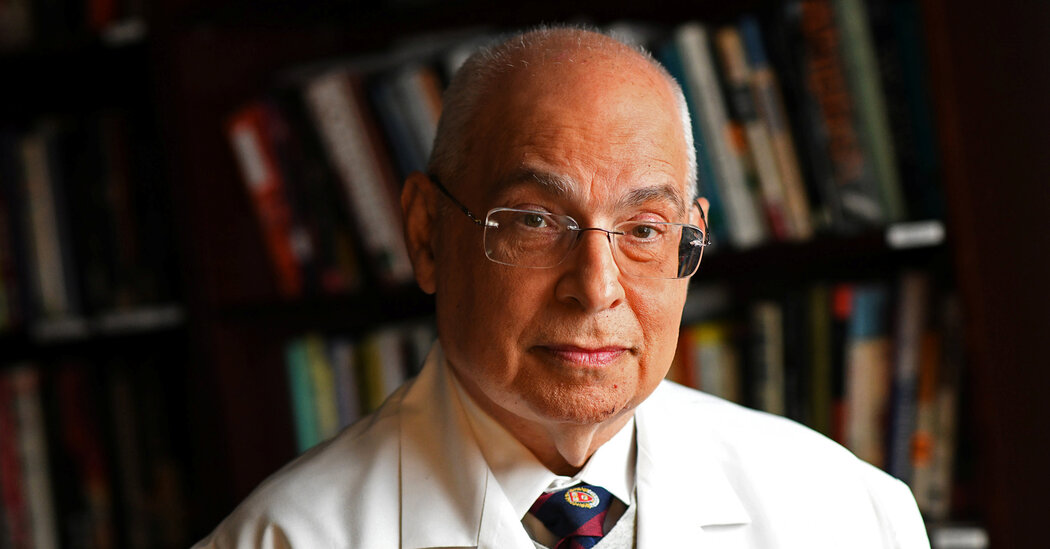
David Egilman, Doctor Who Took On Drug Companies, Dies at 71

His testimony as an expert witness in some 600 trials helped plaintiffs win billions of dollars in cases involving malfeasance by pharmaceutical makers.Dr. David Egilman, a physician and expert witness who, over a 35-year span, gave testimony in some 600 trials involving corporate malfeasance, resulting in billions of dollars in awards for victims and their survivors, died on April 2 at his home in Foxborough, Mass. He was 71.The cause was cardiac arrest, his son Alex said.Many medical experts make a side business in court, offering their informed opinions on the witness stand and helping to validate or undermine plaintiffs’ claims. But few make it a career-long passion in the way Dr. Egilman did. He taught at Brown University and ran a private practice but spent most of his time consulting and testifying in as many as 15 cases a year.He did more than just opine from the stand. A dogged researcher, he dug up incriminating emails and memos showing that, in many cases, drug companies knew the risks involved with putting a new medication on the market but went ahead anyway.He provided critical testimony in a class-action lawsuit against Johnson & Johnson, claiming that it had failed to reveal the health risks involved presented by Johnson’s baby powder and other products containing talc. After years of litigation, the company settled for $8.9 billion in 2023.Dr. Egilman testifying in a trial in Joplin, Mo., in 2004. He helped attorneys strategize for trial and coached them on how to explain complicated medical topics to juries.Pool photo by T. Rob BrownDr. Egilman’s work as an expert witness rubbed some people the wrong way, especially defense lawyers and pharmaceutical company executives, who argued that he was too dogmatic to provide objective analysis. But Dr. Egilman saw things differently.We are having trouble retrieving the article content.Please enable JavaScript in your browser settings.Thank you for your patience while we verify access. If you are in Reader mode please exit and log into your Times account, or subscribe for all of The Times.Thank you for your patience while we verify access.Already a subscriber? Log in.Want all of The Times? Subscribe.

Read more →

Physical activity reduces stress-related brain activity to lower cardiovascular disease risk

New research indicates that physical activity lowers cardiovascular disease risk in part by reducing stress-related signaling in the brain.
In the study, which was led by investigators at Massachusetts General Hospital (MGH), a founding member of the Mass General Brigham healthcare system and published in the Journal of the American College of Cardiology, people with stress-related conditions such as depression experienced the most cardiovascular benefits from physical activity.
To assess the mechanisms underlying the psychological and cardiovascular disease benefits of physical activity, Ahmed Tawakol, MD, an investigator and cardiologist in the Cardiovascular Imaging Research Center at Massachusetts General Hospital, and his colleagues analyzed medical records and other information of 50,359 participants from the Mass General Brigham Biobank who completed a physical activity survey.
A subset of 774 participants also underwent brain imaging tests and measurements of stress-related brain activity.
Over a median follow-up of 10 years, 12.9% of participants developed cardiovascular disease. Participants who met physical activity recommendations had a 23% lower risk of developing cardiovascular disease compared with those not meeting these recommendations.
Individuals with higher levels of physical activity also tended to have lower stress-related brain activity. Notably, reductions in stress-associated brain activity were driven by gains in function in the prefrontal cortex, a part of the brain involved in executive function (i.e., decision making, impulse control) and is known to restrain stress centers of the brain. Analyses accounted for other lifestyle variables and risk factors for coronary disease.
Moreover, reductions in stress-related brain signaling partially accounted for physical activity’s cardiovascular benefit.
As an extension of this finding, the researchers found in a cohort of 50,359 participants that the cardiovascular benefit of exercise was substantially greater among participants who would be expected to have higher stress-related brain activity, such as those with pre-existing depression.
“Physical activity was roughly twice as effective in lowering cardiovascular disease risk among those with depression. Effects on the brain’s stress-related activity may explain this novel observation,” says Tawakol, who is the senior author of the study.
“Prospective studies are needed to identify potential mediators and to prove causality. In the meantime, clinicians could convey to patients that physical activity may have important brain effects, which may impart greater cardiovascular benefits among individuals with stress-related syndromes such as depression.”

Read more →

Specific nasal cells protect against COVID-19 in children

Important differences in how the nasal cells of young and elderly people respond to the SARS-CoV-2 virus, could explain why children typically experience milder COVID-19 symptoms, finds a new study led by researchers at UCL and the Wellcome Sanger Institute.
The study, published in Nature Microbiology, focused on the early effects of SARS-CoV-2 infection on the cells first targeted by the viruses, the human nasal epithelial cells (NECs).
These cells were donated from healthy participants from Great Ormond Street Hospital (GOSH), University College London Hospital (UCLH) and the Royal Free Hospital, including children (0-11 years), adults (30 — 50 years) and, for the first time, the elderly (over 70 years).
The cells were then cultured using specialised techniques, allowing them to regrow into the different types of cells you originally find in the nose. Using single-cell RNA sequencing techniques that enable scientists to identify the unique genetic networks and functions of thousands of individual cells, the team identified 24 distinct epithelial cell types. Cultures from each age group were then either mock infected or infected with SARS-CoV-2.
The researchers found that, after three days, the NECs of children responded quickly to SARS-CoV-2 by increasing interferon (the body’s first line of anti-viral defence) — restricting viral replication. However, this early anti-viral effect became less pronounced with age.
The researchers also found that NECs from elderly individuals not only produced more infectious virus particles, but also experienced increased cell shedding and damage.
The strong antiviral response in the NECs of children could explain why younger people typically experience milder symptoms. In contrast, the increased damage and higher viral replication found in NECs from elderly individuals could be linked to the greater severity of disease observed in older adults.

Project lead, Dr Claire Smith (Associate Professor at UCL Great Ormond Street Institute of Child Health), said: “Our research reveals how the type of cells we have in our nose changes with age, and how this affects our ability to combat SARS-CoV-2 infection. This could be crucial in developing effective anti-viral treatments tailored to different age groups, especially for the elderly who are at higher risk of severe COVID-19.”
Co-Senior author, Dr Kerstin Meyer (Wellcome Sanger Institute), said: “By carrying out SARS-CoV-2 infections of epithelial cells in vitro and studying the responses with single cell sequencing, we get a much more detailed understanding of the viral infection kinetics and see big differences in the innate immune response between cell types.”
Children infected with SARS-CoV-2 rarely progress to respiratory failure, but the risk of mortality in infected people over the age of 85 remains high, despite vaccination and improving treatment options.
The research underscores the importance of considering age as a critical factor in both research and treatment of infectious diseases.
Co-senior author, Dr Marko Nikolic (UCL Division of Medicine), said: “It is fascinating that when we take away immune cells from nasal samples, and are only left with nasal epithelial cells grown in a dish, we are still able to identify age-specific differences in our body’s response to the SARS-CoV-2 between the young and elderly to explain why children are generally protected from severe COVID-19.”
Dr Smith added: “Understanding the cellular differences at the initiation of infection is just the beginning. We now hope to investigate the long-term implications of these cellular changes and test therapeutic interventions using our unique cell culture model. This ‘gold-standard’ system is only possible with the support of our funders and the willingness of participants to provide their samples.”
The team suggest that future research should consider how ageing impacts the body’s response to other viral infections.
This study was funded by UK Research and Innovation (UKRI), and the National Institute for Health and Care Research (NIHR) Great Ormond Street Hospital Biomedical Research Centre, Wellcome and the Chan Zuckerberg Foundation.

Read more →