Enzymes open new path to universal donor blood

The quest to develop universal donor blood has taken a decisive step forward. Researchers at DTU and Lund University have discovered enzymes that, when mixed with red blood cells, are able to remove specific sugars that make up the A and B antigens in the human ABO blood groups. The results have been published in the scientific journal Nature Microbiology.
“For the first time, the new enzyme cocktails not only remove the well-described A and B antigens, but also extended variants previously not recognized as problematic for transfusion safety. We are close to being able to produce universal blood from group B donors, while there is still work to be done to convert the more complex group A blood. Our focus is now to investigate in detail if there are additional obstacles and how we can improve our enzymes to reach the ultimate goal of universal blood production,” says Professor Maher Abou Hachem, who is the study leader at DTU and one of the senior scientists behind the discovery.
He states that the discovery is the result of combining the expertise of DTU researchers in enzymes from the human gut microbiota and Lund University researchers in carbohydrate-based blood groups and transfusion medicine.
High demand for donor blood
Human red blood cells carry specific complex sugars structures (antigens) that define the four ABO blood groups A, B, AB and O. These antigens control compatibility between donors and recipients for safe blood transfusion and organ transplantation. Donor blood is screened for disease markers and the main blood groups. It can then be stored refrigerated for up to 42 days.
The need for donor blood is high due to the elderly making up a larger proportion of the population and more patients undergoing blood-intensive medical procedures. Successfully converting A or B blood types into ABO universal donor blood can markedly reduce the logistics and costs currently associated with storing four different blood types. In addition, the development of universal donor blood will lead to an increased supply of donor blood by reducing the waste of blood approaching its expiry date.
The reason why it is necessary to remove the A and B antigens to create universal donor blood is because they can trigger life-threatening immune reactions when transfused into non-matched recipients.

The concept of using enzymes to generate universal donor blood was introduced more than 40 years ago. Since then, higher efficiency enzymes to remove the A and B antigens were discovered, but researchers are still not able to explain or abolish all immune reactions related to the blood, and therefore these enzymes are still not used in clinical practice.
Enzymes from the gut
The research groups from DTU and Lund University have gone new ways to find enzymes that can remove both the A and B blood antigens and the sugars that block them. The research teams discovered new mixtures of enzymes from the human gut bacterium Akkermansia muciniphila that feeds by breaking down the mucus, which covers the surface of the gut. It turns out that these enzymes are exceptionally efficient, as the complex sugars at the surface of the intestinal mucosa share chemical resemblance with those found at the surface of blood cells.
“What is special about the mucosa is that bacteria, which are able to live on this material, often have tailor-made enzymes to break down mucosal sugar structures, which include blood group ABO antigens. This hypothesis turned out to be correct,” says Maher Abou Hachem.
The researchers in this study tested 24 enzymes, which they used to process hundreds of blood samples.
“Universal blood will create a more efficient utilization of donor blood, and also avoid giving ABO-mismatched transfusions by mistake, which can otherwise lead to potentially fatal consequences in the recipient. When we can create ABO-universal donor blood, we will simplify the logistics of transporting and administering safe blood products, while at the same time minimizing blood waste” says Professor Martin L. Olsson, the leader of the study at Lund University.

The researchers from DTU and Lund University have applied for a patent on the new enzymes and the method for enzyme treatment and expect to make further progress on this in their new joint project over the next three and a half years. If successful, the concept needs to be tested in controlled patient trials before this can be considered for commercial production and clinical use.
The initial research project is funded by the Independent Research Fund Denmark (Technology and Production Sciences, FTP), the Swedish Research Council, ALF grants from the Swedish government and county councils as well as the Knut and Alice Wallenberg Foundation and Research Fund Denmark, Natural Sciences, FNU), while the new continued project is funded by the Novo Nordisk Foundation, Interdisciplinary Synergy Programme.
FACTS:
Donorblood
In Scandinavia, the four main blood types are distributed with approximately 40-45 percent blood type A, the majority of whom are so-called RhD positive and 10-15 percent RhD negative, followed by blood type O with about 40 percent, B with about 10 percent and AB with about 5 percent. Red blood cells from blood group O are the only type that can be used by all receiving patients regardless of ABO type.
Bacterium from the gut
Akkermansia muciniphila is a bacterium found abundantly in the guts of most healthy humans. This bacterium can break down mucus in the gut and produces beneficial compounds such as the short-chain fatty acid propionate, in addition to exerting beneficial effects on body weight and metabolic markers.

Read more →

Possible alternative to antibiotics produced by bacteria

Many bacteria produce substances to gain an advantage over competitors in their highly competitive natural environment. Researchers at the University Hospital Bonn (UKB), the University of Bonn and the German Center for Infection Research (DZIF) have discovered a new so-called lantibiotic, namely epilancin A37. It is produced by staphylococci that colonize the skin and acts specifically against their main competitors there, the corynebacteria. This specificity is presumably mediated by a very special mechanism of action, which the researchers were able to decipher in detail. Their results have now been published in the ISME Journal.
Due to increasing antibiotic resistance in pathogens causing infections, the development of new antibacterial substances is important. Hopes are pinned on a new group of substances produced by gram-positive bacteria, the lantibiotics. These are antimicrobial peptides that often have a very narrow spectrum of activity. “Such compounds are highly interesting from a medical point of view, as they could specifically attack individual groups of organisms without affecting the entire bacterial flora, as is the case with broad-spectrum antibiotics, for example,” says corresponding author Dr. Fabian Grein, until recently head of the DZIF research group “Bacterial Interference” at the Institute of Pharmaceutical Microbiology at the UKB and member of the Transdisciplinary Research Area (TRA) “Life & Health” at the University of Bonn.
Essential competitive advantage over corynebacteria
The UKB research team led by Fabian Grein and Tanja Schneider, together with the team led by Ulrich Kubitscheck, Professor of Biophysical Chemistry at the University of Bonn, have now discovered a new lantibiotic, namely epilancin A37. It is produced by staphylococci, which are typical colonizers of the skin and mucous membranes. Little is known about these antimicrobial peptides. “We were able to show that epilancins are widespread in staphylococci, which underlines their ecological importance,” says first author Jan-Samuel Puls, a doctoral student from the University of Bonn at the Institute of Pharmaceutical Microbiology at the UKB. This is because staphylococci and corynebacteria are important genera of the human microbiota — i.e. the totality of all microorganisms such as bacteria and viruses — in the nose and skin, which are closely linked to health and disease. The need to produce such a compound indicates a pronounced competition between the species. The researchers were able to show that the newly discovered epilancin A37 acts very specifically against corynebacteria, which are among the main competitors of staphylococci within the skin microbiome.
New mode of action in the “bacterial war” decoded
“This specificity is presumably mediated by a very special mechanism of action that we were able to decipher in detail,” says Grein. Epilancin A37 penetrates the corynebacterial cell, initially without destroying it. The antimicrobial peptides accumulate in the cell and then dissolve the cell membrane from the inside, thus killing the corynebacterium. Co-author Dr. Thomas Fließwasser from the Institute of Pharmaceutical Microbiology at the UKB, postdoctoral researcher at the University of Bonn and acting head of the DZIF research group “Bacterial Interference” adds: “Our study shows how a specific mechanism of action can be used to specifically combat a single bacterial species. It therefore serves us as a ‘proof of concept’.”

Read more →

Experimental type 1 diabetes drug shelters pancreas cells from immune system attack

Scientists at Johns Hopkins Medicine say that an experimental monoclonal antibody drug called mAb43 appears to prevent and reverse the onset of clinical type 1 diabetes in mice, and in some cases, to lengthen the animals’ lifespan.
The drug is unique, according to the researchers, because it targets insulin-making beta cells in the pancreas directly and is designed to shield those cells from attacks by the body’s own immune system cells. The drug’s specificity for such cells may enable long-term use in humans with few side effects, say the researchers. Monoclonal antibodies are made by cloning, or making identical replicas of, an animal (including human) cell line.
The findings, reported online recently and in the May issue of Diabetes, raise the possibility of a new drug for type 1 diabetes, an autoimmune condition that affects about 2 million American children and adults and has no cure or means of prevention. Unlike type 2 diabetes, in which the pancreas makes too little insulin, in type 1 diabetes, the pancreas makes no insulin because the immune system attacks the pancreatic cells that make it.
The lack of insulin interferes with the body’s ability to regulate blood sugar levels.
“People with type 1 diabetes face lifelong injections of insulin and many complications, including stroke and eyesight problems if the condition is not managed properly,” says Dax Fu, Ph.D., associate professor of physiology at the Johns Hopkins University School of Medicine and leader of the research team.
Fu says mAb43 binds to a small protein on the surface of beta cells, which dwell in clusters called islets. The drug was designed to provide a kind of shield or cloak to hide beta cells from immune system cells that attack them as “invaders.” The researchers used a mouse version of the monoclonal antibody, and will need to develop a humanized version for studies in people.
For the current study, the researchers gave 64 non-obese mice bred to develop type 1 diabetes a weekly dose of mAb43 via intravenous injection when they were 10 weeks old. After 35 weeks, all mice were non-diabetic. One of the mice developed diabetes for a period of time, but it recovered at 35 weeks, and that mouse had early signs of diabetes before the antibody was administered.

In five of the same type of diabetes-prone mice, the researchers held off giving weekly mAb43 doses until they were 14 weeks old, and then continued dosages and monitoring for up to 75 weeks. One of the five in the group developed diabetes, but no adverse events were found, say the researchers.
In the experiments in which mAb43 was given early on, the mice lived for the duration of the monitoring period of 75 weeks, compared with the control group of mice that did not receive the drug and lived about 18-40 weeks.
Next, the researchers, including postdoctoral fellows Devi Kasinathan and Zheng Guo, looked more closely at the mice that received mAb43 and used a biological marker called Ki67 to see if beta cells were multiplying in the pancreas. They said, after treatment with the antibody, immune cells retreated from beta cells, reducing the amount of inflammation in the area. In addition, beta cells slowly began reproducing.
“mAb43 in combination with insulin therapy may have the potential to gradually reduce insulin use while beta cells regenerate, ultimately eliminating the need to use insulin supplementation for glycemic control,” says Kasinathan.
The research team found that mAb43 specifically bound to beta cells, which make up about 1% or 2% of pancreas cells.
Another monoclonal antibody drug, teplizumab, was approved by the U.S. Food and Drug Administration in 2022. Teplizumab binds to T cells, making them less harmful to insulin-producing beta cells. The drug has been shown to delay the onset of clinical (stage 3) type 1 diabetes by about two years, giving young children who get the disease time to mature and learn to manage lifelong insulin injections and dietary restrictions.

“It’s possible that mAb43 could be used for longer than teplizumab and delay diabetes onset for a much longer time, potentially for as long as it’s administered,” says Fu.
“In an ongoing effort, we aim to develop a humanized version of the antibody and conduct clinical trials to test its ability to prevent type 1 diabetes, and to learn whether it has any off-target side effects,” says Guo.
Other scientists who contributed to the research include Dylan Sarver, G. William Wong and Maria Golson from Johns Hopkins; Shumei Yun from the University of Maryland, Aaron Michels and Liping Yu from the University of Colorado; and Chandan Sona and Matthew Poy from Johns Hopkins All Children’s Hospital.
Funding for the research was provided, in part, by the National Institutes of Health (R01DK125746, P30DK116073, R01DK110183, R01 DK135688 and RO1DK084171).

Read more →

The aspirin conundrum: Navigating negative results, age, aging dynamics and equity

A new study examining the role of aspirin in breast cancer treatment reveals critical issues related to health equity and aging that have broad implications for cancer and other disease intervention trials, say researchers from Georgetown University’s Lombardi Comprehensive Cancer Center. They outline their concerns in an editorial accompanying the study’s findings published April 29 in the JAMA (“The Aspirin Conundrum: Navigating Negative Results, Age, Aging Dynamics and Equity”).
The study, called the Alliance trial, was launched after researchers noted that breast cancer survivors taking aspirin as part of another clinical trial for cardiovascular disease lived longer.
To confirm the observation, a phase 3 clinical trial randomly assigned volunteers with nonmetastatic, high-risk breast cancer to receive either 300mg of aspirin or placebo daily. The outcome was a disappointment. The study was suspended at the first interim safety analysis because the results indicated futility — aspirin did not decrease the risk of cancer recurrence or improve survival.
In their editorial examining the trial, the Georgetown researchers, Jeanne Mandelblatt, MD, MPH; Candace Mainor, MD; and Barry Hudson, PhD, raise several important questions about the outcome.
For example, the authors point out that despite efforts to include various groups in the study, certain subgroups, like racial minorities and those with high exposure to systemic racism, may not have been adequately represented.
“Some individuals from these groups may experience chronic life stressors that affect inflammation, accelerate biological aging and contribute to disparities in cancer risk, recurrence and mortality,” they write, noting that these individuals could potentially benefit from aspirin, an anti-inflammatory drug.
Researchers say another issue with this and other trials is how chronological age and biological age might affect implications for a trial’s design and its results.
The results in the Alliance trial “raises the question of whether aspirin’s lack of benefit could be partly explained by variations in biological age, including heterogeneity in immune and platelet function, inflammatory responses and host-tumor microenvironment interactions,” they write. “Thus, careful consideration of the intersectionality of aging, cancer and disparities will be critical in designing future oncology and other disease trials to advance health equity.”
Finally, the researchers say clinicians may find it challenging to integrate new findings into routine practice because the results suggest a lack of intervention efficacy along with the many unanswered questions.
Despite the fairly definitive negative result for use of aspirin to improve invasive disease-free survival among breast cancer survivors, the researchers say, .” ..oncology and primary care providers may still consider discussing with each other and their patients the potential benefits and harms of aspirin used for other reasons.”

Read more →

CPAP Lawsuits Settled for $1.1 Billion

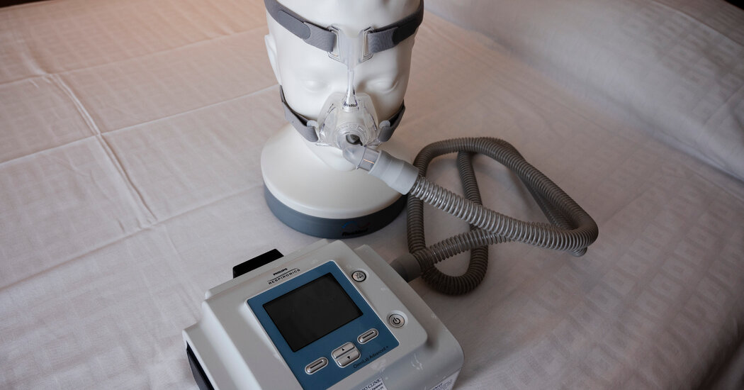
Thousands of people with sleep apnea and other illnesses had sued the company, claiming flawed devices were harming them. Philips Respironics has reached a $1.1 billion settlement over claims that people who used their CPAP and other breathing devices were harmed by noxious gasses and flecks of foam that lodged in their airways, sometimes for years.Thousands of people contended in lawsuits that they had been injured by popular Philips DreamStation machines. The settlement affects CPAP, or continuous positive airway pressure, machines that people with sleep apnea or other respiratory difficulties use at night to improve their breathing, as well as other types of machines used at home and in hospitals.Philips did not admit any fault in the settlement, including whether the devices caused the injuries, according to a financial report issued Monday.The personal injury settlement follows a $479 million settlement reached in September over economic losses to the patients and medical equipment sales companies that financed replacement devices. Philips also agreed to a consent decree earlier this year that forced the company to halt U.S. sales of new devices until certain conditions are met.Monday’s agreement largely settles years of litigation over a problem that was deeply upsetting to patients and doctors, who had to weigh the risk of letting patients’ interrupted breathing go untreated against the use of a machine that might cause harm. Patients flooded lawmakers and the Food and Drug Administration with complaints about a chaotic recall and replacement effort that left many waiting for months or more than a year for an updated device.In a letter to Philips in May 2022, the F.D.A. noted that the company had received reports about the problem as early as 2015, but failed to evaluate the information and address the device’s problems.The recall started in the summer of 2021 amid concerns that the machines blew out potentially cancer-causing gases. The initial recall affected about 15 million breathing machines produced since 2006, though roughly five million were still in circulation in mid-2021.The F.D.A. reported earlier this year that since Philips first warned of the problems, officials had received 116,000 complaints, including 561 reports of deaths, that people or lawyers said were linked to the faulty foam in the device.The company has since tempered its warnings, saying that further testing showed that the gasses were not as toxic as initially believed.Investors recognized the resolution, as the company stock surged by about 33 percent Monday morning, to about $28 per share. The company said that part of the settlement would be covered by insurance.Plaintiffs’ lawyers welcomed the settlement.“Ultimately, these combined agreements accomplish what we sought to achieve when this litigation began — holding Philips accountable by obtaining care for those with physical injuries and compensation for those needing new respiratory devices,” Sandra L. Duggan, Kelly K. Iverson and Christopher A. Seeger, lawyers representing the plaintiffs, said in a statement.

Read more →

Physical Fitness Can Improve Mental Health in Children and Adolescents, Study Suggests

A new study bolsters existing research suggesting that exercise can protect against anxiety, depression and attention challenges.Physical fitness among children and adolescents may protect against developing depressive symptoms, anxiety and attention deficit hyperactivity disorder, according to a study published on Monday in JAMA Pediatrics.The study also found that better performance in cardiovascular activities, strength and muscular endurance were each associated with greater protection against such mental health conditions. The researchers deemed this linkage “dose-dependent”, suggesting that a child or adolescent who is more fit may be accordingly less likely to experience the onset of a mental health disorder.These findings come amid a surge of mental health diagnoses among children and adolescents, in the United States and abroad, that have prompted efforts to understand and curb the problem.In a study, improved performance with activities such as 800-meter runs, curl-ups and standing jumps was linked with lower risk of mental health disorder.Michelle Gustafson for The New York TimesThe StudyThe new study, conducted by researchers in Taiwan, compared data from two large data sets: the Taiwan National Student Fitness Tests, which measures student fitness performance in schools, and the National Insurance Research Databases, which records medical claims, diagnoses prescriptions and other medical information. The researchers did not have access to the students’ names but were able to use the anonymized data to compare the students’ physical fitness and mental health results.The risk of mental health disorder was weighted against three metrics for physical fitness: cardio fitness, as measured by a student’s time in an 800-meter run; muscle endurance, indicated by the number of situps performed; and muscle power, measured by the standing broad jump.Improved performance in each activity was linked with a lower risk of mental health disorder. For instance, a 30-second decrease in 800-meter time was associated, in girls, with a lower risk of anxiety, depression and A.D.H.D. In boys, it was associated with lower anxiety and risk of the disorder.We are having trouble retrieving the article content.Please enable JavaScript in your browser settings.Thank you for your patience while we verify access. If you are in Reader mode please exit and log into your Times account, or subscribe for all of The Times.Thank you for your patience while we verify access.Already a subscriber? Log in.Want all of The Times? Subscribe.

Read more →

‘Seeing the invisible’: New tech enables deep tissue imaging during surgery

Hyperspectral imaging (HSI) is a state-of-the-art technique that captures and processes information across a given electromagnetic spectrum. Unlike traditional imaging techniques that capture light intensity at specific wavelengths, HSI collects a full spectrum at each pixel in an image. This rich spectral data enables the distinction between different materials and substances based on their unique spectral signatures. Near-infrared hyperspectral imaging (NIR-HSI) has attracted significant attention in the food and industrial fields as a non-destructive technique for analyzing the composition of objects. A notable aspect of NIR-HSI is over-thousand-nanometer (OTN) spectroscopy, which can be used for the identification of organic substances, their concentration estimation, and 2D map creation. Additionally, NIR-HSI can be used to acquire information deep into the body, making it useful for the visualization of lesions hidden in normal tissues.
Various types of HSI devices have been developed to suit different imaging targets and situations, such as for imaging under a microscope or portable imaging and imaging in confined spaces. However, for OTN wavelengths, ordinary visible cameras lose sensitivity and only a few commercially available lenses exist that can correct chromatic aberration. Moreover, it is necessary to construct cameras, optical systems, and illumination systems for portable NRI-HSI devices, but no device that can acquire NIR-HSI with a rigid scope, crucial for portability, has been reported yet.
Now, in a new study, a team of researchers, led by Professor Hiroshi Takemura from Tokyo University of Science (TUS) and including Toshihiro Takamatsu, Ryodai Fukushima, Kounosuke Sato, Masakazu Umezawa, and Kohei Soga, all from TUS, Hideo Yokota from RIKEN, and Abian Hernandez Guedes and Gustavo M. Calico, both from the University of Las Palmas de Gran Canaria, has recently developed the world’s first rigid endoscope system capable of HSI from visible to OTN wavelengths. Their findings were published in Volume 32, Issue 9 of Optics Express on April 17, 2024.
At the core of this innovative system lies a supercontinuum (SC) light source and an acoustic-opto tunable filter (AOTF) that can emit specific wavelengths. Prof. Takemura explains, “An SC light source can output intense coherent white light, whereas an AOTF can extract light containing a specific wavelength. This combination offers easy light transmission to the light guide and the ability to electrically switch between a broad range of wavelengths within a millisecond.”
The team verified the optical performance and classification ability of the system, demonstrating its capability to perform HSI in the range of 490-1600 nm, enabling visible as well as NIR-HSI. Additionally, the results highlighted several advantages, such as the low light power of extracted wavelengths, enabling non-destructive imaging, and downsizing capability. Moreover, a more continuous NIR spectrum can be obtained compared to that of conventional rigid-scope-type devices.
To demonstrate their system’s capability, the researchers used it to acquire the spectra of six types of resins and employed a neural network to classify the spectra pixel-by-pixel in multiple wavelengths. The results revealed that when the OTN wavelength range was extracted from the HSI data for training, the neural network could classify seven different targets, including the six resins and a white reference, with an accuracy of 99.6%, reproducibility of 93.7%, and specificity of 99.1%. This means that the system can successfully extract molecular vibration information of each resin at each pixel.
Prof. Takemura and his team also identified several future research directions for improving this method, including enhancing image quality and recall in the visible region and refining the design of the rigid endoscope to correct chromatic aberrations over a wide area. With these further advancements, in the coming years, the proposed HSI technology is expected to facilitate new applications in industrial inspection and quality control, working as a “superhuman vision” tool that unlocks new ways of perceiving and understanding the world around us.
“This breakthrough, which combines expertise from different fields through a collaborative, cross-disciplinary approach, enables the identification of invaded cancer areas and the visualization of deep tissues such as blood vessels, nerves, and ureters during medical procedures, leading to improved surgical navigation. Additionally, it enables measurement using light previously unseen in industrial applications, potentially creating new areas of non-use and non-destructive testing,” remarks Prof. Takemura. “By visualizing the invisible, we aim to accelerate the development of medicine and improve the quality of life of physicians as well as patients.”

Read more →

AI algorithms can determine how well newborns nurse, study shows

A modified pacifier and AI algorithms to analyze the data it produces could determine if newborns are learning the proper mechanics of nursing, a recent study shows.
Specifically, the researchers from the University of California San Diego measured if babies are generating enough suckling strength to breastfeed and whether they are suckling in a regular pattern based on eight independent parameters.
The results, published in the April 18 online edition of IEEE Journal of Translational Engineering in Health and Medicine, give researchers objective data that shows standard assessments can be improved and could potentially prevent surgical interventions.
Currently, to determine if an infant is feeding properly, clinicians rely on two measures. One is objective: is the baby gaining enough weight? The other is more subjective: clinicians put a finger in the baby’s mouth and evaluate how well the baby is sucking on that finger.
“The method we developed with our clinical partners replaces this subjective assessment with objective data,” said James Friend, a professor in the Department of Mechanical and Aerospace Engineering and the Department of Surgery at UC San Diego and one of the paper’s senior authors.
The testing method has two components. One is a device made up of a simple pacifier, connected to a 36-inch-long tube connected in turn to a vacuum sensor and a chip that collects the data from the sensor. The device can connect to any laptop.
“We wanted to keep the technology as simple as possible with off-the-shelf, cost-effective components to facilitate adoption in the clinic,” said Friend, who is a faculty member at the UC San Diego Jacobs School of Engineering.

The second component is software that both displays the data and uses machine learning algorithms to identify abnormalities and outliers. The software records data as an infant sucks on the pacifier and compares that data with information from other infants. Two different machine learning algorithms analyze the data and flag abnormal patterns if they are present.
Prior studies have shown that non-nutritive suckling on a pacifier produces data that can be used to evaluate breastfeeding.
“It’s reassuring for me to rely on scientific data to back up my assessments,” said co-senior author of the study, Erin Walsh, a speech-language pathologist and lactation consultant at UC San Diego Health. “We hope our findings help support parents struggling to breastfeed and improve long-term health outcomes.”
The study’s findings mostly validated clinician conclusions. But the study also showed that subjective evaluation of infants’ suckling ability by a clinician inserting their fingertip into the infants’ mouth, which is currently standard practice, could be improved by data the device generates.
“Early detection of breastfeeding difficulties within the first month is critical, as it coincides with the important phase of milk establishment and susceptibility to breast injury,” said Phuong Truong, the paper’s first author and a Ph.D. student in Friend’s lab.
Although mothers can receive medical assistance, the absence of precise measurement tools means that identifying underlying issues can take longer, potentially contributing to a decline in breastfeeding rates, she said. “Our measurement system aims to offer rapid and precise data on an infant’s suckling ability early on, empowering clinicians to address root causes quickly and potentially mitigate breastfeeding attrition,” Truong said.

Is tongue-tie surgery needed?
An estimated 7% of babies are diagnosed with a condition called tongue-tie, in which the connection between the tongue and the floor of the mouth is too strong and limits tongue movement. The condition presents challenges with breastfeeding and often requires surgery, known as a frenotomy, where the connective tissue between the tongue and the floor of the mouth are cut.
Data from the device showed there was not a change pre and post-surgery for half of the infants examined who had undergone a frenotomy. The other half, whose data patterns were abnormal, and whom the algorithms identified as needing a frenotomy, did benefit from the operation with much improved suckling behavior after the surgery.
These results suggest that, in some cases, surgical interventions could potentially be prevented.
Data from the device also flagged abnormal nursing behavior in five babies, which had not been found during a clinical exam.
These findings are important because frenotomies have had a tenfold increase in less than a decade. “Our data show that frenotomies are not a blanket solution for breastfeeding difficulties,” Walsh added.
How the study was conducted
The proof of concept study was approved by UC San Diego’s Internal Review Board. Parents of healthy full-term infants under 30 days old were recruited from the UC San Diego Center for Voice and Swallowing, UC San Diego Health La Jolla Pediatrics, and the UC San Diego Jacobs Medical Center.
In all, the 91 participants in the proof of concept study were recruited during routine postpartum care with their general pediatrician at UC San Diego Health or while consulting with feeding specialists at their respective locations. Infant inclusion criteria included full-term healthy infants establishing breastfeeding and without significant birth or postpartum complications.
Clinicians were blinded to device data in this study and performed evaluations solely based on standard practice. After clinical assessments, parents were provided the opportunity to introduce the modified pacifier for a 60 second measurement of their infant’s intraoral suckling vacuum.
Next steps
Next steps include conducting a clinical trial outside of UC San Diego Health with the ultimate goal of making both the device and algorithm widely available in pediatric practices, where they could be used during an infant’s first visit.
Friend and Walsh are in the process of starting a company to license the technology from UC San Diego and bring it to the clinic.
The study was funded in part with the Galvanizing Engineering in Medicine initiative at UC San Diego, National Institutes of Health and the Willia H. and Mattie Wattis Harris Foundation and the UC San Diego Krupp Center for Integrative Research.
Application of Statistical Analysis and Machine Learning to Identify Infants’ Abnormal Suckling Behavior
Phuong Truong and James Friend, Medically Advanced Devices Laboratory, Department of Mechanical and Aerospace Engineering, Jacobs School of Engineering and Department of Surgery, School of Medicine, University of California at San Diego
Erin Walsh, Center for Voice and Swallowing, Department of Otolaryngology, School of Medicine, University of California at San Diego
Vanessa P. Scott and Michelle Leff, Department of Pediatrics, School of Medicine, University of California at San Diego, San Diego
Alice Chen, UC San Diego School of Medicine, UC San Diego Health, Department of Family Medicine

Read more →

Cardio-fitness cuts death and disease by nearly 20%

Running, cycling, or swimming — if you regularly exercise, you’re well on track for a long and healthy life, as groundbreaking new research from the University of South Australia finds that an increased cardio fitness level will reduce your risk of death from any cause by 11-17%.
Published in BJSM, the study found that for every 1-MET increase in cardiorespiratory fitness — the amount of energy used for quiet sitting — a person can reduce their risk of death by 11-17%, and specifically, their risk of heart disease by 18%.
Comprising 26 systematic reviews with meta-analysis representing more than 20.9 million observations from 199 unique cohort studies, it is the first study to collate all the scientific evidence that looked at the prospective link between cardiorespiratory fitness and health outcomes among adults.
Senior author, UniSA’s Professor Grant Tomkinson, says that cardiorespiratory fitness is probably the most important type of fitness for good health.
“Cardiorespiratory fitness (or CRF) is your ability to perform physical activity for a long period of time like running, cycling, and swimming,” Prof Tomkinson says.
“In this study we found prolonged cardiorespiratory fitness is strongly and consistently associated with all types of premature death and incident disease — spanning heart failure, depression, diabetes, dementia and even cancer.
“We summarised the evidence linking CRF to numerous health outcomes and found that those with low levels of CRF are far more likely to die early or develop chronic conditions like heart disease later in life.

“Specifically, we found that every 1-MET increase in CRF, which is the amount of energy used when sitting quietly, reduced the risk of early death from any cause and heart failure by 11-17% and 18%, respectively.
“For most people, a 1-MET increase in CRF can be achieved through a regular aerobic exercise program.
“The message is quite simple: if you do a lot of “huff and puff” exercise, then your risk of dying early or developing diseases in the future is reduced. If you avoid exercise your health may suffer.”
Chronic health conditions are an ongoing cause of poor health, disability, and premature death. In Australia, an estimated 11.6 million people (47%) have a chronic and debilitating health conditions, which contributes to two thirds of the burden of disease.
Lead author from the Public Health Agency of Canada and Adjunct Professor at UniSA, Dr Justin Lang, says the study delivers a strong message for public health that cardiorespiratory fitness is an important marker of health status.
“Clearly, cardiorespiratory fitness is as an important factor for good health. If you are already exercising, this is good news; but if you know you need to up your fitness and movement, then this is a timely reminder,” Dr Lang says.
“People can make meaningful improvements through additional moderate physical activity, such as brisk walking, at least 150 minutes a week. And as they improve their fitness, their risk of death and disease will decline.
“But the onus for improvement should not just sit with the individual, it should also be routinely assessed in clinical and public health practice, so that we can support people to improve their health outcomes.
“Through regular assessment, clinicians and exercise professionals could better identify adults at greater risk of early death and initiate exercise programs aimed at increasing CRF through regular physical activity.”

Read more →

Blood samples enhance B-cell lymphoma diagnostics and prognosis

B-cell lymphoma is the most common cancer of the lymphatic system. In roughly 30% of patients with aggressive B-cell lymphoma, the disease relapses. At the moment, risk profiling for the disease derives from clinical estimates, such as a risk classification based on the patient’s age and general condition as well as disease stage. However, these methods are not sufficiently accurate: patients with the highest risk may go undetected, and biological differences between lymphomas remain unexplored. Moreover, poor tissue samples can compromise exact diagnoses.
In a recently completed joint Nordic study, researchers at the University of Helsinki and Helsinki University Hospital investigated how blood samples from lymphoma patients could be utilised to improve the diagnosis and treatment of the disease. From blood samples of 109 patients with aggressive B-cell lymphoma, the researchers analysed the levels of 1,400 proteins, or protein profiles.
The samples were collected before, at the mid-point, and at the end of the treatments. Subsequently, the researchers compared the samples with clinical data on the patients, the characteristics of the tumour tissue, and circulating tumour DNA originating in lymphoma.
In the blood samples, the researchers identified an inflammatory protein profile associated with poor survival, inflamed tumour tissue, and tumour burden. In addition, the researchers found that different subtypes of B-cell lymphoma can be classified based on the protein profiles obtained from blood samples.
The fact that protein data enables the monitoring of patients’ response to treatment in the first place was an important observation.
“We found that the protein profiles of blood samples can help direct care to the patients who will most benefit from it. This technique would significantly boost personalised care, as it takes into account both the characteristics of tumour tissue and the patient’s response to the disease,” says Professor Sirpa Leppä from the University of Helsinki and HUS Helsinki University Hospital.
Disease recurrence observable in blood samples
According to Doctoral Researcher Maare Arffman from the University of Helsinki, protein profiles could be used to increase the accuracy of diagnoses in cases where a tissue specimen alone is not enough.

In addition, protein profiles can help patients in further care and follow-up observation. For example, a blood test could be used to determine whether proteins typical of the disease have reached their normal level at the end of treatment. Further measures could be planned according to patient needs.
“In practice, this could mean that it would be possible to monitor any potential relapses with the help of blood samples instead of imaging,” Arffman says.
Next step: clinical trials
The researchers point out that before the day-to-day utilisation of protein profiles, their feasibility must be investigated in clinical trials.
Professor Leppä’s research group is already planning to carry out a clinical study where they will employ blood samples in profiling lymphoma patients and personalising care.
According to Leppä, the utilisation of liquid biopsies in cancer research is a rapidly developing field.
“The proteins and circulating tumour DNA in blood samples have enormous potential for improving cancer diagnostics, refining patient risk profiling, and guiding treatment decisions,” she sums up.

Read more →