U.S. Suspends Funding for Group at Center of Covid Origins Fight

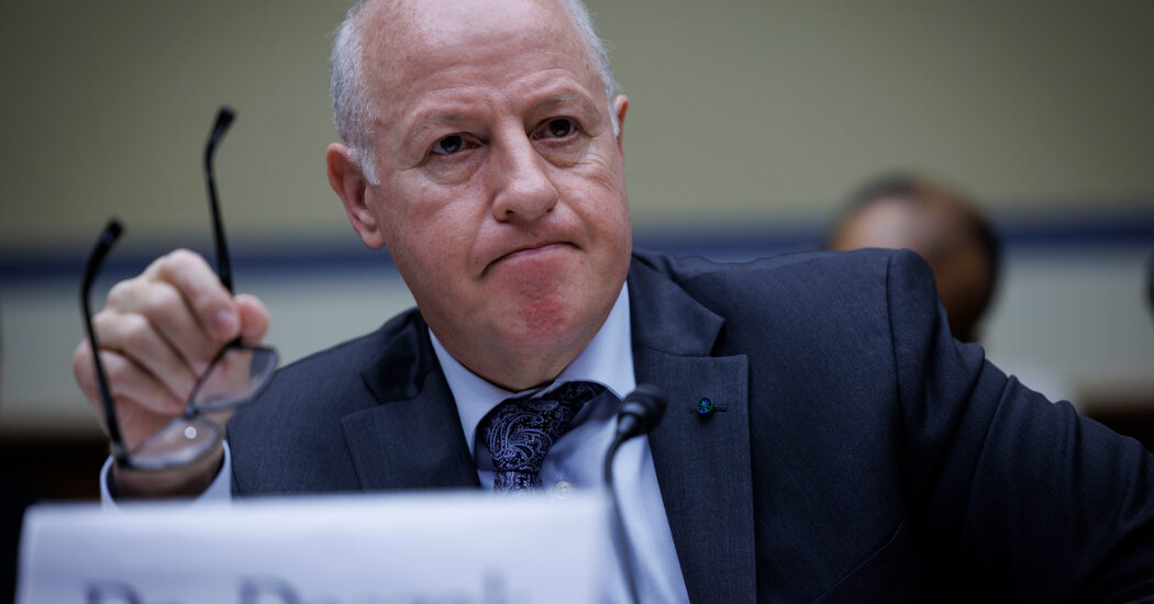
The decision came after a scorching hearing in which lawmakers barraged EcoHealth Alliance’s president with claims of misrepresenting work with Chinese virologists.The Biden administration, under acute pressure from House lawmakers, moved on Wednesday to ban funding for a prominent virus-hunting nonprofit group whose work with Chinese scientists had put it at the heart of theories that Covid leaked from a lab.The decision, announced in a letter from the Department of Health and Human Services, came on the heels of a scorching congressional hearing this month at which lawmakers barraged the group’s president with suggestions that he had misrepresented work with virologists in Wuhan, China, where the pandemic began. Republicans went further, demanding that Peter Daszak, the president of the nonprofit, EcoHealth Alliance, be criminally investigated.For EcoHealth, which relied on federal funding to study the threat of wild animal viruses, the loss of funding is another twist in a saga that has long dominated discussions of how the pandemic began.In April 2020, under orders from the Trump administration, the National Institutes of Health terminated a grant to EcoHealth amid President Donald J. Trump’s feud with China over the origin of the coronavirus. Three years later, an internal federal watchdog agency determined that the N.I.H. had failed to give a proper cause for ending the grant, which supplied an average of roughly $625,000 per year. The N.I.H. restarted a pared-back version of the award.Now, with Republicans stepping up their campaign against EcoHealth, and Democrats joining in the anger, the Biden administration has cut off funding for EcoHealth again.Health officials said they were suspending three active N.I.H. grants to EcoHealth that totaled $2.6 million for last year. And they proposed barring the group from receiving future federal research funding. Such bans, they said, usually last no more than three years, but could be longer or shorter.We are having trouble retrieving the article content.Please enable JavaScript in your browser settings.Thank you for your patience while we verify access. If you are in Reader mode please exit and log into your Times account, or subscribe for all of The Times.Thank you for your patience while we verify access.Already a subscriber? Log in.Want all of The Times? Subscribe.

Read more →

Research sheds light on how proteins linked to Alzheimer’s disease influence neuronal growth

New research has shed light in the complex interplay between cell proteins, and how they impact on neurons in neurodevelopmental disorders and Alzheimer’s disease.
A new study led by the University of Exeter and published in Royal Society Open Biology has discovered the key role that the protein Contactin-4 (encoded by the gene CNTN4) plays in shaping neurons.
The researchers began studying CNTN4 because it was known to have a role in autism, but its functional roles were not well understood. The team explored how CNTN4 functions within the brain, particularly its interactions with proteins involved in neurodegenerative diseases like Alzheimer’s disease.
For the first time, the researchers studied mice who have had the CNTN4 gene knocked out in the cortex, the region of the brain responsible for key functions including memory, thinking and reasoning. They found that neurons developed in a different way in the cortex region.
Researchers have demonstrated for the first time in human cells the interaction between genes CNTN4 and APP, a gene strongly linked to Alzheimer’s disease, revealing a co-dependent relationship that is essential for brain development, and specifically for the healthy growth of neurons. They found that CNTN4 not only contributes to neural elongation in the frontal cortex region of the brain, but also CNTN4 expression is regulated via a relationship with APP.
Using studies in genetically modified human cells, the team also discovered that a complex interaction exists between CNTN4 and APP. If CNTN4 is knocked out, then levels of APP decrease, but not to zero. The scientists believe that APP may compensate for the loss of CNTN4, and vice versa.
The study’s lead author, Dr Rosemary Bamford, of the University of Exeter Medical School, said: “It was quite remarkable to discover that CNTN4, a gene linked to developmental processes, also plays a role in modulating factors involved in Alzheimer’s disease. This intersection of developmental and neurodegenerative pathways offers exciting new insights into the broader implications of these proteins.”
Senior author Dr Asami Oguro-Ando, of the University of Exeter Medical School, said: “Looking ahead, my group is keen to further dissect the molecular mechanisms underpinning the interaction between CNTN4 and APP and explore their wider implications for disorders like Alzheimer’s and Autism Spectrum Disorder. Our next steps involve clarifying how the CNTN4-APP interaction impacts neural activity. Understanding this interaction is crucial as it represents a fundamental step towards a comprehensive grasp of neurodevelopmental and neurodegenerative disorders.”

Read more →

An active agent against hepatitis E

At present, there is no specific active substance against hepatitis E. As the disease kills 70,000 people every year, researchers are actively searching for one. The team from the Department of Molecular and Medical Virology at Ruhr University Bochum, Germany, may have found what they’re looking for. The researchers showed that the compound K11777 prevents host cells from helping the virus out of its shell by cleaving the viral capsid. This means it can no longer infect cells. “The compound is already being tested in clinical trials against other viruses such as Sars-Cov-2,” says lead author Mara Klöhn. “There’s still a lot of work to be done to find out whether it can be used as an active substance against hepatitis E, but it’s a first step.” The researchers published their findings on May, 11, 2024 in the journal Hepatology.
In order to infect an organ, viruses need the help of the host cells. “An effective approach is therefore to identify targets in the host that can be manipulated by drugs so that they no longer perform this helper function,” explains Mara Klöhn.
The researchers became aware of the compound K11777 in a roundabout way: During a control study conducted as part of cell culture studies on the hepatitis C virus with a known active ingredient, they discovered that this active ingredient was also effective against hepatitis E. “However, the drug wasn’t using the same pathway as with the hepatitis C virus, because the hepatitis E virus doesn’t have the target structure that this active substance attacks,” explains Mara Klöhn. This suggested that the drug may have an effect on host cells instead.
The research team narrowed down the possible target structures and turned their attention to cathepsins, which can process proteins, i.e. cleave them. K11777 inhibits many cathepsin types, i.e. blocks their function. Tests in cell culture with human liver cells showed that the compound actually prevents infection with hepatitis E viruses. “In follow-up experiments, we proved our hypothesis that the compound prevents cathepsin L from cleaving and opening up the viral capsid,” says Mara Klöhn. “This means that the virus can no longer infect host cells.”
Hepatitis E
The hepatitis E virus (HEV) is the main cause of acute viral hepatitis. Approximately 70,000 people die from the disease every year. After the first documented epidemic outbreak between 1955 and 1956, more than 50 years passed before researchers began to address the issue in depth. Acute infections usually clear up spontaneously in patients with an intact immune system. In patients with a reduced or suppressed immune system, such as organ transplant recipients or people infected with HIV, HEV can become chronic. HEV also poses a serious threat to pregnant women. There aren’t any vaccines nor specific active substances against the virus.

Read more →

New group training tool for the prevention of dementia

Solving a quiz as a group while moving around the room at the same time — this combination is the basis of a new tool designed to prevent dementia. Researchers developed and evaluated it in the “go4cognition” project with industry partners and brought it to market maturity. Vanessa Lissek and Professor Boris Suchan from Ruhr University Bochum, Germany, and Stefan Orth from the company Ontaris describe how effective the training with the system is in the Journal of Alzheimer’s Disease. The article was published online on April 30, 2024.
“In the project, we’ve developed a completely new system that can be used for dementia prevention training,” says Boris Suchan. “We were specifically interested in designing a tool for group training, as it produces very good results and increases acceptance. Also, all participants benefit and learn from each other.”
Passing the baton from quiz station to quiz station
The new system is designed for groups of seven to ten people. It consists of six stations arranged around the room, each equipped with a tablet. The tablets display different tasks, such as listing German chancellors, memorizing series of numbers or planning a trip around the world to specific locations. In order to complete the tasks, a participant has to move to the relevant station and insert a baton fitted with a microchip into a specific device. “We can then digitally record how long it took to complete the task and whether the answer was correct,” explains Boris Suchan. As everyone has their own baton, their individual performance can be evaluated.
Significant reduction of cognitive impairments through training
The researchers tested the system with 30 volunteers aged between 60 and 89 who had been diagnosed with mild cognitive impairment, i.e. who showed moderate cognitive impairment in neuropsychological tests. “Our participants are not restricted in their everyday activities, but have an increased risk of developing dementia at a later stage,” says Boris Suchan. Mild cognitive impairment occurs in 15 to 20 percent of people above the age of 60.
The participants trained with the go4cognition system two days a week for six weeks. After completing the training, 70 percent of them were no longer diagnosed with mild cognitive impairment.

System already in use in retirement homes
The group training tool is marketed by the company Ontaris. “It’s already used in retirement homes, for example in Oberhausen — also for residents who have not yet been diagnosed with mild cognitive impairment,” says Boris Suchan, and offers an outlook: “In future, we’re also considering testing the system for people with acquired brain damage.”
For people who don’t have access to the go4cognition system but still wish to take steps to prevent dementia, the Bochum-based researcher recommends physical and mental activities and a healthy diet. “All of this helps to prevent dementia or at least delay it for as long as possible,” says Suchan. As part of the go4cognition project, he and his colleagues published a review article on this topic.
About go4cognition
The go4cognition project ran from 2018 to 2023, supported by Leitmarkt Agentur.NRW, the European Union and the state of NRW (funding number ERDF -0801287). Specialists from various institutions developed new tools to prevent cognitive impairment in old age.

Read more →

Two genes of the germline are essential for the development of brain tumors in Drosophila

Every cell in an organism carries an identical set of genetic instructions within its DNA. However, what determines whether a cell develops as a neuron, a muscle, or any other specific cell type lies in the activation or inactivation of specific genes in that particular cell. For instance, in the Drosophila fly, the TrxT and dhd genes, which belong to the germline — that is, they participate in the formation of sperm and eggs, respectively — should be inactive in the brain.
However, scientists led by Dr. Cayetano González, ICREA researcher at IRB Barcelona, have discovered that TrxT and dhd are essential for the development of brain tumours. The inhibition of either of these two genes causes a partial reduction in the size of the tumours but simultaneous inhibition of both leads to a significantly greater decrease.
Published in the journal EMBO Reports, the study reveals that the two genes, TrxT and dhd, which are not normally coexpressed, are unexpectedly activated in brain tumours in the Drosophila fly. This abnormal activation leads to the development of aggressive tumours that invade surrounding tissues. Furthermore, the study uncovers disparities in the influence of genes, depending on whether the fly is male or female.
“This study reveals new germline genes involved in tumour development and underlines the importance of considering sexual dimorphism (or difference between the sexes) in cancer research. Our findings further suggest that collaboration between several genes may offer new therapeutic strategies to address the disease more effectively in the future,” says Dr. González.
The differences between sexes in cancer
Differences in cancer incidence and progression between men and women have been known for decades, but a detailed understanding of sexual dimorphism in relation to cancer biology is still limited.
TrxT and dhd, which are genes specific to the Drosophila germline, are equally expressed in somatic tumours of males and females and their simultaneous inactivation leads to a significant reduction in tumour size, especially in males. This study points to a synergic effect between TrxT and dhd in the initial development of the tumour, while only TrxT appears to be essential for the maintenance of the tumour once established.

“Our results suggest that the initial phases of tumour development and the growth of established tumours are two distinct phenomena that may depend on different molecular mechanisms,” explains Dr. Cristina Molnar, first author of the study.
Germline genes as targets against cancer
Called “cancer-germline” genes (or “cancer-testis” genes due to their initial discovery in the testes), the genes traditionally associated with the formation of sperm and eggs have been identified as key players in the pathogenesis of certain types of cancers, such as melanomas, lung carcinoma, and others.
Given that the function of these genes is limited mainly to the production of sex cells, they are a particularly promising target for cancer treatment. Therefore, blocking them could result in minimal side effects, since the rest of the body’s cells would not be affected.
While the researchers emphasize that this study does not directly lead to new cancer therapies, it offers valuable insight into the biological processes involved in tumour formation and growth that could pave the way for future research and the development of therapeutics.

Read more →

New study links protein secreted by blood vessels to drug-resistant cancer

Cancer is a leading cause of death globally. One of the primary reasons why cancer is such a deadly disease is the ability of cancer cells to become drug-resistant. After decades of medical research, scientists came to understand that malignant tumors often harbor a special population of cells called cancer stem cells (CSCs). Much like normal stem cells, CSCs can self-renew and differentiate into various cell types within a tumor, playing important roles not only in tumor growth and metastasis but also in the development of drug resistance.
Unfortunately, developing therapies targeting CSCs directly has proven quite challenging, given their ability to adapt and repopulate. Thus, some researchers have directed their attention to the blood vessels within the tumor tissue. Over the past few years, researchers have found that certain subpopulations of endothelial cells (cells lining the blood vessels) secrete angiocrine factors, which regulate the proliferation and maturation of stem cells. Understanding which cells produce these factors and their functions in the tumor microenvironment can, in turn, lead to novel cancer therapies.
Against this backdrop, a research team from Japan, including Professor Hiroyasu Kidoya and Dr. Yumiko Hayashi from the Department of Integrative Vascular Biology at the Faculty of Medical Science at the University of Fukui, conducted a study on secreted frizzled-related protein 1 (Sfrp1) — an angiocrine factor — to clarify its role in tumor tissues. Their findings were published online in the journal In Vitro Cellular & Developmental Biology — Animal on April 16, 2024.
“While blood vessels are usually considered merely a supply route for oxygen and nutrients, our research focused on a completely different function of blood vessels, namely the production of angiocrine factors. We conducted this research with the idea that angiocrine factors might also be involved in tumor progression and aimed to elucidate whether Sfrp1 affects CSC maintenance and that of the tumor tissue as a whole,” explains Prof. Kidoya.
To shed light on these issues, the researchers bred Sfrp1-knockout (KO) mice using CRISPR-Cas9 genome editing. Then, they transplanted lung carcinoma tumors into Sfrp1-KO and wild-type mice and observed the effects of Sfrp1 (or the lack thereof) using standard techniques, such as immunostaining, flow cytometry, and quantitative genetic expression analysis.
Initial experiments revealed that Sfrp1 is produced in a small subset of vascular endothelial cells in tumor tissue and that its presence is important for tumor growth. Not only was the tumor growth suppressed in Sfrp1-KO mice, but transplanted tumor cells overexpressing Sfrp1 gave rise to faster growing tumors.
Interestingly, researchers found that Sfrp1-deficient tumors could not maintain substantial CSC populations during the late stages of tumor growth, even though these tumors had a higher initial percentage of CSCs. This finding is particularly important as it hints at one of the biological roles of Sfpr1 in the tumor microenvironment and its involvement in cancer pathology. “Some CSCs in tumor tissues exist in a state of arrested cell proliferation, and their presence contributes to tumor growth and resistance to anticancer drugs,” explains Prof. Kidoya. “Our results indicate that Sfrp1 may regulate CSC self-renewal and transient malignant growth, and act to maintain a dormant state.”
Further cementing these results, the researchers noted that Sfrp1 did not affect the structure of blood vessels within the tumor, implying that the observed effects on tumor growth were unrelated to vasculature. Instead, genetic expression analyses revealed that Sfrp1 contributes to CSC maintenance by modulating the well-preserved Wnt signaling pathway (an evolutionarily conserved pathway that regulates crucial aspects of cell fate determination, cell migration, and organogenesis during embryonic development).
Overall, the newfound knowledge provided by this work could pave the way for the development of new cancer therapies targeting mechanisms that help maintain CSCs. “Targeting specialized vascular endothelial cells involved in producing angiocrine factors could help destroy the CSC niche, serving as a potential approach for inhibiting tumor growth with minimal side effects,” concludes Prof. Kidoya. “I believe this could lead to the development of treatments for intractable cancer patients whose tumors exhibit resistance to anticancer drugs as well as therapeutic agents to suppress cancer recurrence and metastasis.”
Further research building on these findings will serve as a stepping stone to effective therapies for drug-resistant cancer.

Read more →

Exploring the mechanism behind drug eruptions in the skin

Although medications can often help patients find a cure or respite from their condition, millions of people worldwide suffer from unpredictable drug toxicities every year. In particular, drug eruptions which manifest through symptoms such as redness, blisters, and itching on the skin, are quite common. Severe drug eruptions can become life-threatening and can have long-lasting consequences. Thus, understanding how and why drug eruptions occur is an important area of research in medical science.
To this end, previous studies have identified specific variants of certain genes as potential causal agents of drug eruptions. Scientists believe that the genes encoding the human leukocyte antigen (HLA), a protein expressed on the surface of leucocytes known to play an important role in the immune system, are involved in the onset of drug eruption. However, current theories cannot explain why HLA-related drug eruptions typically manifest on the skin rather than in multiple organs throughout the body.
To address this knowledge gap, a research team including Lecturer Shigeki Aoki, Kousei Ito, and Akira Kazaoka from the Graduate School of Medical and Pharmaceutical Sciences, Chiba University, conducted an in-depth study on the link between HLA and drug eruptions. Their findings were published in PNAS Nexus on April 2, 2024.
The researchers first conducted a series of experiments on keratinocytes from mice, which are the primary type of cells found in the skin. These keratinocytes were engineered to express a specific variant of the HLA gene called HLA-B*57:01, which specifically bind to the antiviral drug abacavir. Then, they validated these findings in genetically modified mice expressing HLA-B*57:01, that were exposed to abacavir.
The researchers found that HLA-B*57:01 expressing keratinocytes that were exposed to abacavir exhibited endoplasmic reticulum (ER) stress responses, such as immediate release of calcium into the cytosol and elevated expression of heat shock protein 70 (HSP70). They also observed an increased production of cytokines and immune cell migration. Abacavir exposure triggered HLA misfolding in the ER, leading to ER stress. Moreover, the researchers observed that the ER stress could be reduced by using 4-phenylbutyrate (4-PB). By alleviating this stress, they managed to suppress the onset of severe drug eruption symptoms. This newfound knowledge could form the basis for innovative treatment options for management of drug eruptions.
But how does this new information contrast with what was already known about HLA? “HLA molecules are an integral component of our immune system, that typically present foreign antigens to white blood cells, which judge these antigens as self or non-self. In this established role, HLAs are usually secondary players,” explains Dr. Aoki. “However, our research highlights a novel function of the HLA molecule within skin cells. We revealed that a specific HLA genotype in keratinocytes can recognize certain drugs as foreign, triggering an endoplasmic reticulum stress response.”
Taken together, the findings of this study uncover a new role of HLA proteins in sensing and responding to potential threats in skin cells. Thus, their functions may extend well beyond mere antigen presentation for the immune system. Moreover, considering that the variant of HLA possessed by an individual can be determined through genetic testing, this study could help develop preventive measures and diagnostics against severe adverse drug reactions. According to Dr. Aoki, this is in line with current research directions and trends in medical science. “In 10 years, we anticipate entering the ‘whole genome era,’ where personalized medicine based on individual genomes will become a standard practice,” he comments. He further adds, “Building on the findings of this study, we believe that a comprehensive understanding of the mechanism underlying HLA-dependent adverse drug reactions will enable the delivery of safe medical care, allowing patients to avoid unnecessary suffering due to side effects.”
Overall, future investigations in this research area might minimize the occurrence of drug eruptions and save people from potentially fatal adverse drug reactions.
About Dr. Shigeki Aoki Dr. Shigeki Aoki is a lecturer at the Graduate School of Pharmaceutical Sciences, Chiba University, Japan. His research focuses mainly on cancer metabolism and drug toxicity. He has authored multiple papers published in reputed journals. Dr. Aoki is a member of various professional bodies in Japan. He has also received several awards for his research, including the Award for Young Scientists conferred by The Pharmaceutical Society of Japan.

Read more →

Longer sprint intervals can improve muscle oxygen utilization compared to shorter intervals

Physical activities like jogging, walking, cycling, and sprinting are activities known to engage the musculoskeletal system and result in the utilization of energy. Sprint interval training (SIT) is a type of sprinting exercise that involves cycles of intense exercise followed by a short duration of rest. How the durations of exercise and rest are structured can affect the impact of SIT on physiological responses. In recent years, the field of sports physiology has witnessed increased interest in optimizing SIT protocols. This surge can enhance the recognition of SIT’s efficacy in improving athletic performance and overall well-being, highlighting its versatility as a tool for promoting health and fitness.
Now, in an effort to highlight the benefits of SIT, a team of researchers from Japan, comprising Dr. Takaki Yamagishi from the Department of Sport Science and Research, Japan Institute of Sports Sciences and Human Performance Laboratory at the Comprehensive Research Organization at Waseda University, and Professor Yasuo Kawakami who directs the Human Performance Lab and is from the Faculty of Sport Sciences, Waseda University, conducted SIT experiments with healthy human volunteers in a recent study. This study was published online in Medicine & Science in Sports & Exercise journal on March 7, 2024.
Explaining the motivation behind their research work, Yamagishi says, “Establishing a minimum dose of exercise training to bring about training benefits, such as aerobic fitness, has been one of my main research interests. Due to the support from Prof. Kawakami and other co-authors, along with the collaboration with Waseda University, this unique research work using a multifaceted approach was possible.”
The team of researchers matched two different sprint interval exercises (SIE) for the total duration of sprint and sprint-to-rest ratio. They analyzed the influence of SIE on physiological and metabolic responses by examining pulmonary oxygen uptake (V̇O2) levels and changes in tissue oxygenation index (∆TOI) in thigh muscles. Additionally, they utilized the T2-weighted magnetic resonance imaging (MRI) technique to assess the activation of thigh muscles.
The researchers highlight that SIE20, comprising of two 20-second sprints with 160-second recovery, outperformed SIE10, which involved four 10-second sprints with 80-second recovery. While both the SIE protocols significantly increased whole-body and peripheral oxidative metabolism and the activation of the major thigh muscles, as indicated by the increases in V̇O2 levels, ∆TOI and MRI T2 values, respectively, greater peripheral oxidative metabolism was achieved with SIE20. They also observed that successive sprint repetitions in SIE10 did not correlate with greater oxidative metabolism.
Elaborating on the real-life applications and impact of the present research, Yamagishi says, “In today’s fast-moving world, lack of time is a major hindrance to regular physical activity. However, the exercise modalities employed in our study require less than 15 minutes to complete and provide considerable health benefits.”
In summary, the findings from this study can address important research gaps in SIT, such as the effects of minimum sprint duration and repetitions on aerobic and metabolic responses in humans. In-depth studies on low-volume SIT have the potential to improve training programs and exercise regimens. Sharing his concluding thoughts, Yamagishi says, “Exercise guidelines proposed by major organizations such as the American College of Sports Medicine are updated every 5 to 10 years, and we are hopeful that our study can be a part of it. Future studies on SIE can build on our findings to establish the dose-response relationship between exercise volume or intensity and the degree of training adaptations.”

Read more →

Fighting fat and inflammation: Scientists develop powerful new compounds

Obesity and inflammatory diseases are increasing in prevalence and contribute to the growing burden of lifestyle disorders such as diabetes and hypertension. There is a lack of naturally derived alternatives to tackle these issues. Researchers from Tokyo University of Science have synthesized novel amino acid derivatives of menthol and studied its properties. The menthyl esters showed exceptional anti-inflammatory and anti-obesity activities during preclinical studies and can be developed as therapeutic compounds with further research.
Modified derivatives of natural products have led to significant therapeutic advances and commercial success in recent times. Menthol is a naturally occurring cyclic monoterpene alcohol found in various plants, particularly in members of the mint family such as peppermint and spearmint. It is a common ingredient found in a wide range of confectionaries, chewing gums and oral care products. Interestingly, menthol also has high medicinal value due to its analgesic, anti-inflammatory, and anti-cancer effects.
In a recent study, a team of researchers led by Professor Gen-ichiro Arimura from the Department of Biological Science and Technology, Tokyo University of Science, Japan, developed and investigated menthyl esters of valine (MV) and isoleucine (MI), which are derived from menthol by replacing its hydroxyl group with valine and isoleucine, respectively.
Their research findings were published in the Immunology journal on May 08, 2024. Sharing the motivation behind the present work, Prof. Arimura says, “The functional components of plants that contribute to human health have always intrigued me. Discovering new molecules from natural materials inspired our research team to develop these amino acid derivatives of menthol.”
The researchers began by synthesizing menthyl esters of six amino acids characterized by less-reactive side chains. Subsequently, they assessed the properties of these esters using in vitro cell line studies. Finally, they conducted experiments in mice to explore the effects of these compounds under induced disease conditions. The exceptional anti-inflammatory profiles of MV and MI was determined by assessing the transcript levels of tumor necrosis factor-α (Tnf) in stimulated macrophage cells. Remarkably, both MV and MI outperformed menthol in the anti-inflammatory assay. RNA sequencing analysis revealed that 18 genes involved in inflammatory and immune responses were effectively suppressed.
Elated with their findings, the researchers went a step further and investigated the mechanism of action of the menthyl esters. They discovered that liver X receptor (LXR) — an intracellular nuclear receptor, had an important role in the anti-inflammatory effects and this was independent of the cold-sensitive transient receptor TRPM8, which primarily detects menthol. Delving deeper into the LXR-dependant activation of MV and MI, they found that Scd1 gene — central to lipid metabolism was upregulated by LXR. Moreover, in mice with induced intestinal colitis, the anti-inflammatory effects were further validated with suppressed transcript levels of Tnf and Il6 genes by MV or MI, in an LXR-dependent manner.
Driven by the discovery of LXR-SCD1 intracellular machinery, Prof. Arimura and his team hypothesized the menthyl esters to possess anti-obesity properties. They found that these esters inhibited adipogenesis-fat accumulation, specifically at the mitotic clonal expansion stage in 3T3-L1 adipocyte cells. During animal studies, the diet-induced obesity in mice was ameliorated and adipogenesis was suppressed.
Menthyl esters possess unique advantages compared to other anti-inflammatory or anti-obesity compounds currently being researched or used. Their specific mechanisms of action, that contribute to their dual anti-inflammatory and anti-obesity effects sets them apart from other compounds and may make them particularly effective in addressing both inflammatory conditions and metabolic disorders. They could benefit specific populations like individuals with chronic inflammatory conditions, metabolic syndrome, or obesity-related complications.
“Although this study focused on their functions and mechanisms of action in diseases modeled after inflammation and obesity, we expect that these compounds will also be effective against a wide range of lifestyle-related diseases caused by metabolic syndrome, such as diabetes and hypertension, as well as allergic symptoms,” says Prof. Arimura optimistically.
In conclusion, this study underscores the importance and value of multi-faceted molecules derived from naturally occurring substances. Future research involving these novel and superior menthyl esters may result in therapeutic compounds to tackle the ever-growing health concerns of obesity and inflammatory conditions.

Read more →

New cardiac research will save women’s lives by improving detection of heart failure

An important new study has advanced how heart failure is detected in women — meaning more female patients can be diagnosed and at an earlier stage.
Researchers led by teams from the Universities of East Anglia (UEA), Sheffield and Leeds, have been able to fine-tune how magnetic resonance imaging (MRI) is used to detect heart failure in women’s hearts, making it more accurate.
Lead author Dr Pankaj Garg, of the University of East Anglia’s Norwich Medical School and a consultant cardiologist at the Norfolk and Norwich University Hospital, said: “By refining the method for women specifically, we were able to diagnose 16.5pc more females with heart failure.
“This could have huge impact in the NHS, which diagnoses around 200,000 patients with heart failure each year.
“This improved method will increase early detection, meaning more women can get life-saving treatment sooner.”
In 2022, UEA and the University of Sheffield published research which showed how using MRI scans could be used to detect heart failure and which led to this technique being widely employed by medics.
When a heart starts to fail, it is unable to pump blood out effectively, and so the pressure in the heart rises.

Co-author Dr Gareth Matthews, of the University of East Anglia’s Norwich Medical School, said: “Currently one of the best ways of diagnosing heart failure is to measure pressures inside the heart with a tube called a catheter.
“While this is very accurate, it is an invasive procedure, and therefore carries risks for patients, which limits its use.
“For this reason, doctors tend to use echocardiograms, which are based on ultrasound, to assess heart function, but this is inaccurate in up to 50 per cent of cases. Using MRI, we can get much more accurate images of how the heart is working.”
The team was able to create an equation which allowed them to non-invasively derive the pressure in the heart using an MRI scanner.
However, previous use of this method wasn’t as accurate as the researchers would have liked in diagnosing heart failure in women, especially in early or borderline disease.
Co-author Professor Andy Swift, of the University of Sheffield’s School of Medicine and Population Health, said: “Women’s hearts are biologically different to men’s.

“Our work suggests that in heart failure women’s hearts may respond differently in response to increases in pressure.”
Heart failure can be classed differently, depending on the amount of blood squeezed out of the main chamber of the heart with every beat, known as the heart’s ejection fraction.
Women suffer disproportionately from a type of heart failure where the pumping function of the heart is preserved but the ability of the heart to relax and fill with blood is impaired.
Echocardiography really struggles to diagnose this type of heart failure. The improvements in diagnosis from this new work will enable more of this particular cohort to be diagnosed more accurately and hopefully drive better treatments.
Co-author Dr Peter Swoboda, of the University of Leeds’ Faculty of Medicine and Health, said: “The symptoms of heart failure, like breathlessness and fatigue, can have a devastating effect on patients’ quality of life.
“We are increasingly recognising the importance of early diagnosis and, early treatment can improve symptoms and life expectancy.
“This research will help diagnose heart failure in women more quickly and get them established on life-saving treatments sooner.”
The Government’s Health and Social Care Secretary, Victoria Atkins, said: “Heart failure is a devastating condition affecting hundreds of thousands of women in the UK, so this research is a hugely positive development that could make it possible for thousands of people to get diagnosed and treated at an earlier stage.
“For the second year of our Women’s Health Strategy for England, I have been clear that we need more research to look at the differences between how conditions affect men and women.
“I am delighted that this government-backed research has met this challenge so that we can get life-saving treatment to women faster.”
The research was a collaboration between the University of East Anglia, the University of Leeds, the University of Sheffield, the Norfolk and Norwich University Hospital NHS Foundation Trust, the National Heart Research Institute Singapore, Duke-NUS Medical School in Singapore, Queen Mary University of London, the National Institute for Health and Care Research’s Sheffield Biomedical Research Centre, the University of Amsterdam and Kocaeli City Hospital in Turkey.
It was funded by the National Institute for Health and Care Research (NIHR) Sheffield Biomedical Research Centre, the Wellcome Trust, and the National Medical Research Council (NMRC).

Read more →