My feet and hands were amputated after sepsis – MP

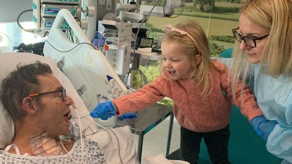
Published15 minutes agoShareclose panelShare pageCopy linkAbout sharingThis video can not be playedTo play this video you need to enable JavaScript in your browser.By Helen Catt, Isabella Allen & Kate Whannel BBC NewsConservative MP Craig Mackinlay is due to return to Parliament for the first time after suffering a life-threatening episode of sepsis which led to the amputation of his hands and feet. The South Thanet MP recalled the shock of waking from an induced coma to find his limbs had turned completely black. He says they were “like plastic…you could almost knock them…they were black, desiccating, clenched”.”They managed to save above the elbows and above the knees,” he added. “So you might say I’m lucky.”Speaking to the BBC, he said he now he wants to be known as the first “bionic MP”, after he was fitted with prosthetic legs and hands.’A very strange blue’It was on 27 September, when Mr Mackinlay, 57, began feeling unwell. He didn’t think much of it, took a Covid test (which came back negative) and had an early night.During the night he was badly sick but still didn’t think it was anything serious.However, as the night wore on, his wife Kati – a pharmacist – began to get worried and tested his blood pressure and temperature.By the morning, she noticed that his arms felt cold and she couldn’t feel a pulse. After ringing for an ambulance, Mr Mackinlay was admitted to hospital.Within half an hour he had turned what he calls “a very strange blue”. “My whole body, top to bottom, ears, everything, blue,” he says.He had gone into septic shock. The MP was put into an induced coma that would last for 16 days.His wife was told she should prepare for the worst, with staff describing her husband as “one of the illest people they’d ever seen”. His chances of survival stood at just 5%. Image source, Craig Mackinlay At his wife’s insistence, Mr Mackinlay was transported from his local hospital in Medway, Kent, to St Thomas’ in central London, directly opposite his workplace, the Houses of Parliament. He remembers little of this – but what he can remember is the strange dreams which he thinks were brought on by morphine. As he came to, the grim reality set in.On waking up, he remembers hearing discussions about his arms and legs. “By then they had turned black…you could almost knock them,” he says, likening them to the plastic of a mobile phone.He says he wasn’t surprised when he was told they might have to be amputated.”I haven’t got a medical degree but I know what dead things look like. I was surprisingly stoic about it… I don’t know why I was. It might have been the various cocktail of drugs I was on.”‘A sombre Christmas’The operation – for all four amputations – was on 1 December. He remembers waking up following the procedures feeling strangely alert. So alert, he wondered if the amputations had actually happened at all. “But I woke up and I looked down and you obviously realise that they had.”Christmas was “sombre”, spent with his family, including his four-year-old daughter Olivia. “She adapted to it very easily,” says Mr Mackinlay. “Probably better than anybody else frankly. I think children are just so remarkably adjustable.”Image source, Craig Mackinlay Olivia has had to adapt to her father’s new prosthetic legs – one he has nicknamed Albert, after the dummy used by war camp prisoners in the 1950s film, Albert R.N.Learning to walk with his prosthetics has taken time. First, he had to re-build the muscles which had wasted away.”My legs have never been big – I always say I’ve got chicken legs, but now they are sparrow legs. “There was no muscle on them at all, it was quite horrible. You picked up your leg and you can see a bone and a bit of sort of hanging.”Once his prosthetic legs were attached he gradually relearnt how to walk.”After a really quite quick time you think ‘I can do this’.”On 28 February – five months after first feeling ill – he was able to walk his first 20 steps unaided. Inevitably progress was stop-start. He got painful blisters in areas where his skin had broken down and had to stop for a bit. “That was very frustrating – for me walking was my sign of success,” he says.Image source, Craig MackinlayWhat is sepsis?Sepsis is a rare but serious condition that develops when the body’s immune system overreacts to an infection and starts attacking its own tissues and organs. Symptoms can include,

Read more →

New tool may help prioritize high-risk infants for RSV immunization

On the heels of a shortage of nirsevimab for infant respiratory syncytial virus (RSV) lower respiratory tract infection (LRTI) prevention, a new tool may help identify newborns at highest risk for developing serious RSV LRTI, according to research published at the ATS 2024 International Conference.
“Timely identification of infants at highest risk of RSV-related morbidity is key to prevention,” said lead author Brittney M. Snyder, PhD, assistant professor, Division of Allergy, Pulmonary and Critical Care Medicine, Vanderbilt University Medical Center. “Our personalized risk prediction tool may have applications in allocating expensive and/or limited immunoprophylaxis (immunization with nirsevimab or palivizumab) to achieve the greatest benefit and in promoting RSV prevention among families with high-risk infants.”
More than half of RSV LRTIs are among healthy, term infants who are generally considered low risk. These infants are, in fact, at risk for requiring intensive care unit -level care, and some may die from their illness. Early immunization with nirsevimab is recommended for all infants by the Centers for Disease Control & Prevention, yet in October 2023 nirsevimab was in short supply and the CDC recommended giving it only to high-risk infants who weren’t eligible for immunization with palivizumab.Both products, which prevent RSV LRTI in newborns and young children, are monoclonal antibodies (nirsevimab is long-acting and only requires one dose, while palivizumab is short-acting and requires monthly injections during RSV season).
In the population-based study by Dr. Snyder and colleagues including children insured by the Tennessee Medicaid Program, the researchers assessed infants who did not receive RSV immunoprophylaxis in the first year of life. They gathered demographic and clinical data from administrative health care encounters and linked birth certificates. “To predict whether these infants developed severe RSV LRTI requiring ICU admission during the first year of life, we developed a multivariable logistic regression model. The model includes demographic and clinical variables collected at or shortly after birth-19 variables in all, such as prenatal smoking, delivery method, maternal age and assisted breathing (ventilation) during birth hospitalization,” said lead biostatistician Tebeb Gebretsadik, MPH, Department of Biostatistics, Vanderbilt University Medical Center.
Among 429,365 infants in the study, 713 had severe RSV LRTI requiring ICU admission. The tool had good predictive accuracy and internal validation that indicated a good fit.
“Our objective was to develop a personalized tool for use in all newborns using readily available birth and postnatal data to predict risk of RSV LRTI requiring ICU admission, useful for prioritizing RSV prevention products with limited availability,” said Principal Investigator Tina V. Hartert, MD, MPH, professor of medicine and pediatrics at Vanderbilt University Medical Center. Even though the recent nirsevimab shortage has, fortunately, eased up, it is not known whether shortages will occur in the future. “This tool may be particularly helpful in prioritizing which infants should be immunized during times of limited availability of RSV prevention medicines. Using the tool to identify if their infant is at high risk for RSV infection requiring ICU care may also persuade vaccine-hesitant families to accept RSV immunoprophylaxis, by showing them their newborn is at high risk,” she added.
“To ensure compatibility with nirsevimab and maternal vaccination, our tool was developed for use in all infants,” concluded co-author Niek Achten, MD, postdoctoral fellow in pediatrics, Erasmus University Medical Center, Rotterdam, Netherlands, who imagined the need for such a tool. “In addition to use in the United States during times of limited availability, our tool may prove useful in countries with budgetary constraints needing to prioritize administration to the highest risk infants.”
The authors note that next steps to ensure optimal usefulness include validation of the tool in external populations, further cost-effectiveness analyses and decision curve analyses.

Read more →

Body lice may be bigger plague spreaders than previously thought

A new laboratory study suggests that human body lice are more efficient at transmitting Yersinia pestis, the bacterium that causes plague, than previously thought, supporting the possibility that they may have contributed to past pandemics. David Bland and colleagues at the United States’ National Institute of Allergy and Infectious Diseases present these findings in the open-access journal PLOS Biology on May 21.
Y. pestis has been the culprit behind numerous pandemics, including the Black Death of the Middle Ages that killed millions of people in Europe. It naturally cycles between rodents and fleas, and fleas sometimes infect humans through bites; thus, fleas and rats are thought to be the primary drivers of plague pandemics. Body lice — which feed on human blood — can also carry Y. pestis, but are widely considered to be too inefficient at spreading it to contribute substantially to outbreaks. However, the few studies that have addressed lice transmission efficiency have disagreed considerably.
To help clarify the potential role of body lice in plague transmission, Bland and colleagues conducted a series of laboratory experiments in which body lice fed on blood samples containing Y. pestis. These experiments involved the use of membrane feeders, which simulate warm human skin, enabling scientists to study transmission potential in a laboratory setting.
They found that the body lice became infected with Y. pestis and were capable of routinely transmitting it after feeding on blood containing levels of the pathogen similar to those found in actual human plague cases.
They also found that Y. pestis can infect a pair of salivary glands found in body lice known as the Pawlowsky glands, and lice with infected Pawlowsky glands transmitted the pathogen more consistently than lice whose infection was limited to their digestive tract. It is thought that Pawlowsky glands secrete lubricant onto the lice’s mouthparts, leading the researchers to hypothesize that, in infected lice, such secretions may contaminate mouthparts with Y. pestis, which may then spread to humans when bitten.
These findings suggest that body lice may be more efficient spreaders of Y. pestis than previously thought, and they could have played a role in past plague outbreaks.
The authors add, “We have found that human body lice are better at transmitting Yersinia pestis than once appreciated and achieve this in more than one way. We describe a new bite-based mechanism in which a set of accessory salivary glands unique to lice, termed the Pawlowsky glands, become infected with Y. pestis and secrete lubricant containing plague bacilli onto the insect’s mouthparts prior to blood feeding.”

Read more →

Inherited genes play a larger role in melanoma risk than previously believed

When it comes to skin cancer, most people think of warnings about sunburn and tanning beds. Thoughts of “cancer genes” or inherited risks are reserved for diseases like breast cancer or colon cancer. A new study challenges this status quo by showing that genetics play a larger role in melanoma risk than recognized.
Physicians rarely order genetic screens to assess risk factors for patients with a family history of melanoma because, according to the previous, limited studies, only 2-2.5% of all cases are genetic. For the same reason, insurance companies rarely cover these tests outside of the most extreme situations. In the medical field, genetic testing is generally not offered for cancers that don’t meet a threshold of 5%.
A study from researchers and clinicians led by Cleveland Clinic’s Joshua Arbesman, MD, and Stanford Medicine’s Pauline Funchain, MD (formerly Cleveland Clinic), suggests that melanoma more than meets that threshold. Their results, published in the Journal of the American Academy of Dermatology, report that up to 15% (1 in 7) of patients who received melanoma diagnoses from Cleveland Clinic physicians between 2017 and 2020 carried mutations in cancer susceptibility genes. The research team, which includes Cleveland Clinic Center for Immunotherapy and Precision Oncology’s Ying Ni, PhD, and Claudia Marcela Diaz, PhD, analyzed international patient databases and found similar results.
“Hereditary cancers can wreak havoc through families and leave devastation in their wake. Genetic testing lets us proactively identify, screen and even treat these families to equip them with the tools they need to get the best healthcare possible,” says Dr. Arbesman. “I would recommend physicians and insurance companies expand their criteria when it comes to offering genetic testing to individuals with family histories of melanoma, because inherited predisposition to it isn’t nearly as rare as we think it is.”
Dr. Arbesman, who runs a lab in Cleveland Clinic Lerner Research Institute’s Cancer Biology, also says his findings support an increasingly popular opinion amongst cancer biologists: there are risk factors beyond sun exposure that can influence an individual’s chances of developing melanoma.
“Not all of my patients had inherited mutations that made them more susceptible to the sun,” he says. “There’s clearly something more going on here and more research is needed.”
Dr. Arbesman and his team are studying many of the genes that came up in his patients’ genetic tests to learn more about how melanoma develops and how it can be treated. For example, he is working to determine if some of his patients and their families who show inherited mutations may benefit more from immunotherapy compared to those who don’t carry inherited mutations. His lab is also working to determine how other patients’ genes contributed to the development and the severity of their melanoma.

Read more →

Scientists create tailored drug for aggressive breast cancer

Scientists have used breast cancer cells’ weakness against themselves by linking a tumour-selective antibody with a cell-killing drug to destroy hard-to-treat tumours.
The research, published today in Clinical Cancer Research by a team from King’s College London and funded by Breast Cancer Now, marks a new method in cancer treatment.
The discovery is particular to triple negative breast cancer, which makes up 15% of all diagnosed breast cancer. This type of breast cancer is typically aggressive, resistant to chemotherapy, has a lower survival rate and is more common in women under 40.
Usual treatment involves surgery, chemotherapy and radiotherapy, however this type of cancer can evade the drugs and return to spread again.
The scientists conducted data analysis using over 6000 breast cancer samples to investigate the properties of breast cancer cells that are associated with aggressive and chemotherapy-resistant cancers.
They studied the cancer’s biology, what is expressed in the tumour and the cell surface, and the cell’s insides to understand how the cancer cells escape from cancer drugs. They established the presence of the cancer cell surface marker EGFR along with oncogenic molecules cyclin-dependent kinases (CDK), which are responsible for cell division and proliferation.
They used this knowledge against the cancer cells to link cetuximab, a tumour-selective antibody that targets the EGFR protein expressed in this type of cancer, with a CDK-blocking drug to create a tailored drug for breast cancer. Because the antibody drug conjugate specifically targets the cancer cell, it may be possible to administer a lower inhibitor dose than usual which means it’s less toxic for the patient.

Lead author Professor Sophia Karagiannis, from King’s College London, said: “We were on the hunt for cancer’s vulnerabilities and now we’ve found out how we can guide our therapies to one of these. We combined these two drugs to create a tailored antibody drug conjugate for patients with this aggressive cancer. The antibody guides the toxic drug directly to the cancer cell which offers the possibility for a lower dose and less adverse side effects to be experienced.
“More work needs to be done before this therapy can reach the clinic, but we expect that this can offer new treatment options for cancers with unfavourable prognosis. Beyond this antibody drug conjugate, we hope that our concept will lead the way for new antibody drug conjugates of this type to be tailored to patient groups likely to benefit.”
Lead research scientist Dr Anthony Cheung from King’s College London said: ”Triple negative breast cancer represents a molecularly and clinically diverse disease. By exploiting EGFR overexpression and dysregulated cell cycle molecules in selected patient groups, the antibody drug conjugate, but not the antibody alone, could stop the cancer cell from dividing and engender cytotoxic functions specifically against the cancer cells.”
Dr Simon Vincent, director of services, support and influencing at Breast Cancer Now, which funded this research, said: “Each year, around 8,000 women in the UK are diagnosed with triple negative breast cancer, which is typically more aggressive than other breast cancers and more likely to return or spread following treatment.
“This exciting research has not only improved our understanding of the properties of aggressive breast cancer cells that are resistant to chemotherapy but has also brought us closer to developing a targeted therapy that destroys these cancer cells while minimising side effects for patients.
“While further research is needed before this treatment can be used in people, this is an exciting step forward in developing targeted therapies for triple negative breast cancer, and we look forward to seeing how these findings could lead to new and effective ways of tackling this devastating disease.”

Read more →

Dr. Paul Parkman, Who Helped to Eliminate Rubella, Dies at 91

He also identified the virus, which can cause infants to be born with severe physical and mental impairments as well as causing miscarriages and stillbirths.Dr. Paul D. Parkman, whose research was instrumental in identifying the virus that causes rubella and developing a vaccine that has prevented an epidemic of the disease in the United States for more than 50 years, died on May 7 at his home in Auburn, N.Y., about 60 miles east of Rochester in the Finger Lakes region. He was 91.The cause was lymphoblastic leukemia, his niece Theresa M. Leonardi said.Rubella, also known as German measles because German scientists classified it in the 19th century, is a moderate illness for most patients, identified by a spotty and often itchy red rash. But in pregnancies, it can cause infants to be born with severe physical and mental impairments and can also cause miscarriages and stillbirths.When Dr. Parkman was a pediatric medical resident in the 1950s at the State University Health Science Center (now the SUNY Upstate Medical University) in Syracuse, he once recalled, he anguished over a showing a new mother her stillborn baby whose rash, he would learn later, probably resulted from the mother’s infection with rubella during pregnancy.In 1964 and 1965, rubella — an epidemic that struck every six to nine years — caused about 11,000 pregnancies to be miscarried, 2,100 newborns to die and 20,000 infants to be born with birth defects.After the discovery of a vaccine for rubella, also known as German measles, displays educated the public about vaccination and the potential transmission of the virus from mother to fetus.Smith Collection/Gado, via Getty ImagesThat was the worst outbreak in three decades — and the last epidemic in the United States. The disease was declared eliminated in the Americas in 2015, although the virus has not yet been eradicated in Africa or Southeast Asia.We are having trouble retrieving the article content.Please enable JavaScript in your browser settings.Thank you for your patience while we verify access. If you are in Reader mode please exit and log into your Times account, or subscribe for all of The Times.Thank you for your patience while we verify access.Already a subscriber? Log in.Want all of The Times? Subscribe.

Read more →

Drug-like inhibitor shows promise in preventing flu

Currently available flu medications only target the virus after it has already established an infection, but what if a drug could prevent infection in the first place? Now, scientists at Scripps Research and the Albert Einstein College of Medicine have designed drug-like molecules to do just that, by thwarting the first stage of influenza infection.
The drug-like inhibitors block the virus from entering the body’s respiratory cells — specifically, they target hemagglutinin, a protein on the surface of type A influenza viruses. The findings, published in the Proceedings of the National Academy of Sciences on May 16, 2024, represent an important step forward in developing a drug that can prevent influenza infection.
“We’re trying to target the very first stage of influenza infection since it would be better to prevent infection in the first place, but these molecules could also be used to inhibit the spread of the virus after one’s infected,” says corresponding author Ian Wilson, DPhil, the Hansen Professor of Structural Biology at Scripps Research.
The inhibitors will need to be further optimized and tested before they can be assessed as antivirals in humans, but the researchers say that these molecules ultimately have the potential to help prevent and treat seasonal flu infections. And, unlike vaccines, the inhibitors likely wouldn’t need to be updated yearly.
The researchers had previously identified a small molecule, F0045(S), with a limited capacity to bind and inhibit H1N1 type A influenza viruses.
“We began by developing a high-throughput hemagglutinin binding assay that allowed us to rapidly screen large libraries of small molecules and found the lead compound F0045(S) with this process,” says corresponding author Dennis Wolan, PhD, senior principal scientist at Genentech and former associate professor at Scripps Research.
In this study, the team aimed to optimize F0045(S)’s chemical structure to design molecules with better drug-like properties and more specific binding ability to the virus. To start, the Wolan lab used “SuFEx click-chemistry,” which was first developed by two-time Nobel laureate and co-author K. Barry Sharpless, PhD, to generate a large library of candidate molecules with various tweaks to F0045(S)’s original structure. When they screened this library, the researchers identified two molecules — 4(R) and 6(R) — with superior binding affinity compared to F0045(S).

Next, Wilson’s lab produced X-ray crystal structures of 4(R) and 6(R) bound to the flu hemagglutinin protein so that they could identify the molecules’ binding sites, determine the mechanisms behind their superior binding ability, and identify areas for improvement.
“We showed that these inhibitors bind much more tightly to the viral antigen hemagglutinin than the original lead molecule did,” says Wilson. “By using click-chemistry, we basically extended the compounds’ ability to interact with influenza by making them target additional pockets on the antigen surface.”
When the researchers tested 4(R) and 6(R) in cell culture to verify their antiviral properties and safety, they found 6(R) was non-toxic and had more than 200-times improved cellular antiviral potency compared to F0045(S).
Finally, the researchers used a targeted approach to further optimize 6(R) and develop compound 7, which proved to have even better antiviral ability.
“This is the most potent small-molecule hemagglutinin inhibitor developed to date,” says corresponding author Seiya Kitamura, who worked on the project as a postdoctoral fellow at Scripps Research and is now an assistant professor at the Albert Einstein College of Medicine.
In future studies, the team plans to continue to optimize compound 7 and to test the inhibitor in animal models of influenza.
“In terms of potency, it will be hard to improve the molecule any further, but there are many other properties to consider and optimize, for example, pharmacokinetics, metabolism and aqueous solubility,” says Kitamura.
Because the inhibitors developed in this study only target H1N1 strains of influenza, researchers are also working to develop equivalent drug-like inhibitors to target other strains of influenza such as H3N2 and H5N1.
This work was supported by the NIH, the Nathan Shock Institute of Aging Research, and Einstein-Montefiore.

Read more →

PFAS exposure in men linked to the health of their offspring

Wayne State University researchers are reporting new findings that demonstrate a link between exposure to per- and polyfluorinated alkyl substances (PFAS) in males and health issues in their offspring.
The study, “Mixtures of per- and polyfluoroalkyl substances (PFAS) alter sperm methylation and long-term reprogramming of offspring liver and fat transcriptome,” published recently in Environment International, assessed the effect of PFAS mixtures on the sperm methylome and transcriptional changes in offspring metabolic tissues such as in the liver and fat.
“PFAS research is important, especially in Michigan,” said Michael C. Petriello, Ph.D., assistant professor in the Institute of Environmental Health Sciences and the Department of Pharmacology. “It has been recently in the news, since the EPA is finally starting to regulate PFAS chemicals and include them as part of the Clean Water Act. All over the country, communities will have standards they will have to meet. PFAS are associated with many chronic diseases and can impact inflammation and the immune system, for instance. This work is focused on reproductive outcomes, fertility and offspring metabolism. The idea that exposure of the father could affect the health of offspring is entirely new.”
“Dr. Petriello’s prior work has shown that PFAS exposure has effects on cardio-metabolic health,” said J. Richard Pilsner, Ph.D., M.P.H., professor of obstetrics and gynecology, associate director of the C.S. Mott Center for Human Growth and Development and the Robert J. Sokol, M.D. Endowed Chair of Molecular Obstetrics and Gynecology. “What my research has done is examine paternal exposures and how they may affect the next generation through sperm-related markers. The burden has always been on maternal health during pregnancy in regard to the health of offspring. This research shows that environmental health prior to conception also is a key factor that affects offspring health and development.”
The team’s results demonstrate that exposure to a mixture of legacy and newly emerging PFAS chemicals in adult male mice result in aberrant sperm methylation and altered gene expression of offspring liver and fat in a sex-specific manner. These data indicate that preconception PFAS exposure in males can be transmitted to affect phenotype in the next generation.
“I hope these findings promote an appreciation of male health on their offspring’s development,” said Pilsner. “In addition to female partners, clinical doctors advising male partners that their pre-conception health impacts their children’s health would be a significant change to positively impact future generations.”
“This cutting-edge research may have a significant impact on how individuals look at harmful chemicals in their communities, and ultimately how medical professionals advise their patients,” said Ezemenari M. Obasi, Ph.D., vice president for research at Wayne State University. “Our researchers are playing a crucial role in investigating new methods to improve the well-being of people locally, nationally and beyond, and are an excellent example of how Wayne State is empowering health in our neighborhoods, as well as fueling innovation with creative solutions to benefit the public.”

Read more →

Ruptured Achilles tendon shows faster repair amid plasma irradiation treatment

What is the largest ligament in the human body? It might surprise some people that it is the Achilles tendon. Even though it is also considered the toughest ligament, the Achilles tendon can rupture, with many such injuries involving sports enthusiasts in their 30s or 40s. Surgery might be required, and a prolonged period of rest, immobilization, and treatment can be difficult to endure.
Seeking to shorten the recovery time, a research team led by Osaka Metropolitan University Graduate School of Medicine’s Katsumasa Nakazawa, a graduate student in the Department of Orthopedic Surgery, Associate Professor Hiromitsu Toyoda, and Professor Hiroaki Nakamura, and Graduate School of Engineering Professor Jun-Seok Oh has focused on non-thermal atmospheric-pressure plasma as a treatment method.
This study is the first to show that such plasma irradiation can accelerate tendon repair. The team ruptured then sutured the Achilles tendon of lab rats. For one group of rats, the sutured area was irradiated with a helium plasma jet. The plasma-irradiated group exhibited faster tendon regeneration and increased strength at two, four, and six weeks after surgery compared to the untreated group.
“We have previously discovered that irradiation of non-thermal atmospheric-pressure plasma has the effect of promoting bone regeneration. In this study, we discovered that the technology also promotes tendon regeneration and healing, showing that it has applications for a wide range of fields,” Professor Toyoda declared. “Combined with current tendon treatments, it is expected to contribute to more reliable tendon regeneration and shorter treatment time.”

Read more →

How immune cells recognize the abnormal metabolism of cancer cells

When cells become tumor cells, their metabolism changes fundamentally. Researchers at the University of Basel and the University Hospital Basel have now demonstrated that this change leaves traces that could provide targets for cancer immunotherapies.
Cancer cells function in turbo mode: Their metabolism is programmed for rapid proliferation, whereby their genetic material is also constantly copied and translated into proteins. As researchers led by Professor Gennaro De Libero from the University of Basel and the University Hospital Basel now report, this turbo metabolism leaves traces on the surface of tumor cells that can be read by specific immune cells. The research team’s findings have been published in the journal Science Immunology.
The immunologists working with De Libero discovered the immune cells in question, known as MR1T cells, around ten years ago. This previously unknown type of T cell can attack and eliminate tumor cells. Since then, the team has been researching these cells as a potential tool for novel immunotherapies against a variety of different types of cancer.
Modified DNA and RNA building blocks
The team has now been able to decipher exactly how the T cells recognize the degenerated cells: The altered metabolism of the cancer cells produces a certain type of molecule that appears on the surface of these degenerated cells. “These molecules are chemically modified DNA and RNA building blocks that are the result of changes in three important metabolic pathways,” explains De Libero.
“The fact that cancer cells have a profoundly altered metabolism makes them recognizable to MR1T cells,” adds Dr. Lucia Mori, who was involved in the research. In previous work, the researchers had already discovered that these T cells recognize a surface protein found on all cells, known as MR1. It acts as the proverbial silver platter and presents metabolic products from inside the cell on the cell surface so that the immune system can then check to see whether the cell is healthy or not.
“Several metabolic pathways are altered in cancer cells. This produces particularly suspicious metabolic products and thus alert the MR1T cells,” explains Dr. Alessandro Vacchini, the study’s first author.
As a next step, the researchers intend to examine in more detail just how these telltale metabolites interact with the MR1T cells. The long-term vision: Within the framework of future therapies, a patient’s T cells could be reprogrammed and optimized to recognize and attack these cancer-typical molecules.

Read more →