Abortion Pills May Become Controlled Substances in Louisiana

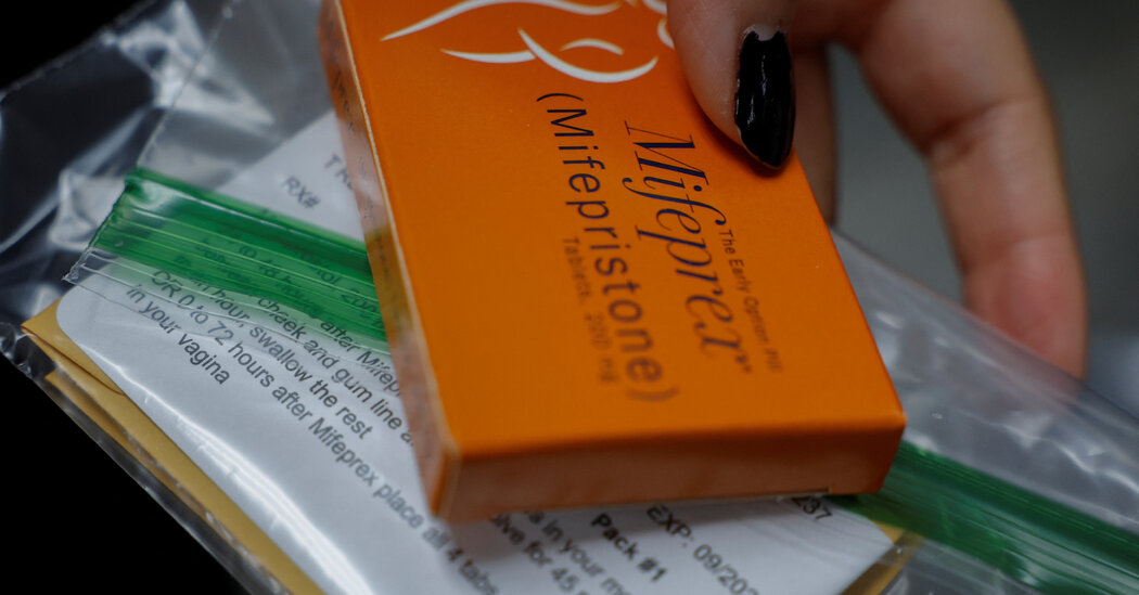
A bill that is expected to pass would impose prison time and thousands of dollars in fines on people possessing the pills without a prescription.Louisiana could become the first state to classify abortion pills as dangerous controlled substances, making possession of the pills without a prescription a crime subject to jail time and fines.A bill that would designate the abortion pills mifepristone and misoprostol as Schedule IV drugs — a category of medicines with the potential for abuse or dependence — passed the state’s Republican-controlled House of Representatives on Tuesday by a vote of 63 to 29. Should the Senate follow suit, Gov. Jeff Landry, a Republican and a vocal opponent of abortion, is likely to sign the legislation into law. The measure — which would put abortion pills in the same category as Xanax, Ambien and Valium — contradicts the way the federal government classifies mifepristone and misoprostol. The federal Food and Drug Administration does not consider abortion pills to be drugs with the potential for dependence or abuse, and decades of medical studies have found both to be overwhelmingly safe. Under the legislation, possession of mifepristone or misoprostol without a prescription in Louisiana could be punishable with thousands of dollars in fines and up to five years in jail. Pregnant women would be exempt from those penalties; most abortion bans and restrictions do not punish pregnant women because most voters oppose doing so. “These drugs are increasingly being shipped from outside our state and country to women and girls in our state,” Attorney General Liz Murrill, a Republican, said in a statement on social media. “This legislation does NOT prohibit these drugs from being prescribed and dispensed in Louisiana for legal and legitimate reasons.”Louisiana already bans most abortions, except when women’s lives or health are in danger or fetuses have some fatal conditions. As a result, abortion rights advocates and legal scholars said that in practice, the measure might not prevent many abortions among Louisiana women. Since the state imposed its strict abortion ban after the Supreme Court overturned Roe v. Wade, many patients have traveled to states where abortion is legal or have obtained pills under shield laws from doctors or nurses in other states who prescribe and mail the medications to Louisiana. Such circumstances would not be affected by the new bill, experts say. We are having trouble retrieving the article content.Please enable JavaScript in your browser settings.Thank you for your patience while we verify access. If you are in Reader mode please exit and log into your Times account, or subscribe for all of The Times.Thank you for your patience while we verify access.Already a subscriber? Log in.Want all of The Times? Subscribe.

Read more →

A Second Dairy Worker Has Contracted Bird Flu, C.D.C. Reports

The new case, in a Michigan farmworker, did not suggest that bird flu was widespread in people, health officials said, adding that the risk to the general public remained low.A farmworker in Michigan has been diagnosed with bird flu, state officials announced on Wednesday, making it the second human case associated with the outbreak in cows.Officials said that the individual became infected with the virus, called H5N1, after exposure to infected livestock. The individual had only mild symptoms and has fully recovered, officials said. They did not provide additional details in order to protect the privacy of the farm and farmworker, they said.In 2022, a person in Colorado with direct exposure to infected poultry became the first confirmed human case of H5N1 in the United States. Last month, the Centers for Disease Control and Prevention reported an H5N1 infection in a Texas dairy farm worker.The detection of this latest case did not suggest that bird flu was widespread in people, officials said, adding that the risk to the general public remained low.“This virus is being closely monitored, and we have not seen signs of sustained human-to-human transmission at this point,” Dr. Natasha Bagdasarian, Michigan’s chief medical executive, said in a statement.According to the C.D.C., a nasal swab from the individual had tested negative for the H5N1 virus, but an eye swab sent to the agency tested positive. Like the infected person in Texas, the Michigan patient only reported eye symptoms, the C.D.C. said.We are having trouble retrieving the article content.Please enable JavaScript in your browser settings.Thank you for your patience while we verify access. If you are in Reader mode please exit and log into your Times account, or subscribe for all of The Times.Thank you for your patience while we verify access.Already a subscriber? Log in.Want all of The Times? Subscribe.

Read more →

Century-old vaccine protects type 1 diabetics from infectious diseases

In new research, investigators at Massachusetts General Hospital (MGH) show that the 100-year-old Bacillus Calmette-Guérin (BCG) vaccine, originally developed to prevent tuberculosis, protects individuals with type 1 diabetes from severe COVID-19 disease and other infectious diseases.
Two back-to-back randomized double-blinded placebo-controlled trials found that the BCG vaccine provided continuous protection for nearly the entire COVID-19 pandemic in the US, regardless of the viral variant.
“Individuals with type 1 diabetes are highly susceptible to infectious diseases and had worse outcomes when they were infected with the SARS-CoV-2 virus,” said senior author Denise Faustman, MD, PhD, director of the Immunobiology Laboratory at MGH and an Associate Professor of Medicine at Harvard Medical School. “Published data from other investigators show mRNA COVID-19 vaccines are not very effective in this group of vulnerable patients. But we’ve shown that BCG can protect type 1 diabetics from COVID-19 and other infectious diseases.”
The 18-month Phase III trial, published in iScience, was conducted late in the US pandemic when the highly transmissible Omicron variant was circulating. A 15-month Phase II trial was conducted early in the pandemic; results of that trial were published in Cell Reports Medicine.
During the COVID-19 pandemic, several international trials tested if BCG as a single shot, or booster, given to previously BCG-vaccinated adults protected them from infection and COVID-19. This research expanded the large global clinical trial database showing that BCG administered to newborns works as a platform for all infectious disease, maybe for decades. But results from these COVID-19 booster trials in people previously vaccinated with BCG were mixed, with five randomized trials showing efficacy and seven trials showing no benefit.
The MGH Phase II and Phase III clinical trials testing BCG differed from other BCG trials in important ways. Instead of receiving one dose of BCG, participants received five or six doses of a particularly potent strain of BCG vaccine. The US participants were followed for a total of 36 months instead of weeks or months. “We know that in people who are naïve to BCG vaccine, the off-target effects can take at least two years to achieve full protection,” said Faustman. “Giving multiple doses of the vaccine may speed up that process.”
And importantly, the US population had never received BCG vaccines, so these clinical trials were not booster trials. “The Phase II and Phase III trials conducted at MGH were unique in that they were the only COVID trials in the world in which the study population had never received a BCG vaccine and was never exposed to TB,” said Faustman. “Trials conducted in countries where participants had previously received BCG vaccine as newborns or who had previous exposure to tuberculosis may have obscured any benefit from a BCG booster.”
The MGH trials enrolled 141 participants with type 1 diabetes; 93 people in the treatment group received five or six doses of BCG vaccine and the 48 individuals in the placebo group received sham vaccine and were followed for 36 months to capture diverse COVID-19 genetic variants and many infectious disease exposures.

During the earlier Phase II trial (January 2020 to April 2021) when the virus was more lethal but less transmissible, the BCG vaccine’s efficacy was 92%, comparable to the efficacy of the Pfizer and Moderna COVID-19 vaccines in healthy adults.
Over the full 34 months of the US COVID-19 pandemic, the BCG vaccine had a significant efficacy of 54.3%. The investigators also found that the BCG-treated participants had lower rates of viral, bacterial, and fungal infections as well as COVID-19 disease itself.
The BCG vaccine confers an immunity that likely lasts decades, a clear advantage to the COVID-19 vaccine and vaccines against other infectious diseases, such as influenza, where the duration of effectiveness is only two or three months. “The BCG vaccine offers the prospect of near-lifelong protection against every variant of COVID-19, the flu, respiratory syncytial virus, and other infectious diseases,” said Faustman.
Some of the BCG-treated participants also received the commercially available COVID-19 vaccines during the Phase III trial. The investigators observed that the Pfizer, Moderna, and Johnson & Johnson vaccines did not protect people with type 1 diabetes against COVID-19. “Our study showed that the BCG vaccine neither increased the efficacy of the COVID-19 vaccine, nor was it harmful to those who received the COVID-19 vaccine,” said Faustman. “As the pandemic continues to evolve it will interesting to see if we can work with the FDA to allow access to BCG vaccine for type 1 diabetics, who appear to be particularly at risk for all infectious diseases.”
This research was supported by The Iacocca Foundation, Boston, MA.
Other contributing authors include Willem M. Kühtreiber, Emma R. Hostetter, Grace E. Wolfe, Maya S. Vayshnaw, Rachel Goldstein, Emily R. Bulczynski, Neeshi S. Hullavarad, Joan E. Braley, and Hui Zheng.

Read more →

Sweet move: a modified sugar enhances antisense oligonucleotide safety and efficacy

Diseases that affect the brain and spinal cord can be particularly devastating, and finding new and more effective ways to treat these conditions is an important goal for researchers and clinicians alike. Now, a research group from Japan reports that slightly modifying an existing treatment for central nervous system (CNS) disease dramatically increases its effectiveness.
In a study published recently in Molecular Therapy Nucleic Acids, researchers from Tokyo Medical and Dental University (TMDU) and Osaka University have revealed that adding a modified sugar to antisense oligonucleotides (ASOs), a recently developed treatment strategy for CNS disease caused by toxic proteins, greatly decreases disease symptoms.
Multiple ASOs have been approved for clinical use, including gapmer ASOs, which are small pieces of genetic material binding to RNA messenger molecules that produce mutant disease-causing proteins to flag them for degradation. Modifying the chemical composition of these gapmer ASOs can both increase their ability to target molecules for degradation and decrease the toxic side effects of the treatment.
“We recently developed a new chemical modification called BNAP-AEO,” says lead author of the study Taiki Matsubayashi. “While ASOs carrying BNAP-AEO are expected to be highly effective, their biological efficacy and toxicity have not been investigated.”
To characterize gapmer ASOs modified by the inclusion of BNAP-AEO, the researchers first evaluated the stability of gapmer ASO binding to target molecules at different temperatures. They then tested the ability of these modified gapmer ASOs to block the production of disease-causing proteins in brain cancer cells and in mice.
“The results were very exciting,” explains Takanori Yokota, senior author. “Not only did ASOs carrying BNAP-AEO have a higher binding affinity for the target than those without BNAP-AEO, they also induced more efficient gene silencing in vitro and strongly suppressed gene expression in the mouse brain.”
Furthermore, modifying gapmer ASOs with BNAP-AEO decreased their toxic side effects in mice, possibly by changing their interactions with receptors on the surface of cells in the brain and spinal cord.
“Our findings highlight the efficient gene-silencing effect of ASOs incorporating BNAP-AEO, as well as an unexpected role for this modification in decreasing CNS toxicity,” says Matsubayashi.
In addition to providing a new way to increase the efficacy and safety of gapmer ASOs, the results from this study suggest that ASOs that bind stably at high temperatures are not necessarily more potent, as is commonly thought. This could be explained by the BNAP-AEO chemical modification affecting other features of the ASOs, such as which cell compartment they localize to.

Read more →

Flicker stimulation shines in clinical trial for epilepsy

Biomedical engineer Annabelle Singer has spent the past decade developing a noninvasive therapy for Alzheimer’s disease that uses flickering lights and rhythmic tones to modulate brain waves. Now she has discovered that the technique, known as flicker, also could benefit patients with a host of other neurological disorders, from epilepsy to multiple sclerosis.
Previously, Singer and her collaborators demonstrated that the lights and sounds, delivered to patients through goggles and headphones, have beneficial effects. Flicker has been successful in animal studies and in early human feasibility trials, where it was tested for safety, tolerance, and patient adherence.
Now, thanks to a clinical trial for people with epilepsy, the researchers quantified flicker’s effects with unprecedented precision. They also made an unexpected, but encouraging, discovery: The treatment reduced interictal epileptiform discharges (IEDs) in the brain.
These large, intermittent electrophysiological events are observed between seizures in people with epilepsy. They appear as sharp spikes on an EEG readout.
“What’s interesting about these IEDs is that they don’t just occur in epilepsy,” said Singer, McCamish Foundation Early Career Professor in the Wallace H. Coulter Department of Biomedical Engineering at Georgia Tech and Emory University. “They occur in autism, multiple sclerosis, Alzheimer’s, and other neurological disorders, too.” And IEDs disrupt normal brain function, causing memory impairment.
Singer and her team published their findings recently in Nature Communications.
The Rhythm in Our Heads
Inside the brain are elaborate symphonies of electrical activity: brain waves, or oscillations, that compose our memories, thoughts, and emotions. Singer wants to modulate those oscillations for therapeutic purposes.

At specific frequencies of light and sound, the flicker treatment can induce gamma oscillations in mice. This helps the brain recruit microglia, cells responsible for removing beta amyloid, which is believed to play a central role in Alzheimer’s pathology. Part of the work is in recording what’s happening in the brain during treatment to verify how it’s working.
The patients in the trial were under the care of physician Jon Willie at the Emory University Hospital Epilepsy Monitoring Unit. (Willie, co-corresponding author of the study with Singer, is now at Washington University in St. Louis.) They were awaiting surgery to remove an area of the brain where seizures occur. Before that could happen, they had to undergo intracranial seizure monitoring — recording electrodes are placed in the brain to pinpoint the seizure onset zone and determine exactly which tissue should be removed. Then, patients and their care team wait for a seizure to happen. It can take days.
“In human studies, we’ve used noninvasive methods like functional MRI or scalp EEG, but they have real downsides in terms of resolution,” Singer said. “Working with these patients was a game changer. These are people with treatment-resistant epilepsy, which means that drugs aren’t working for them.”
Pathway to Healing
Singer’s team recruited 19 patients. Lead author of the study, Lou Blanpain, a former Ph.D. student in Singer’s lab and now a medical student at Emory, went from patient to patient with the flicker stimulation and recording equipment.
“Because these patients already had recording probes implanted for clinical reasons, we were able to record directly from the brain,” Singer said. “We’ve never been able to get recordings of this quality during flicker treatment before.”
As the researchers expected, flicker modulated the visual and auditory brain regions that respond strongly to stimuli. But it also reached deeper, into the medial temporal lobe and prefrontal cortex, brain regions crucial for memory. And across the brain, in regions Singer hadn’t fully explored before, she found IEDs were decreasing.
“That has important implications for whether flicker is therapeutically relevant for people with Alzheimer’s, but also in general if we want to target anything beyond the primary sensory regions,” she said. “All of this points to the potential use of flicker in a lot of different contexts. Going forward, we’re definitely going to look at other conditions and other potential implications.”

Read more →

Dispelling severe ‘morning sickness’ myths: It’s not normal or harmless, but prevention and treatment might be on the way

Late last year, geneticist Marlena Fejzo and colleagues made the discovery that morning sickness’s most serious presentation, hyperemesis gravidarum (HG), is caused by the hormone GDF15, not human chorionic gonadotropin as previously thought. In a peer-reviewed opinion article publishing May 22 in the journal Trends in Molecular Medicine, Fejzo dispels common morning sickness myths and discusses potential treatments, including sensitizing people to GDF15 prior to pregnancy, similar to the way we treat allergies.
“HG can be life threatening and is associated with adverse outcomes that need to be taken seriously,” says Fejzo of the Keck School of Medicine of the University of Southern California. “Now that we know that GDF15 is the most likely cause of HG, we are on the cusp of having treatments that target this hormonal pathway and end the suffering.”
Myth 1: Severe morning sickness is harmless and normal
Pregnant people with HG are essentially starving, Fejzo says, and an increasing number of studies have demonstrated that this has serious short- and long-term clinical implications for both the parent and child. HG is a top predictor of postnatal depression, and 26% of pregnant people with HG report suicidal ideation while 18% meet the full criteria for post-traumatic stress disorder.
For the child, HG is associated with preterm birth, low birth weight, and later in life, autism spectrum disorder, ADHD, depression, social problems, in addition to an increased risk of childhood cancer and respiratory and cardiovascular disease. Still, pregnant people with the condition are often dismissed by their clinicians and families.
“It really is like a teratogen in pregnancy, a factor which interferes with normal fetal development, but it’s still not taken seriously by a lot of medical professionals,” Fejzo says. “A lot of people are brushed off and told, ‘oh that’s normal, it’s okay, just don’t take your pre-natal vitamins; you don’t need them.'”
At its most extreme, individuals with HG can develop Wernicke encephalopathy, a life-threatening swelling of the brain due to thymine (vitamin B1) deficiency. Since individuals with HG can have trouble even swallowing vitamins, the American College of Obstetricians and Gynecologists currently recommends that they replace broad spectrum prenatal vitamins with folic acid, but Fejzo warns that this is likely insufficient, and that thiamine supplementation is also warranted for individuals with HG.

“I believe all women who have hyperemesis should be given vitamin B1 to avoid this serious brain swelling that can lead to permanent brain damage and often leads to fetal death,” Fejzo says.
Myth 2: Morning sickness is caused by human chorionic gonadotropic hormone (hCG) or is psychosomatic
Though it was long thought that morning sickness is caused by hCG, the recent breakthrough has shown that HG’s main cause is actually the hormone GDF15, which is part of a normal stress response. Usually, GDF15 is expressed only in very small amounts, but during early pregnancy it spikes by a huge amount, then wanes, and finally rises again during the third trimester.
A recent Nature study co-authored by Fejzo showed that individuals who suffer from HG can have genetic variants that causes them to have lower levels of circulating GDF15 prior to pregnancy, which makes them extra sensitive when they become pregnant and are suddenly exposed to high levels. This finding has clinical implications for preventing and treating HG, since preliminary research suggests that it might be safe to manipulate GDF15 during or even prior to pregnancy.
“GDF15 may be safe to manipulate in pregnancy or even prior to pregnancy,” says Fejzo. “If we can increase levels of GDF15 before someone becomes pregnant, that might desensitize them, similar to how we try to desensitize people to allergens who have severe allergies,” says Fejzo. “And during pregnancy, we may be able to minimize or get rid of symptoms by blocking GDF15 or its receptors in the brain stem.”
Myth 3: Only humans experience morning sickness
Nausea and appetite loss during gestation is not a uniquely human trait — these symptoms have been observed throughout the animal kingdom, from monkeys, dogs, and cats, to chickens, vipers, and octopuses.

“I always think it’s interesting that the recommendation for cats is that if they’re unable to eat for a day, you should contact your veterinarian, but we don’t have that recommendation out there for women with hyperemesis,” says Fejzo. “If you call your doctor’s office and say you haven’t eaten for a day, they’ll say, ‘that’s normal’ and won’t do anything. There’s more proactive care for cats than humans.”
In addition to preventing ingestion of harmful foods, Fejzo speculates that pregnancy-induced nausea likely evolved to prevent dangerous foraging trips.
“This condition likely evolved because it was probably beneficial to avoid going out searching for food during pregnancy,” says Fejzo. “That may still be true for animals, but people don’t need this anymore, so let’s end the suffering once and for all if we can.”
Now, Fejzo is working toward developing and testing the proposed GDF15-based treatments. She also plans to investigate other genes and variants of GDF15 that might contribute to HG.

Read more →

Gentler cell therapies for blood cancer

Researchers have developed an approach to “deleting” a blood system affected by leukemia while simultaneously building up a new, healthy system with donor blood stem cells. Writing in the journal Nature, the team reports on the promising results obtained in animal experiments and with human cells in the laboratory.
In aggressive cases of leukemia, the only chance for a cure is to replace the diseased blood system with a healthy one. Although the transplantation of donor blood stem cells is a well-established form of treatment, it is an onerous process for patients. First, chemotherapy is used to remove the body’s own blood stem cells as well as most of the blood cells. Only then do the attending physicians intravenously administer the stem cells from a suitable donor to the patient. This procedure is associated with side effects and potential complications.
The team led by Professor Lukas Jeker from the Department of Biomedicine at the University of Basel has taken a different approach. Writing in the journal Nature, the team describes how all blood cells can be removed from a leukemia sufferer in a targeted manner while a new blood system is built up at the same time. The results represent the successful completion of a project funded by the European Research Council with a Consolidator Grant of EUR 2.4 million.
Mixing console for blood systems
The system established by the researchers in Jeker’s team can be imagined as a mixing console, where a DJ gradually fades down the level of the first song while raising the volume of the second until the first track dies away completely and only the second is audible.
The fading-down process works as follows: Specific antibodies coupled to a cytotoxic drug recognize all blood cells in the patient’s body based on a surface structure. This marker is common to all the different types of blood cell (both healthy and diseased) but does not appear on other cells of the body. Bit by bit, the antibody-drug conjugate therefore recognizes and destroys all cells of the diseased blood system.
While this is taking place, the second song also starts — that is, the patient receives a transplant of new, healthy blood cells from a suitable donor. To prevent the antibody-drug conjugates from also attacking the new blood stem cells, or the blood cells they produce, the researchers use genetic engineering techniques to modify the donor stem cells in a targeted manner. Specifically, they introduce a small change in the surface molecule so that the antibodies don’t recognize the new blood cells. The researchers refer to this targeted modification of the donor stem cells as “shielding,” because it acts like a protective shield against the cancer treatment.

Elaborate search for suitable regulators
The two first authors of the study, Simon Garaudé and Dr. Romina Matter-Marone, worked with an interdisciplinary team of bioinformaticians, biochemists, genetic engineering specialists, and clinicians from academia and industry to select the best-suited target structure — and the best protective modification for the fading-down process — from the multitude of surface molecules on blood cells. The chosen molecule, known as CD45, proved extremely promising in trials on mice and human cells in the laboratory.
“We needed a surface molecule that appeared with approximately the same frequency on all blood cells if possible, including the leukemia cells, but that wasn’t present on other cells in the body,” explains Jeker. CD45 met this requirement and, at the same time, was also suitable for “shielding” — in other words, it could be modified on the donor blood stem cells in such a way that these cells were protected from the cancer treatment but the function of CD45 remained completely normal.
Applications beyond cancer
“The new approach could pave the way for new treatment options for patients whose state of health is incompatible with the chemotherapy needed for stem cell transplantation,” says joint first author Romina Matter-Marone. Although further tests and optimization are needed, the aim is for initial clinical trials to begin in just a few years’ time.
The “mixing console for blood systems” also opens up further possibilities, as joint first author Simon Garaudé explains: “We show how cells that are ‘invisible’ to a blood cell remover can be used to swap out the entire blood system.” This, he says, is an important step toward a programmable blood system that could also assume functions on demand — for example, to correct a serious genetic defect or to impart resistance to specific viruses such as HIV.

Read more →

Adhesive coatings can prevent scarring around medical implants

When medical devices such as pacemakers are implanted in the body, they usually provoke an immune response that leads to buildup of scar tissue around the implant. This scarring, known as fibrosis, can interfere with the devices’ function and may require them to be removed.
In an advance that could prevent that kind of device failure, MIT engineers have found a simple and general way to eliminate fibrosis by coating devices with a hydrogel adhesive. This adhesive binds the devices to tissue and prevents the immune system from attacking it.
“The dream of many research groups and companies is to implant something into the body that over the long term the body will not see, and the device can provide therapeutic or diagnostic functionality. Now we have such an ‘invisibility cloak,’ and this is very general: There’s no need for a drug, no need for a special polymer,” says Xuanhe Zhao, an MIT professor of mechanical engineering and of civil and environmental engineering.
The adhesive that the researchers used in this study is made from cross-linked polymers called hydrogels, and is similar to a surgical tape they previously developed to help seal internal wounds. Other types of hydrogel adhesives can also protect against fibrosis, the researchers found, and they believe this approach could be used for not only pacemakers but also sensors or devices that deliver drugs or therapeutic cells.
Zhao and Hyunwoo Yuk SM ’16, PhD ’21, a former MIT research scientist who is now the chief technology officer at SanaHeal, are the senior authors of the study, which will appear in Nature. MIT postdoc Jingjing Wu is the lead author of the paper.
Preventing fibrosis
In recent years, Zhao’s lab has developed adhesives for a variety of medical applications, including double-sided and single-sided tapes that could be used to heal surgical incisions or internal injuries. These adhesives work by rapidly absorbing water from wet tissues, using polyacrylic acid, an absorbent material used in diapers. Once the water is cleared, chemical groups called NHS esters embedded in the polyacrylic acid form strong bonds with proteins at the tissue surface. This process takes about five seconds.

Several years ago, Zhao and Yuk began exploring whether this kind of adhesive could also help keep medical implants in place and prevent fibrosis from occurring.
To test this idea, Wu coated polyurethane devices with their adhesive and implanted them on the abdominal wall, colon, stomach, lung, or heart of rats. Weeks later, they removed the device and found that there was no visible scar tissue. Additional tests with other animal models showed the same thing: Wherever the adhesive-coated devices were implanted, fibrosis did not occur, for up to three months.
“This work really has identified a very general strategy, not only for one animal model, one organ, or one application,” Wu says. “Across all of these animal models, we have consistent, reproducible results without any observable fibrotic capsule.”
Using bulk RNA sequencing and fluorescent imaging, the researchers analyzed the animals’ immune response and found that when devices with adhesive coatings were first implanted, immune cells such as neutrophils began to infiltrate the area. However, the attacks quickly quenched out before any scar tissue could form.
“For the adhered devices, there is an acute inflammatory response because it is a foreign material,” Yuk says. “However, very quickly that inflammatory response decayed, and then from that point you do not have this fibrosis formation.”
One application for this adhesive could be coatings for epicardial pacemakers — devices that are placed on the heart to help control the heart rate. The wires that contact the heart often become fibrotic, but the MIT team found that when they implanted adhesive-coated wires in rats, they remained functional for at least three months, with no scar tissue formation.

Mechanical cues
The researchers also tested a hydrogel adhesive that includes chitosan, a naturally occurring polysaccharide, and found that this adhesive also eliminated fibrosis in animal studies. However, two commercially available tissue adhesives that they tested did not show this antifibrotic effect because the commercially available adhesives eventually detached from the tissue and allowed the immune system to attack.
In another experiment, the researchers coated implants in hydrogel adhesives but then soaked them in a solution that removed the polymers’ adhesive properties, while keeping their overall chemical structure the same. After being implanted in the body, where they were held in place by sutures, fibrotic scarring occurred. This suggests that there is something about the mechanical interaction between the adhesive and the tissue that prevents the immune system from attacking, the researchers say.
“Previous research in immunology has been focused on chemistry and biochemistry, but mechanics and physics may play equivalent roles, and we should pay attention to those mechanical and physical cues in immunological responses,” says Zhao, who now plans to further investigate how those mechanical cues affect the immune system.
Yuk, Zhao, and others have started a company called SanaHeal, which is now working on further developing tissue adhesives for medical applications.
“As a team, we are interested in reporting this to the community and sparking speculation and imagination as to where this can go,” Yuk says. “There are so many scenarios in which people want to interface with foreign or manmade material in the body, like implantable devices, drug depots, or cell depots.”
The research was funded by the National Institutes of Health and the National Science Foundation.

Read more →

Dermatologists find ultraviolet irradiation increases appetite but prevents body weight gain

Obesity and metabolic disorders are increasingly significant global public health issues. In a novel study, a team of dermatologists evaluated the effect of ultraviolet (UV) exposure on appetite and weight regulation. They found that UV exposure raises norepinephrine levels, decreases leptin levels, and induces the browning of subcutaneous fat, thereby increasing energy expenditure. These results potentially pave the way for new approaches to prevent and treat obesity and metabolic disorders. Their findings appear in the Journal of Investigative Dermatology, published by Elsevier.
UV radiation is a common environmental factor that has multifaceted effects on the skin, which encompasses a substantial surface area of the body. UV radiation induces detrimental effects such as sunburn, photoaging, and skin cancer; however, it is also associated with beneficial effects such as vitamin D synthesis.
Co-first authors Qing-Ling Quan, MD, PhD, and Eun Ju Kim, PhD, Department of Dermatology, Seoul National University Hospital, explained, “Recent evidence has suggested that UV exposure limits body weight gain in mouse models of obesity. Subcutaneous fat is a critical organ in regulating energy homeostasis. Alongside previous studies on the effects of UV exposure on obesity and metabolic disorders, our team was inspired by our prior discovery that, although UV rays do not directly reach subcutaneous fat when exposed to the skin, they can regulate the metabolism of subcutaneous fat. This led us to hypothesize that skin exposure to UV rays could play a significant role in systemic energy homeostasis, prompting this research.”
Investigators discovered that when exposed to UV radiation consistently, mice fed a normal diet and those on a high-fat diet exhibited increased appetite due to a decrease in leptin, a key hormone in appetite regulation. However, there was no weight increase. They found that UV radiation inhibits weight gain by enhancing secretion of the neurotransmitter norepinephrine, which not only decreases leptin but also increases energy expenditure through the “browning” of subcutaneous fat.
The increased energy intake, driven by heightened appetite, is converted to heat and burned before it can accumulate in subcutaneous fat, thus preventing weight gain.
This research provides new insights into the impact of UV exposure on appetite and weight regulation, opening possibilities for novel approaches in the prevention and treatment of obesity and metabolic disorders. Specifically, uncovering the mechanism by which UV radiation prevents weight gain could offer new approaches to dietary regulation and weight loss, providing innovative insights into health and obesity management that could positively impact human health.
Lead investigator Jin Ho Chung, MD, PhD, Department of Dermatology, Seoul National University Hospital, Seoul National University College of Medicine, explained, “This study elucidates the mechanism by which UV exposure can increase appetite while inhibiting weight gain. These findings contribute significantly to understanding the effects of UV radiation on energy metabolism and homeostasis and open new avenues for exploring prevention and treatment strategies for obesity and metabolic disorders. Notably, the fact that UV radiation lowers leptin levels and increases norepinephrine, thereby promoting the browning of subcutaneous fat and increasing energy expenditure, provides a groundbreaking clue for the development of obesity treatment strategies. This research demonstrates that UV exposure not only affects the skin but also plays a deep role in our body’s energy metabolism and homeostasis processes. However, further research is needed on the long-term effects and safety of UV exposure, and there should be significant interest in developing new therapeutic approaches that utilize the efficacy of UV radiation.”
However, as co-corresponding author Dong Hun Lee, MD, PhD, Institute of Human-Environment Interface Biology, Seoul National University, noted, “Because UV exposure can accelerate skin aging and promote skin cancer, it is advisable to minimize UV exposure and protect the skin with sunscreen. Thus, our research team plans to conduct follow-up studies to develop new strategies that could mimic the effects of UV radiation for obesity and metabolic regulation.”

Read more →