Malaria Vaccine Rollout to Africa Is a Cautionary Tale

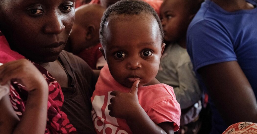
The first vaccine for malaria received major regulatory approval in 2015.It didn’t become part of vaccination programs in Africa until 2024.What if it had come faster?What if the shots had arrived9 years ago?143,000.That’s how many children’s deaths could have been averted.July 5, 2024Nurses in countries from Sierra Leone to Cameroon are packing a new vaccine into the coolers they tote to villages for immunization clinics: a shot to protect against malaria, one of the deadliest diseases for children.Babies and toddlers in eight countries in the region recently started to get the vaccine as part of their routine childhood shots. Seven other African countries are eagerly awaiting its arrival.This is a milestone in global health.But it’s also a cautionary tale about a system that is ill equipped to deliver critical tools to the people who need them most.It took decades and at least a billion dollars to reach this point. Even now, only a fraction of the children whose lives are at risk will get the vaccine this year, or next year, or the year after.It’s been clear for some time what went wrong, but almost none of those issues have been fixed. That means that the next desperately needed vaccine stands every chance of running into those same problems.Take, for example, a new vaccine for tuberculosis that started clinical trials a few months ago. If it works as well as hoped, it could save at least a million lives a year. We’ll know by 2028 if it stops tuberculosis infections. But if it follows the same trajectory, it will be at least 2038 before it’s shipped to clinics.“Children are receiving the vaccine, and for that, I am the happiest man in the world. But on the other hand, I cannot avoid being dismayed at this inexcusably long delay.”— Dr. Joe Cohen, co-inventor of the first malaria vaccineThe U.S. Army started work on a malaria vaccine back in the 1980s, hoping to protect soldiers deployed to the tropics. It teamed up with the drug company GlaxoSmithKline, and together they produced promising prototypes. But the military lost interest after a few years, and that left GSK with a problem.The people who desperately needed a malaria vaccine were in villages in sub-Saharan Africa. They would not be able to pay for a product that would cost millions of dollars to develop.GSK needed an altruistically minded partner. It found one in the nonprofit global health agency PATH, and by the late 1990s they had a vaccine to test. The Bill & Melinda Gates Foundation put up more than $200 million to test it.The clinical trials were complex, because this was a whole new type of vaccine — the first ever against a parasite — delivered to children in places with limited health systems. The process took more than a decade.Finally, in 2014, results showed this vaccine cut severe malaria cases by about a third.This was a successful result, but not as much protection as scientists had hoped to see. Still, GSK and PATH planned a production facility to make millions of doses. Gavi, the organization that procures vaccines for low- and middle-income countries, with funds from donors, would buy them.Then the Gates Foundation pulled its support.There was a shake-up in the malaria division, and the leadership reoriented toward a new goal: eliminating the disease.The new malaria team said the vaccine didn’t work well enough to justify pouring millions more dollars into it. It would be better, they said, to wait for a more effective shot in the future, and in the meantime to fund other strategies, such as genetically modifying mosquitoes.“If you go from very enthusiastic to very unenthusiastic and you’re the Gates Foundation, people pay attention.”— Dr. Robert Newman, former director, Global Malaria Program, W.H.O.The decision was driven by researchers who were looking at data. They didn’t factor in that the idea of a vaccine, even one with limited efficacy, would be so important to African parents — and African governments, which would come to see this as a classic example of a paternalistic donor ignoring their priorities. More than 300,000 children died of malaria that year.The foundation’s announcement shoved the vaccine into limbo — in ways the foundation today says it did not anticipate.“In hindsight, we could have communicated more often and more clearly about our decisions and listened more clearly to what the impact of those might have been on other institutions and their decisions.”— Dr. Chris Elias, president of global development at the Bill & Melinda Gates Foundation

.css-1ggt3fz {
padding-left: 1rem;
border-left: 0px solid var(–color-stroke-quaternary, #FFFFFF);
}

.css-7ad88g {
border-top: 1.3px solid #dc0000;
}

/* TOP AD CODE BELOW */
#top-wrapper, sponsor-wrapper { display: none; }
We are having trouble retrieving the article content.Please enable JavaScript in your browser settings.Thank you for your patience while we verify access. If you are in Reader mode please exit and log into your Times account, or subscribe for all of The Times.Thank you for your patience while we verify access.Already a subscriber? Log in.Want all of The Times? Subscribe.

Read more →

Scientists discover new T cells and genes related to immune disorders

Researchers led by Yasuhiro Murakawa at the RIKEN Center for Integrative Medical Sciences (IMS) and Kyoto University in Japan and IFOM ETS in Italy have discovered several rare types of helper T cells that are associated with immune disorders such as multiple sclerosis, rheumatoid arthritis, and even asthma. Published July 4 in Science, the discoveries were made possible by a newly developed technology they call ReapTEC, which identified genetic enhancers in rare T cell subtypes that are linked to specific immune disorders. The new T cell atlas is publicly available and should help in the development of new drug therapies for immune-mediated diseases.Helper T cells are kind of white blood cell that make up a large part of the immune system. They recognize pathogens and regulate the immune response. Many immune-mediated disease are caused by abnormal T cell function. In autoimmune diseases like multiple sclerosis, they mistakenly attack parts of the body as if they were pathogens. In the case of allergies, T cells overreact to harmless substances in the environment like pollen. We know of several common T cells, but recent studies have shown that rare and specialized types of T cells exist, and they might be related to immune-mediated diseases.Within all cells, including T cells, there are regions of DNA called “enhancers”. This DNA does not code for proteins. Instead, it codes for small pieces of RNA, and enhances the expression of other genes. Variations in T cell enhancer DNA therefore lead to differences in gene expression, and this can affect how T cells function. Some enhancers are bidirectional, which means that both strands of the DNA are used as templates for enhancer RNA. The researchers from several different laboratories at RIKEN IMS, as well as colleagues at other institutes, teamed up to develop the new ReapTEC technology and look for connections between bidirectional T cell enhancers and immune diseases.After analyzing about a million human T cells, they found several groups of rare T cell types, accounting for less than 5% of the total. Applying ReapTEC to these cells identified almost 63,000 active bidirectional enhancers. To figure out if any of these enhancers are related to immune diseases, they turned to genome-wide association studies (GWAS), which have reported numerous genetic variants, called single-nucleotide polymorphisms, that are related to various immune diseases.When the researchers combined the GWAS data with the results of their ReapTEC analysis, they found that genetic variants for immune-mediated diseases were often located within the bidirectional enhancer DNA of the rare T cells that they had identified. In contrast, genetic variants for neurological diseases did not show a similar pattern, meaning that the bidirectional enhancers in these rare T cells are related specifically to immune-mediated diseases.Going even deeper into the data, the researchers were able to show that individual enhancers in certain rare T cells are related to specific immune diseases. Overall, among the 63,000 bidirectional enhancers, they were able to identify 606 that included single-nucleotide polymorphisms related to 18 immune-mediated diseases. Lastly, the researchers were able to identify some of the genes that are the targets of these disease-related enhancers. For example, when they activated an enhancer that contained a genetic variant related to inflammatory bowel disease, the resulting enhancer RNA triggered upregulation of the IL7R gene.“In the short-term, we have developed a new genomics method that can be used by researchers around the world,” says Murakawa. “Using this method, we discovered new types of helper T cells as well as genes related to immune disorders. We hope that this knowledge will lead to a better understanding of the genetic mechanisms underlying human immune-mediated diseases.”In the long-term, the researchers believe follow-up experiments will be able to identify new molecules that can be used to treat immune-mediated diseases.

Read more →

Scientists map how deadly bacteria evolved to become epidemic

Pseudomonas aeruginosa — an environmental bacteria that can cause devastating multidrug-resistant infections, particularly in people with underlying lung conditions — evolved rapidly and then spread globally over the last 200 years, probably driven by changes in human behaviour, a new study has found.
P. aeruginosa is responsible for over 500,000 deaths per year around the world, of which over 300,000 are associated with antimicrobial resistance (AMR). People with conditions such as COPD (smoking-related lung damage), cystic fibrosis (CF), and non-CF bronchiectasis, are particularly susceptible.
How P. aeruginosa evolved from an environmental organism into a specialised human pathogen was not previously known. To investigate this, an international team led by scientists at the University of Cambridge examined DNA data from almost 10,000 samples taken from infected individuals, animals, and environments around the world. Their results are published today in Science
By mapping the data, the team was able to create phylogenetic trees — ‘family trees’ — that show how the bacteria from the samples are related to each other. Remarkably, they found that almost seven in ten infections are caused by just 21 genetic clones, or ‘branches’ of the family tree, that have rapidly evolved (by acquiring new genes from neighbouring bacteria) and then spread globally over the last 200 years. This spread occurred most likely as a result of people beginning to live in densely-populated areas, where air pollution made our lungs more susceptible to infection and where there were more opportunities for infections to spread.
These epidemic clones have an intrinsic preference for infecting particular types of patients, with some favouring CF patients and other non-CF individuals. It turns out that the bacteria can exploit a previously unknown immune defect in people with CF, allowing them to survive within macrophages. Macrophages are cells that ‘eat’ invading organisms, breaking them down and preventing the infection from spreading. But a previously-unknown flaw in the immune systems of CF patients means that once the macrophage ‘swallows’ P. aeruginosa, it is unable to get rid of it.
Having infected the lungs, these bacteria then evolve in different ways to become even more specialised for a particular lung environment. The result is that certain clones can be transmitted within CF patients and other clones within non-CF patients, but almost never between CF and non-CF patient groups.
Professor Andres Floto, Director of the UK Cystic Fibrosis Innovation Hub at the University of Cambridge and Royal Papworth Hospital NHS Foundation Trust, and senior author of the study said: “Our research on Pseudomonas has taught us new things about the biology of cystic fibrosis and revealed important ways we might be able to improve immunity against invading bacteria in this and potentially other conditions.

“From a clinical perspective, this study has revealed important information about Pseudomonas. The focus has always been on how easily this infection can spread between CF patients, but we’ve shown that it can spread with worrying ease between other patients, too. This has very important consequences for infection control in hospitals, where it’s not uncommon for an infected individual to be on an open ward with someone potentially very vulnerable.
“We are incredibly lucky at Royal Papworth Hospital where we have single rooms and have developed and evaluated a new air-handling system to reduce the amount of airborne bacteria and protect all patients.”
Dr Aaron Weimann from the Victor Phillip Dahdaleh Heart & Lung Research Institute at the University of Cambridge, and first author on the study, said: “It’s remarkable to see the speed with which these bacteria evolve and can become epidemic and how they can specialise for a particular lung environment. We really need systematic, pro-active screening of all at risk patient groups to detect and hopefully prevent the emergence of more epidemic clones.”
The research was funded by Wellcome and the UK Cystic Fibrosis Trust.

Read more →

Cuts to processed meat intake bring a range of health benefits

Reducing consumption of processed meat by around one-third could prevent more than 350,000 cases of diabetes in the US over 10 years, a study suggests.
Cutting US adults’ processed meat intake by 30 per cent — the equivalent of around 10 slices of bacon a week — would also lead to tens of thousands of fewer cases of cardiovascular disease and colorectal cancer, researchers say.
A team from the University of Edinburgh’s Global Academy of Agriculture and Food Systems together with the University of North Carolina, Chapel Hill, has developed a simulation tool to estimate the health impacts of reducing consumption of processed meat and unprocessed red meat.
While many studies have identified links between high levels of processed meat consumption and chronic disease, few have evaluated the impact on multiple health outcomes. Some previous research also suggests unprocessed red meat may contribute to chronic disease risk but evidence is still limited.
The researchers used data from a Centers for Disease Control and Prevention (CDC) national health survey to create a simulated, representative sample of the US adult population — a so-called microsimulation.
Their microsimulation is the first to estimate the effects of reducing processed meat and unprocessed red meat consumption — from between 5 and 100 per cent — on multiple health outcomes in the US.
The team estimated how changes in meat consumption affect adults’ risk of diabetes, cardiovascular disease, colorectal cancer and death. The effects were evaluated in the overall population and separately based on age, sex, household income and ethnicity.

As well as preventing more than 350,000 cases of diabetes, cutting processed meat intake by 30 per cent would lead to 92,500 fewer cardiovascular disease cases and 53,300 fewer colorectal cancer cases over a decade, researchers say.
In this scenario, white males and those with an annual household income between $25,000 and $55,000 were found to experience the greatest health benefits.
Researchers also analysed the impacts of reducing unprocessed red meat intake alone and cutting consumption of both processed meat and unprocessed red meat.
Reducing consumption of both by 30 per cent resulted in 1,073,400 fewer diabetes cases, 382,400 fewer cardiovascular disease cases and 84,400 fewer colorectal cancer cases.
Cutting unprocessed red meat intake alone by 30 per cent — which would mean eating around one less quarter-pound beef burger a week — resulted in more than 732,000 fewer diabetes cases. It also led to 291,500 fewer cardiovascular disease cases and 32,200 fewer colorectal cancer cases.
The finding that more disease cases were prevented by reducing unprocessed red meat compared to processed meat is partly due to the average daily intake of unprocessed red meat being higher than processed meat, at 47g a day versus 29g a day, respectively.
As less is known about the effect of eating unprocessed red meat on chronic disease risk, the team says these estimates should be interpreted with caution and that more research is needed.
The study, published in The Lancet Planetary Health journal, was funded by The Wellcome Trust.
Professor Lindsay Jaacks, Personal Chair of Global Health and Nutrition at the University of Edinburgh, and one of the authors of the study, said “Cutting consumption of meat has been recommended by national and international organisations to reduce greenhouse gas emissions, including the Climate Change Committee here in the UK and the United Nations Intergovernmental Panel on Climate Change or IPCC. Our research finds that these changes in diets could also have significant health benefits in the US, and so this is a clear win-win for people and planet.”

Read more →

Deep machine-learning speeds assessment of fruit fly heart aging and disease, a model for human disease

Drosophila — commonly known as fruit flies — are a valuable model for human heart pathophysiology, including cardiac aging and cardiomyopathy. However, a choke point in evaluating fruit fly hearts is the need for human intervention to measure the heart at moments of its largest expansion or its greatest contraction, measurements that allow calculations of cardiac dynamics.
Researchers at the University of Alabama at Birmingham now show a way to significantly cut the time needed for that analysis while utilizing more of the heart region, using deep learning and high-speed video microscopy for each heartbeat in the fly.
“Our machine learning method is not just fast; it minimizes human error because you don’t have to manually mark each heart wall under systolic and diastolic conditions,” said Girish Melkani, Ph.D., associate professor in the UAB Department of Pathology, Division of Molecular and Cellular Pathology. “Furthermore, you can run the analyses of several hundred hearts and look at the analyses when done for all the hearts.”
This can expand the ability to test how different environmental or genetic factors affect heart aging or pathology. Melkani envisions using deep learning-assisted studies to explore cardiac mutation models and other small animal models, such as zebrafish and mice. “Additionally, our techniques could be adapted for human heart models, providing valuable insights into cardiac health and disease. Incorporating uncertainty quantification methods could further enhance the reliability of our analyses. Moreover, the machine learning approach can predict cardiac aging with high accuracy.”
The fruit fly model has already been tremendously powerful for understanding the pathophysiological bases for several human cardiovascular diseases, Melkani says. Cardiovascular disease continues to be one of the leading causes of death and disability in the United States.
Melkani and UAB colleagues assessed their trained model on heart performance both in fruit fly cardiac aging and in a fruit fly model of dilated cardiomyopathy caused by the knockdown of a pivotal TCA cycle enzyme, oxoglutarate dehydrogenase. These automated assessments were then validated against existing experimental datasets. For example, for aging of fruit flies at one week versus five weeks of age, which is about halfway through a fruit fly’s life span, the UAB team used 54 hearts for model training and then validated their measurements against an experimental aging model with 177 hearts. Their trained model was able to reconstruct expected trends in cardiac parameters with aging.
Melkani says his team’s model can be applied to readily available consumer hardware, and his team’s code can provide calculated statistics including diastolic and systolic diameters/intervals, fractional shortening, ejection fraction, heart period/rate, and quantified heartbeat arrhythmicity.
“To our knowledge, this innovative platform for deep learning-assisted segmentation is the first of its kind to be applied to standard high-resolution high-speed optical microscopy of Drosophila hearts while also quantifying all relevant parameters,” Melkani said.
“By automating the process and providing detailed cardiac statistics, we pave the way for more accurate, efficient and comprehensive studies of heart function in Drosophila. This method holds tremendous potential — not only for understanding aging and disease in fruit flies — but also for translating these insights into human cardiovascular research.”

Read more →