Too much, too little sleep linked to elevated heart risks in people free from disease

People who clock six to seven hours of sleep a night had the lowest chance of dying from a heart attack or stroke when compared with those who got less or more sleep, according to a study being presented at the American College of Cardiology’s 70th Annual Scientific Session. This trend remained true even after the research team accounted for other known conditions or risk factors for heart disease or stroke.
The study, according to researchers, is the first to explore the association between baseline cardiovascular risk and duration of sleep and adds to mounting evidence that sleep — similar to diet, smoking and exercise — may play a defining role in someone’s cardiovascular risk.
“Sleep is often overlooked as something that may play a role in cardiovascular disease, and it may be among the most cost-effective ways to lower cardiovascular risk,” said Kartik Gupta, MD, resident, Division of Internal Medicine, Henry Ford Hospital in Detroit and lead author of the study. “Based on our data, sleeping six to seven hours a night is associated with more favorable heart health.”
For the study, Gupta and his team included data from 14,079 participants in the 2005-2010 National Health and Nutrition Examination Survey. Participants were followed for a median duration of 7.5 years to determine if they died due to heart attack, heart failure or stroke. Those surveyed were 46 years old on average, half were women and 53% were non-white. Less than 10% of participants had a history of heart disease, heart failure or stroke.
Researchers divided participants into three groups based on answers to a survey question about their average length of sleep — less sleep (seven hours). Researchers then assessed participants’ atherosclerotic cardiovascular disease (ASCVD) risk scores and levels of C-reactive protein (CRP), a key inflammatory marker known to be associated with heart disease.
The ASCVD risk score, which accounts for age, gender, race, blood pressure and cholesterol, is widely used to predict how likely someone is to have a heart attack or stroke or die from atherosclerosis, a hardening of the arteries, in the next 10 years. An ASCVD risk score less than 5% is considered low risk.

Read more →

Prediabetes may not be as benign as once thought

People with prediabetes were significantly more likely to suffer a heart attack, stroke or other major cardiovascular event when compared with those who had normal blood sugar levels, according to research being presented at the American College of Cardiology’s 70th Annual Scientific Session. Researchers said the findings should serve as a wake-up call for clinicians and patients alike to try to prevent prediabetes in the first place.
“In general, we tend to treat prediabetes as no big deal. But we found that prediabetes itself can significantly boost someone’s chance of having a major cardiovascular event, even if they never progress to having diabetes,” said Adrian Michel, MD, internal medicine resident at Beaumont Hospital-Royal Oak, MI, and lead author of the study, which he said is one of the largest to date. “Instead of preventing diabetes, we need to shift focus and prevent prediabetes.”
Prediabetes is a condition in which the average amount of sugar in the blood is high but not high enough to be diagnosed as Type 2 diabetes. While Type 2 diabetes is a well-known, leading risk factor for heart attack, stroke and blockages in the heart’s arteries, the role of prediabetes has been less clear. Yet prediabetes is fairly common. The U.S. Centers for Disease Control and Prevention estimates that 34 million Americans — just over 1 in 10 — have diabetes, and another 88 million — approximately 1 in 3 — have prediabetes.
This study revealed that serious cardiovascular events occurred in 18% of people with prediabetes compared with 11% of people with normal blood sugar levels over a median of five years follow-up. The relationship between higher blood sugar levels and cardiovascular events remained significant even after taking into account other factors that could play a role, such as age, gender, body mass index, blood pressure, cholesterol, sleep apnea, smoking and peripheral artery disease.
“Based on our data, having prediabetes nearly doubled the chance of a major adverse cardiovascular event, which accounts for 1 out of 4 deaths in the U.S.,” Michel said. “As clinicians, we need to spend more time educating our patients about the risk of elevated blood sugar levels and what it means for their heart health and consider starting medication much earlier or more aggressively, and advising on risk factor modification, including advice on exercise and adopting a healthy diet.”
Of particular concern was the finding that even when patients in the prediabetes group were able to bring their blood sugar level back to normal, the risk of having a cardiovascular event was still fairly high. Events occurred in just over 10.5% of these patients compared with 6% of those with no diabetes or prediabetes.
“Even if blood sugar levels went back to normal range, it didn’t really change their higher risk of having an event, so preventing prediabetes from the start may be the best approach,” Michel said.
This single-center, retrospective study included data from 25,829 patients treated within the Beaumont Health System in Michigan between 2006 and 2020. Patients were then split into either the prediabetes or control group based on at least two A1C levels five years apart; the control group included patients who maintained a normal hemoglobin A1C during the study. A total of 12,691 patients and 13,138 were included in the prediabetes and control groups, respectively. Participants ranged in age from 18 to 104 years. All patients were followed for the 14-year study period and researchers used international classification of disease codes or diagnostic codes to determine whether a major adverse cardiovascular event occurred.
The relationship between prediabetes and events were strongest among males, Blacks and people with a family history of cardiovascular disease or personal risk factors for heart disease. People who were overweight had the highest rates of cardiovascular events among all patients, even more than those who were obese, which is something Michel said needs to be studied further.
Prediabetes is thought to play a role in heart health because elevated glucose levels in the blood can damage and cause inflammation within the vessels. This causes injury to the vessels in the body and can lead to narrowing of the vessels and ultimately cardiovascular injury, Michel said.
The study findings are an important reminder for adults to know their blood sugar numbers, especially as prediabetes usually has no symptoms. As with diabetes, prediabetes is diagnosed based on results from blood sugar tests, including an A1C, which reflects someone’s average blood sugar for the past two to three months; a fasting plasma glucose test, which measures your blood sugar after not eating or drinking for at least eight hours beforehand; and/or an oral glucose tolerance test, which checks how well the body processes sugar after drinking a sweet drink given by the clinician. Prediabetes is suspected with an A1C between 5.7-6.4%, fasting blood sugar of 100-125 mg/dl, or an oral glucose tolerance test of 140-199 mg/dl, according to the American Diabetes Association.
More research is needed to validate these findings.

Read more →

New guidelines for schools recommend against food bans

Schools and child care centres should train staff on food allergies and have epinephrine available to treat anaphylaxis, but new guidelines do not recommend food bans.
The recommendations come from an international team, led by McMaster University, which has developed practice guidelines for the prevention and management of allergic reactions to food at child care centres and schools. The guidelines were published today in the Journal of Allergy and Clinical Immunology.
“The management of food allergy is a sensitive topic,” said Susan Waserman, chair of the guidelines panel, an allergist and professor of medicine at McMaster University.
“Our goal is to help the school community understand the risk of allergic reactions — and offer evidence-informed guidance for managing it.”
The guidelines recommend that child care and school personnel receive training on how to prevent, recognize, and respond to allergic reactions. The guidelines also suggest that unassigned epinephrine autoinjectors, which are sold under the brand names ALLERJECT, Emerade, and EpiPen, be stocked on site.
The guidelines recommend there be no site-wide food prohibitions, such as ‘nut-free’ schools, or allergen-restricted zones, such as ‘milk-free’ tables, except in limited special circumstances.

Read more →

One bone fracture increases risk for subsequent breaks in postmenopausal women

Current guidelines for managing osteoporosis specifically call out hip or spine fractures for increasing the risk for subsequent bone breaks. But a new UCLA-led study suggests that fractures in the arm, wrist, leg and other parts of the body should also set off alarm bells.
A fracture, no matter the location, indicates a general tendency to break a bone in the future at a different location, said Dr. Carolyn Crandall, the study’s lead author and a professor of medicine at the David Geffen School of Medicine at UCLA.
“Current clinical guidelines have only been emphasizing hip and spine fractures, but our findings challenge that viewpoint,” Crandall said. “By not paying attention to which types of fractures increase the risk of future fractures, we are missing the opportunity to identify people at increased risk of future fracture and counsel them regarding risk reduction.
“Postmenopausal women and their physicians may not have been aware that even a knee fracture, for example, is associated with increased risk of future fractures at other locations of the body.”
The study will be published May 5 in the peer-reviewed journal EClinicalMedicine.
The researchers examined records from 1993 through 2018 for more than 157,000 women aged 50 through 79. Data was sourced from the Women’s Health Initiative, a national study funded by the National Heart, Lung, and Blood Institute.
The researchers found that among postmenopausal women, initial fractures of the lower arm or wrist, upper arm or shoulder, upper leg, knee, lower leg or ankle, and hip or pelvis were associated with an approximately three- to six-fold increase in risk for subsequent fractures. That finding held for all of the age groups studied, with higher risks being more pronounced among non-Hispanic Black, Hispanic or Latina, and Asian Pacific Islander women than among non-Hispanic White women.
The authors noted some limitations to the study, including the fact that the fractures were self-reported by participants. However, earlier research has demonstrated that statistics for self-reported fractures is fairly accurate compared with statistics from medical records.
Also, the researchers did not have information about broken ribs, which may have led them to underestimate the risk for other fractures — it’s possible that the true effect could be even more pronounced than the results show — and bone mineral density was measured for only a subset of participants, so the researchers could not investigate whether the risk for future fractures was associated with bone density.
Although there is a need for more studies to understand why women of some ethnicities have a greater risk for a subsequent fracture following an initial bone break, the researchers write that their findings “indicate that aggressive follow-up of postmenopausal women who experience initial fracture is indicated. Our results will inform counseling, future guidelines, and the design of intervention trials regarding the selection of appropriate candidates for pharmacotherapy.”

Read more →

New map reveals genes that control the skeleton

Research led by the Garvan Institute of Medical Research has for the first time mapped the unique genetic profile of the skeleton’s ‘master regulator’ cells, known as osteocytes.
The study published today in Nature Communications outlines the genes that are switched on or off in osteocytes, a type of bone cell that controls how other types of cells make or break down parts of the skeleton to maintain strong and healthy bones.
“This new information provides a kind of genetic shortlist we can look to when diagnosing bone diseases that have a genetic component,” says the study’s first author Dr Scott Youlten, Research Officer in the Bone Biology Lab. “Identifying this unique genetic pattern will also help us find new therapies for bone disease and better understand the impacts of current therapies on the skeleton.”
A first look at the osteocyte transcriptome
The skeleton is a highly dynamic structure that changes shape and composition throughout a person’s life. Osteocytes are the most abundant cell type in bone but have proved difficult to study because they are embedded within the hard mineral structure of the skeleton.
Inside the bone, osteocytes form a network similar in scale and complexity to the neurons in the brain (with over 23 trillion connections between 42 billion osteocytes) that monitors bone health and responds to ageing and damage by signalling other cells to build more bone or break down old bone. Diseases such as osteoporosis and rare genetic skeletal disorders arise from an imbalance in these processes.

Read more →

New MRI technique can detect early dysfunction of the blood-brain barrier with small vessel disease

Collaborative research between the University of Kentucky and the University of Southern California (USC) suggests that a noninvasive neuroimaging technique may index early-stage blood-brain barrier (BBB) dysfunction associated with small vessel disease (SVD). Cerebral SVD is the most common cause of vascular cognitive impairment, with a significant proportion of cases going on to develop dementia. BBB dysfunction represents a promising early marker of SVD because the BBB regulates a number of important metabolic functions, including clearance of toxic brain substances.
Advanced BBB dysfunction can be detected with neuroimaging measures such as positron emission tomography (PET) scanning and dynamic contrast-enhanced (DCE) MRI. However, these methods require exposure to radiation or contrast agents and may only detect moderate to advanced stages of BBB tissue disruption. The UK-USC study used a novel, noninvasive MRI method called diffusion-prepared arterial spin labeling (DP-ASL), which was developed by Xingfeng Shao, Ph.D., and Danny Wang, Ph.D., at USC. The DP-ASL method indexes subtle BBB dysfunctions associated with altered water exchange rate across the BBB.
In the study, healthy older adults (67-86 years old) without cognitive impairment were scanned with the DP-ASL sequence at UK’s Magnetic Resonance Imaging and Spectroscopy Center. In addition, study participants volunteered for lumbar cerebrospinal fluid (CSF) draw as part of their enrollment in the study at UK’s Sanders-Brown Center on Aging (SBCoA). The study focused on CSF levels of amyloid-beta (Aβ), which are abnormally low when this protein is not adequately cleared from the brain into the CSF.
Results indicated that low CSF levels of Aβ were associated with a low BBB water exchange rate assessed with the DP-ASL method. “Our results suggest that DP-ASL may provide a noninvasive index of BBB clearance dysfunction prior to any detectable cognitive impairment,” said Brian Gold, Ph.D., professor in the UK Department of Neuroscience and SBCoA.
Gold is the lead author of the article, which appears in a recent issue of Alzheimer’s & Dementia: The Journal of the Alzheimer’s Association. Wang, a professor of Neurology and Radiology at USC, the study’s senior author, said, “Our data indicate the important role of BBB water exchange in the clearance of amyloid-beta, and the potential for using DP-ASL to noninvasively assess BBB water exchange in clinical trials of SVD.”
In addition to Gold, several others from UK contributed to the research including Dr. Gregory Jicha, professor in the Department of Neurology and SBCoA, Donna Wilcock, Ph.D., professor in the Department of Physiology and SBCoA, Tiffany Sudduth and Elayna Seago.
Results from the UK-USC study also support growing evidence that BBB dysfunction may represent a link between SVD and clinical diagnosis of Alzheimer’s disease (AD). Excess accumulation of Aβ is a hallmark feature of individuals who receive a clinical diagnosis of AD. However, Aβ pathology is also seen in many cases of SVD. Results from the UK-USC study are consistent with theories suggesting that insufficient clearance of Aβ through the BBB may impair BBB function which, in turn, may further accelerate the accumulation of Aβ in the brain. Gold noted that “an important topic for future research is why some individuals with BBB dysfunction and impaired Aβ clearance may develop cognitive declines associated with AD while others develop more vascular-like cognitive declines.”
Story Source:
Materials provided by University of Kentucky. Original written by Hillary Smith. Note: Content may be edited for style and length.

Read more →

How accurate are virtual assessments of cognitive function?

Virtual care provided through telephone or videoconference has been broadly implemented in recent months because of the COVID-19 pandemic. A new analysis of published studies has examined the accuracy and reliability of virtual compared with in-person cognitive assessments for diagnosing dementia or mild cognitive impairment.
The analysis, which is published in the Journal of the American Geriatrics Society, included 121 studies. Three studies comparing videoconference with in-person cognitive assessments demonstrated good reliability and accuracy of virtual cognitive assessments in diagnosing dementia. Investigators did not identify any studies comparing telephone with in-person cognitive assessments.
The analysis also allowed the researchers to identify virtual cognitive test cut-offs suggestive of dementia or mild cognitive impairment, as well as barriers to implementing cognitive assessments for older adults.
“Our results highlight serious knowledge gaps and challenges associated with implementing virtual care for older adults — especially when you consider that the majority of older adults continue to access virtual care via the telephone,” said lead author Jennifer A. Watt, MD, PhD, of St. Michael’s Hospital-Unity Health Toronto, in Canada.
Story Source:
Materials provided by Wiley. Note: Content may be edited for style and length.

Read more →

Operating Rooms Go Under the Knife

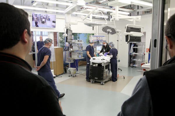
Hospitals are bringing together surgeons, anesthesiologists and nurses with architects, engineers and administrative staff to rethink the modern operating room.This article is part of our new series on the Future of Health Care, which examines changes in the medical field.If you ask Dr. Scott T. Reeves, operating rooms resemble an airplane cockpit. There is sophisticated equipment, tight spaces, blinking lights and a cacophony of sound.On top of that, “they’re often cluttered, people can trip, surgeons and nurses can stick themselves with needles, and side infections from dust and other contaminations are a growing problem,” said Dr. Reeves, chair of the department of anesthesia and perioperative medicine at the Medical University of South Carolina.When he became involved with the design of the operating rooms at the R. Keith Summey Medical Pavilion, part of the children’s hospital of the university, Dr. Reeves focused on how to make surgical suites more accommodating for technology — including imaging machines and robots — as well as staff and patients.Dr. Reeves’s actions are part of an increasing recognition that hospitals are “human centered,” said Anjali Joseph, the director of the Center for Health Facilities Design and Testing at Clemson University, who worked on the design for the ambulatory center that opened in 2019. “We cannot think of patient safety without thinking about the health of everyone in the room. They are interlinked.”Their goal: to rethink the layout as well as plan for the future, and the South Carolina team is not alone. The problem of squeezing people and a variety of machines — not to mention robots — into surgical suites designed decades ago is forcing a change.A full-scale simulated pediatric operating room at the Medical University of South Carolina allowed staff members to evaluate what would work best.Sarah Pack/MUSCFrom increasing in size to reorienting the layout, hospitals — especially those that are part of large university medical centers — are bringing together surgeons, anesthesiologists and nurses with architects, engineers and administrative staff to rethink the modern operating room. But even older community hospitals, with more limited budgets, are getting creative, since surgeries are an all-important source of revenue.While new construction is more straightforward than retrofitting an older building, not every hospital has the financing or the space to begin anew. Building a new operating room alone can cost from $1 million to $3 million per surgical suite, Dr. Reeves said. The cost of a new hospital can exceed $1 billion.Configuring new surgical suites in existing buildings requires creativity, said Joan Saba, a health care architect and partner with NBBJ, an architecture and design firm.For example, older operating rooms may have ceiling heights as low as 10 feet, while 12-16 feet is now considered optimum, to house electronics, cables and ductwork, she said. Some have captured space from the floor above to gain the extra height. Where that is not an option, some hospitals have repurposed adjoining rooms to house electronics and other infrastructure.New equipment and new surgical techniques are largely driving the redesigns. Those designing operating rooms even 20 years ago could not have foreseen the explosion in technology, which often requires more space.“Imaging management” is the biggest challenge that operating rooms have, said Mary Hawn, the chair of the department of surgery at Stanford University, which opened a new hospital in November 2019 (Stanford’s new children’s hospital opened in 2017). “Twenty years ago we would operate on exactly what we were looking at, possibly magnifying it with loupes,” the specialized glasses that augment a surgeon’s vision. Now, monitors provide high definition to guide the surgeon.In addition, for very complicated surgeries, hospitals hope to have equipment like CT scans and other imaging machines in the operating room. This not only saves time but lessens the risk of infection.“Patients need not be closed up, taken out for imaging, see that you missed something and then bring them back to the operating room and open them up again,” Ms. Saba said.Of all the imaging equipment, the only one generally unsuitable for the operating room is that needed for magnetic resonance imaging — commonly known as an M.R.I. — because of its size. As a result, Ms. Saba said, some hospitals essentially are stationing them adjacent to an operating room in case an M.R.I. is needed. A separate space has an added efficiency benefit, because the equipment can be used for nonsurgical patients as well.Ceilings are not overlooked. Freeing up valuable floor space, monitors are often affixed to ceiling-mounted booms, which can have several arms and may also serve as a conduit for gases needed for anesthesia. Ultraviolet cleaning systems, which eliminate bacteria and viruses, can be anchored in the ceilings, to assist with disinfection. And the space above the ceiling is often larger to house a range of cables and other electronic equipment, in addition to ductwork with sophisticated air filtration systems.Access to the space above the ceiling, as well as behind the walls, has become important, so that any technical problems can be investigated and remedied within hours, rather than shutting a room down for lengthy repairs. Some hospitals, for example, are now considering stainless steel prefabricated wall systems for their surgical suites because they are both easier to clean and easier to take out if the electronics hidden behind break, Ms. Saba said.Other important factors are lighting and noise. When it comes to increasingly common laparoscopic surgery, monitors that guide surgeons are lit but overhead lights may be turned off to reduce glare, Dr. Hawn said.That “can be somewhat dangerous because it can be quite dark and people run into things or trip over things,” she added. “We now have green lighting, which allows us to be able to see a sharp image on the monitors without the glare that you get from the white light.”Noise is distracting at best, but with physical repercussions, like hypertension, especially for staff exposed for long periods. High decibel levels are “associated with increased difficulty in communication, which is the largest source of preventable errors in the hospital environment,” John Medina, an affiliate associate professor at the University of Washington department of bioengineering, said in an email.At the Loma Linda University Medical Center in California, which is expected to open a new hospital on its campus this year, the operating room walls are built to mitigate outside noise as well as vibrations, and air duct silencers are being used as well, said Allison Ong, the head of campus transformation.A renovated operating room at the R. Keith Summey Medical Pavilion gives staff the option to move equipment, lights and monitors around the operating table during surgeries.Sarah Pack/MUSCHospital construction — whether for new buildings or even renovated pre-existing spaces, takes years, from inception to opening and can cost hundreds of millions of dollars. Before the spaces are put into use, all the staff — from the surgeons to the orderlies — need to practice in the new configuration. Dress rehearsals are common, in spaces like warehouses or even parking lots that are mocked up with cardboard walls to resemble the finished surgical suite.A run-through can be elaborate, bringing together surgeons, anesthesiologists and nurses. Several days of full hospital rehearsals, for example, are in the works at the Loma Linda center. The planning for the 500 or more people who will attend each day has itself taken months, Ms. Ong said.Evaluating the finished space before the first patient arrives can also help the medical staff make important choices. The Medical University of South Carolina, was considering a specialized piece of imaging equipment for its children’s hospital that would have permitted a fluoroscopy during surgery, Dr. Reeves said. But the machine had a big footprint, so a group taped it out on the floor.“What we realized by doing that was that it greatly decreased the functionality of the room for routine cardiac surgical patients,” he said. “It was great for the 10-15 patients a year we would potentially need it for, but it substantially became a burden for everyone else.” The hospital decided against installing the equipment in the operating room itself.Over all, perhaps the biggest question in these renovations is how to “future proof” the operating rooms, in addition to the overall hospital. It is a particularly challenging exercise with technology changing so rapidly.At Loma Linda, Ms. Ong said, “We had to decide very early on what the future of health care was going to look like. How many I.C.U. beds, how many medical surgical beds and how many O.R’s. You make your best guess.”Part of that is adopting a more modular approach, to allow flexibility for new equipment. As Dr. Reeves said: “The takeaway from Covid is how rigid many operating rooms are. I think you’ll see a lot of architecture firms be more nimble in their designs. And while that comes with an increased cost, it’s a question of either pay me now or pay me later.”

Read more →

Why Exercise Can Be So Draining for People With Rheumatoid Arthritis

Even a gentle session of leg lifts set off an exaggerated nervous system reaction in older women with rheumatoid arthritis.Exercise can feel more difficult and draining than usual if you have rheumatoid arthritis, and it’s not just because of the stiff and painful joints caused by this autoimmune disorder. In a groundbreaking new experiment involving older women and exercise, researchers found that even a gentle session of leg lifts set off an exaggerated nervous system reaction in those with rheumatoid arthritis. Light exercise also negatively affected the inner workings of their muscles and blood vessels.The findings build on earlier research about rheumatoid arthritis and the nervous system and raise pressing new questions about the best and safest ways for people with this disorder or similar autoimmune diseases to become and remain active.Anyone who has rheumatoid arthritis or is close to someone who has it knows the havoc it creates in the body. Immune cells mistakenly attack healthy tissue, especially in joints, causing swelling, pain and deterioration, along with full-body inflammation and fatigue. Rheumatoid arthritis also often results in cardiovascular disease, which initially puzzled doctors, since the misguided immune cells do not directly target the heart or arteries.But in recent years, researchers discovered that people with rheumatoid arthritis tend to have unusually twitchy sympathetic nervous systems. The sympathetic nervous system is the portion of our internal wiring that stimulates the fight-or-flight response, biochemically alerting our brains, heart, muscles and other bodily systems to brace ourselves for impending danger. The opposing parasympathetic nervous system, the Matthew McConaughey of our internal biology, lulls us, sending signals that quiet the sympathetic upsets.But in rheumatoid arthritis patients, researchers found, the sympathetic system seems stuck in overdrive, keeping people’s internal operations constantly on edge. A result is a high risk for elevated blood pressure and heart rate, even when people are resting quietly, which contributes over time to cardiovascular disease.Few of those earlier studies, though, looked at exercise, which also raises blood pressure and heart rates and changes nervous system reactions. Some past studies — and considerable anecdotal evidence — had indicated that people with rheumatoid arthritis feel more fatigue during and after activity than other exercisers. Their heart rates and blood pressures also remain stubbornly elevated for longer after workouts. But what might be going on inside their nerves and muscles leading to these reactions has been mostly unclear.So, for the new study, which was published in February in The Journal of Physiology, scientists at the University of São Paulo in Brazil decided to ask people with rheumatoid arthritis to do a little resistance training. Turning to patients at the university’s rheumatology clinic, they recruited 33 older women with rheumatoid arthritis and another 10 older women without the condition, to serve as controls. Most of them, in both groups, were on various medications.They invited all of their volunteers to the lab, drew blood, asked about their current pain levels, tested blood pressure and other health markers, and gently embedded tiny sensors beneath the skin in one leg to measure nervous system activity. Finally, they asked each woman to complete leg lifts with that leg, using a standard weight machine set to a low resistance. The women were supposed to lift repeatedly for three minutes — although some quit earlier than that — while the researchers tracked their blood pressures, nervous system reactions, and markers of muscular response, during and immediately afterward.What they found when they compared results was that “the women with R.A. showed greater blood pressure and sympathetic responses” to the light workout than those in the control group, says Tiago Peçanha, a postdoctoral research associate at the University of São Paulo who was a co-author of the new study with his doctoral adviser Hamilton Roschel, the director of the university’s Laboratory of Assessment and Conditioning in Rheumatology, and others.Their nerves seemed especially sensitive to the buildup of certain substances in the working muscles, the researchers concluded, which prompted the nerves to send urgent messages to nearby blood vessels, ordering them to contract. The result was lingering high blood pressure, during and after the workout.These reactions were most marked among the rheumatoid arthritis patients with the highest levels of inflammatory activity in their blood before the exercise, the researchers found.Taken as a whole, the findings indicate that physical activity can be extra difficult for people with rheumatoid arthritis, because their nervous systems may overreact to relatively minor changes inside the muscles.But the findings do not suggest that those with the autoimmune disorder should avoid exercise, Dr. Roschel says. “Physical activity is highly recommended for people with R.A,” he points out. “But these individuals may require additional attention and support to engage in physical activity programs.”If you have been diagnosed with rheumatoid arthritis, talk with your physician or an exercise physiologist about how best to exercise, he says. And if you begin a new routine, start slowly and perhaps keep a log of how you feel during workouts.Of course, this study focused on older women with rheumatoid arthritis and a single session of very light resistance training. It is unknown whether the results apply equally to younger women or men with the condition, or whether other types of exercise, such as walking, may produce a similar response. It is also unknown how those with different autoimmune diseases or related conditions might be affected.Dr. Roschel and his colleagues are looking into all of those questions, though. “We have also been conducting some exercise studies with patients who have recovered from Covid-19 in our lab, and they also present abnormal cardiorespiratory responses to exercise,” he says. They hope to publish additional studies soon.

Read more →

Doctors investigate mystery brain disease in Canada

SharecloseShare pageCopy linkAbout sharingimage copyrightCourtesy Steve EllisDoctors in Canada have been coming across patients showing symptoms similar to that of Creutzfeldt-Jakob disease, a rare fatal condition that attacks the brain. But when they took a closer look, what they found left them stumped. Almost two years ago, Roger Ellis collapsed at home with a seizure on his 40th wedding anniversary. In his early 60s, Mr Ellis, who was born and raised around New Brunswick’s bucolic Acadian peninsula, had been healthy until that June, and was enjoying his retirement after decades working as an industrial mechanic. His son, Steve Ellis, says after that fateful day his father’s health rapidly declined. “He had delusions, hallucinations, weight loss, aggression, repetitive speech,” he says. “At one point he couldn’t even walk. So in the span of three months we were being brought to a hospital to tell us they believed he was dying – but no one knew why.” Roger Ellis’ doctors first suspected Creutzfeldt-Jakob disease [CJD]. CJD is a human prion disease, a fatal and rare degenerative brain disorder that sees patients present with symptoms like failing memory, behavioural changes and difficulties with co-ordination. One widely known category is Variant CJD, which is linked to eating contaminated meat infected with mad cow disease. CJD also belongs to a wider category of brain disorders like Alzheimer’s, Parkinson’s and ALS, in which protein in the nervous system become misfolded and aggregated. How BSE crisis shook our faith in foodBut Mr Ellis’ CJD test came back negative, as did the barrage of other tests his doctors put him through as they tried to pinpoint the cause of his illness. His son says the medical team did their best to alleviate his father’s varying symptoms but were still left with a mystery: what was behind Mr Ellis’s decline? image copyrightGetty ImagesIn March of this year, the younger Mr Ellis came across a possible – if partial – answer. Radio-Canada, the public broadcaster, obtained a copy of a public health memo that had been sent to the province’s medical professionals warning of a cluster of patients exhibiting an unknown degenerative brain disease. “The first thing I said was: ‘This is my dad,'” he recalls. Roger Ellis is now believed to be one of those afflicted with the illness and is under the care of Dr Alier Marrero. The neurologist with Moncton’s Dr Georges-L-Dumont University Hospital Centre says doctors first came across the baffling disease in 2015. At the time it was one patient, an “isolated and atypical case”, he says. But since then there have been more patients like the first – enough that now doctors been able to identify the cluster as a different condition or syndrome “not seen before”. The province says it’s currently tracking 48 cases, evenly split between men and women, in ages ranging from 18 to 85. Those patients are from the Acadian Peninsula and Moncton areas of New Brunswick. Six people are believed to have died from the illness. Most patients began experiencing symptoms recently, from 2018 on, though one is believed to have had them as early as 2013.Dr Marrero says the symptoms are wide ranging and can vary among patients.At first, there can be behavioural changes like anxiety, depression and irritability, along with unexplained pain, muscle aches and spasms in previously healthy individuals. Frequently, patients develop difficulties sleeping – severe insomnia or hypersomnia – and memory problems. There can be fast-advancing language impairments that make it difficult to communicate and hold a fluent conversation – issues like stuttering or word repetition. Another symptom is rapid weight loss and muscle atrophy, as well as visual disturbances and co-ordination problems, and involuntary muscle twitching. Many patients need the assistance of walkers or wheelchairs. Some develop disturbing hallucinatory dreams, or waking auditory hallucinations. Several patients have presented with transient “Capgras delusion”, a psychiatric disorder in which a person believes someone close to them has been replaced by an impostor. “It’s quite disturbing because, for instance, a patient would tell his wife: ‘Sorry ma’am you cannot get in bed, I’m a married man’ and even if the wife gives her name, he’d say: ‘You’re not the real one,'” Dr Marrero says. The Moncton based neurologist is leading the investigation into the condition, with help from a team of researchers and the federal public health agency.Suspected patients undergo prion disease tests and tests for genetic conditions, panels looking at autoimmune disorders or forms of cancer, and screenings for things like viruses, bacteria, fungus, heavy metals and abnormal antibodies. They are asked about environmental factors, lifestyle exposures, travel, medical history and food and water sources. They undergo spinal taps to test for various possible infections and disorders. There’s no treatment, beyond helping to alleviate the discomfort of some of the symptoms. For now, the theory is that the disease is acquired, not genetic. “Our first common idea is that there’s a toxic element acquired in the environment of this patient that triggers the degenerative changes,” says Dr Marrero. image copyrightGetty ImagesUniversity of British Columbia neurologist Dr Neil Cashman is one of the researchers trying to unravel the medical mystery. Despite patients showing no trace of any known prion diseases, it hasn’t been completely ruled out as a cause, he says. Another theory is chronic exposure to what’s called an “excitotoxin” like domoic acid, which was linked to a 1987 food poisoning incident from mussels contaminated with the toxin from the nearby province of Prince Edward Island. Along with gastrointestinal upset, about a third of those affected had symptoms like memory loss, dizziness, confusion. Some patients fell into a coma, and four died. Dr Cashman says they are also looking at another toxin – beta-methylamino-L-alanine (BMAA) – which has been implicated as an environmental risk in the development of diseases like Alzheimer’s and Parkinson’s.BMAA is produced by cyanobacteria, commonly known as blue-green algae. Some researchers also believe BMAA is linked to a neurodegenerative disease documented in an indigenous population in the Pacific US island territory of Guam during the mid-20th Century, and found in seeds that were part of their diet. Dr Cashman cautions the current list of theories “is not complete”. “We have to go back to first principles, go back to square one,” he says. “At this point basically nothing can be excluded.” So how many more people may be affected by this disease? image copyrightCourtesy Steve EllisDr Marrero says it’s possible it’s a wider phenomenon found outside the two regions – the Acadian peninsula, with its fishing communities and sandy beaches, and Moncton, a city centre – where patients have currently been identified. “Are we seeing the tip of the iceberg? Maybe,” he says. “I’m hoping we can capture this very fast so we can stop this.” While those living in the affected communities are understandably concerned, Dr Marrero urges people to “work with hope not with fear. Fear paralyses.” Roger Ellis’ condition has stabilised since the early rapid progression, his son says. He’s in a specialised care home and needs some assistance with daily activities, and struggles with speech and sleep issues. Steve Ellis, who runs a Facebook support group for families affected by the condition, says he wants government officials to commit to transparency about the illness. Most of all, he wants to know what made his father fall sick. “I know they’re working on that, but how did this happen?” he says. “As a family, we’re very cognisant of the fact he’s likely going to die from this, and we just hope that whether it happens before he dies or after that they are answers – and accountability if it’s something that could have been prevented.”

Read more →