How Partisanship Affects Pandemic Thinking
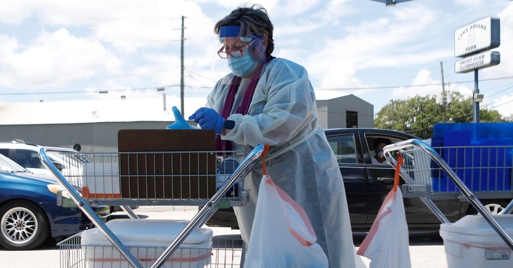
What political scientists and pollsters say about how the Covid wave might affect public behavior.There were no open I.C.U. beds on Wednesday in Alabama, or in parts of Florida, Louisiana, Mississippi and Texas, as hospitals across the South buckled under the weight of a coronavirus surge that could have been mitigated. Cases, hospitalizations and deaths are increasing nationwide. Every day, on average, more than 800 Americans are dying from Covid-19.It is a humanitarian catastrophe, and yet many Americans see it through a political lens. The South has some of the nation’s lowest vaccination rates, driven partly by Republican reluctance. Some governors — including Gov. Greg Abbott of Texas, who is infected with the coronavirus himself — have forbidden local officials to impose mask requirements. Gov. Ron DeSantis of Florida, who has not changed his approach to follow public health guidelines, has claimed falsely that the surge is a result of President Biden’s border policies.The divisions extend beyond policies to general attitudes about the pandemic: While nearly 60 percent of Americans overall said in a recent Quinnipiac poll that they were concerned about the Delta variant, more than 60 percent of Republicans said they weren’t. And research indicates that many people are looking at Covid policies they don’t like and blaming whichever party they’re not part of.It’s enough to make one despair about the American public’s ability to deal in a nonpartisan manner with, well, anything.But that may not quite be right.I talked to several political scientists and pollsters about how the current Covid wave might affect public opinion and, more important, public behavior. Here’s what they said.The bad news: Partisanship is really hard to overcome.Partisanship — more specifically negative partisanship, which is animosity toward the other party as opposed to, or in addition to, a positive allegiance to your own — is an extraordinarily powerful force in American politics. It has become only stronger in recent years as partisanship has become increasingly intertwined with religious and racial identities.When people look at the pandemic or Afghanistan or any other issue, “you’re doing so through this lens of the identity you have and preserving a self-esteem about that identity,” said Julie Wronski, an associate professor at the University of Mississippi who studies political psychology and behavior. “You’re trying to think about the people who are on ‘my team’: Are they good people? Are they winners? And the people on the other team are ‘bad people’ or ‘losers.’”Some of what we’re seeing now in response to the pandemic was baked in very early on, as soon as elected officials — most prominently President Donald J. Trump — began to politicize basic public health measures, leading people to see support for masks or vaccines as partisan.“That didn’t necessarily have to happen, but once it did, you’re not necessarily talking about the science,” Professor Wronski said. “It’s about who they are and who they consider themselves to be.”One group of researchers had an unusual opportunity to study how partisan identity shaped people’s views on Covid, because in 2019, they surveyed more than 3,300 people about their political predispositions for an unrelated project. Once the pandemic began, they went back to the same people, and about 2,500 responded to follow-up questions.They found, in research published in peer-reviewed journals in August and November 2020, that highly partisan Republicans took their initial cues from leaders like Trump and then stuck to them no matter what — even if Covid cases and deaths surged in their state, even if people around them got sick, said one of the five researchers, Yanna Krupnikov, a professor of political science at Stony Brook University.Another of the five, Samara Klar, an associate professor at the University of Arizona’s School of Government and Public Policy, said the crucial element appeared to be not party affiliation alone, but active animosity toward the opposite side.“We’re seeing the gap mostly among those people who personally dislike the other party, and that’s weird,” Professor Klar said. “It’s weird for your views on a public health crisis to be guided by your personal feelings toward members of the other party, but that is in fact what we’re finding.”The good news: Not everyone is rigidly partisan.Most people aren’t the sort of intense partisans described above. The exact percentage varies depending on the questions you ask, but generally, Professor Krupnikov said, only 25 to 30 percent of people fall into the “hyperpolarized” category.And as the pandemic hit closer to home, she said, less-partisan Republicans “actually started to look very much like Democrats” in their personal precautions and the Covid-related policies they supported.In other words, Democrats tended to take the pandemic seriously from the start, but once case counts spiked in the home counties of Republicans who weren’t extremely partisan, they began to take it seriously, too.This reaffirms a longstanding belief of political science, Professor Klar said: “When an issue becomes really threatening and really important to you, then partisanship weakens its grip on your decision making.”It is, at least, a moderately reassuring thought.“There’s often so much focus on people whose partisanship seems to surpass their care even for their own health, or care for others,” Professor Krupnikov said. “But I do think it’s important to highlight that there are, at least in our data, a lot of people for whom politics was in fact tremendously secondary to the health crisis happening around them.”So what’s next?What this means practically for the future of the pandemic is less clear, especially because we don’t have much reliable polling conducted since the Delta surge spun out of control.The limited polling we do have shows that a majority of Americans are worried about the Delta variant and support the C.D.C. recommendation that people wear masks indoors regardless of their vaccination status — and that pattern holds across regions, including the South, said Mary Snow, a polling analyst at Quinnipiac University. But there are still deep partisan divides in that data.President Biden’s approval rating also seems to have taken some damage, but that may not be because of the surge itself. Rather, it may be “because we were told that we were out of the woods at the beginning of the summer, and that hasn’t happened,” said Patrick Murray, the director of the Monmouth University Polling Institute. “And that’s a reflection of messaging as much as anything else: ‘Why did you tell us you had this under control when you didn’t?’”Ultimately, especially in the face of such a contagious variant, it takes only a small minority of Americans to derail epidemiological progress — and the most partisan Republicans are taking their cues from leaders who have no political incentive to give different ones.In a state like Mississippi, the governor has more to fear politically from a far-right primary challenger than from a Democrat in a general election, Professor Wronski noted.And while even partisans’ opinions could change if people they were close to started dying, she said, it would be a psychologically difficult shift.“For the past couple years, your identity has been built upon a certain perception of what you think Covid is, who you think the good guys are, your lack of trust in political elites,” she said. “And now, if you’re starting to see death at your doorstep, that’s a cognitive dissonance that you have to reconcile.“How many deaths is it going to take? I don’t have that answer.”On Politics is also available as a newsletter. Sign up here to get it delivered to your inbox.Is there anything you think we’re missing? Anything you want to see more of? We’d love to hear from you. Email us at onpolitics@nytimes.com.

Read more →

Scientists harness human protein to deliver molecular medicines to cells

Researchers from MIT, the McGovern Institute for Brain Research at MIT, the Howard Hughes Medical Institute, and the Broad Institute of MIT and Harvard have developed a new way to deliver molecular therapies to cells. The system, called SEND, can be programmed to encapsulate and deliver different RNA cargoes. SEND harnesses natural proteins in the body that form virus-like particles and bind RNA, and it may provoke less of an immune response than other delivery approaches.
The new delivery platform works efficiently in cell models, and, with further development, could open up a new class of delivery methods for a wide range of molecular medicines — including those for gene editing and gene replacement. Existing delivery vehicles for these therapeutics can be inefficient and randomly integrate into the genome of cells, and some can stimulate unwanted immune reactions. SEND has the promise to overcome these limitations, which could open up new opportunities to deploy molecular medicine.
“The biomedical community has been developing powerful molecular therapeutics, but delivering them to cells in a precise and efficient way is challenging,” said CRISPR pioneer Feng Zhang, senior author on the study, core institute member at the Broad Institute, investigator at the McGovern Institute, and the James and Patricia Poitras Professor of Neuroscience at MIT. “SEND has the potential to overcome these challenges.” Zhang is also an investigator at the Howard Hughes Medical Institute and a professor in MIT’s Departments of Brain and Cognitive Sciences and Biological Engineering.
Reporting in Science, the team describes how SEND (Selective Endogenous eNcapsidation for cellular Delivery) takes advantage of molecules made by human cells. At the center of SEND is a protein called PEG10, which normally binds to its own mRNA and forms a spherical protective capsule around it. In their study, the team engineered PEG10 to selectively package and deliver other RNA. The scientists used SEND to deliver the CRISPR-Cas9 gene editing system to mouse and human cells to edit targeted genes.
First author Michael Segel, a postdoctoral researcher in Zhang’s lab, and Blake Lash, second author and a graduate student also in the group, said PEG10 is not unique in its ability to transfer RNA. “That’s what’s so exciting,” said Segel. “This study shows that there are probably other RNA transfer systems in the human body that can also be harnessed for therapeutic purposes. It also raises some really fascinating questions about what the natural roles of these proteins might be.”
Inspiration from within
The PEG10 protein exists naturally in humans and is derived from a “retrotransposon” — a virus-like genetic element — that integrated itself into the genome of human ancestors millions of years ago. Over time, PEG10 has been co-opted by the body to become part of the repertoire of proteins important for life.

Read more →

Patients helping researchers to advance treatments for prostate cancer

Researchers at Monash University have established one of the world’s largest collections of living tumours from prostate cancer patients, accelerating the testing of new treatments for prostate cancer and leading to faster patient benefit.
One of the most common cancers, prostate cancer is also one of the most difficult to study in the laboratory, with the frequently used models derived more than 40 years ago. With the establishment of the Melbourne Urological Research Alliance (MURAL), hundreds of Victorian men have generously donated samples of their cancer tissue, enabling the team to study a greater diversity of live tumours and test the efficacy of a larger variety of therapies for their ability to stop tumour growth.
The PDX collection (patient-derived xenografts), developed by a multidisciplinary consortium and led by Professor Gail Risbridger and Associate Professor Renea Taylor at the Monash Biomedicine Discovery Institute (BDI), now comprises 59 tumours, collected from 30 patients between 2012 — 2020 and is now one of the largest collections of prostate cancer models in the world.
Full characterisation of the PDX collection is published in Nature Communications.
MURAL PDXs are an enduring resource of new cancer models that can be shared with other academic investigators or pharmaceutical companies. The patients and their families are directly embedded in this venture, including the EJ Whitten Foundation who have been pivotal over the last 10 years in providing over $1M in donations enabling this resource to be developed and the program to come to the forefront of the international field.
“This project begins and ends with patients like EJ Whitten. We take patient tissue — do testing in the laboratory — and the discoveries then advance treatment for patients,” said Professor Risbridger. “Our new models of prostate cancer have attracted interest from scientists and the pharmaceutical industry worldwide.”
Ted Whitten, Executive Director and Founder of the E.J.Whitten Foundation congratulates the Monash University Biomedicine Discovery Institute on its recent findings in regards to Prostate Cancer research. “We believe that Monash University is a leader of Prostate Cancer Research and we have been delighted to have been able to financially support many of their important programs over the past ten years.”
Dr Mitchell Lawrence, also from Monash BDI and a senior author, says: “This resource provides an opportunity to link the molecular changes in prostate cancer to pathology, grow organoids and test functional responses to therapies, which have rarely been applied to prostate cancer given the lack of suitable models.”
The success of this program is based on collaboration between scientists and clinicians such as surgeons and oncologists at Monash, Cabrini Institute and Peter MacCallum Cancer Centre, as well as patients and their families who generously donate cancer tissues. Other organisations who supported the PDX program include Victorian Cancer Agency, Prostate Cancer Foundation of Australia and Movember.
Story Source:
Materials provided by Monash University. Note: Content may be edited for style and length.

Read more →

How ions get their electrons back

Very unusual atomic states are produced at TU Wien: Ions are created by removing not just one but 20 to 40 electrons from each atom. These “highly charged ions” play an important role in current research.
For a long time, people have been investigating what happens when such highly charged ions hit solid materials. This is important for many areas of application in materials research. Therefore it is crucial to know how the charge state of the ions change when they penetrate a material — but this is exactly what has been impossible to observe directly until now. New measurements at TU Wien (Vienna) show that the ions obey remarkably simple laws.
Probing materials layer by layer
When highly charged ions penetrate a solid material, they can retrieve the missing electrons from the material and thus become electrically neutral. But how and where this happens exactly is difficult to investigate, because it happens inside the material.
“We knew that this process must be very fast, because even a fairly thin layer of material is enough to completely neutralise ions,” says Anna Niggas, first author of the present study. She is currently working on her dissertation in Prof. Richard Wilhelm’s group at the Institute of Applied Physics at TU Wien.
Visually observing the processes inside the material may be nearly impossible, but novel 2D materials such as graphene, which consists of only a single layer of carbon atoms, now give scientists a chance to get to the bottom of these phenomena for the first time: “Graphene layers can be stacked on top of each other, so that thicker and thicker samples are created — you can assemble a solid body layer by layer,” says Richard Wilhelm. “We have studied single, double and triple graphene layers. That way, we can see step by step, atomic layer by atomic layer, how the highly charged ions change.”
In this way, you can study a transition, from a single atomic layer to an ordinary three-dimensional material. Graphite, the material pencil leads are made of, is nothing more than a large number of graphene layers stacked on top of each other.

Read more →

New study sheds light on how an autoimmune disorder may underlie male infertility

Investigators have found that the absence of autoimmune regulator (Aire) in mice results in fertility problems similar to those affecting men with autoimmune polyendocrine syndrome type I (APS-1). Aire-dependent central tolerance plays a critical role in maintaining male fertility by preventing autoimmune attack against multiple reproductive targets, they report in The American Journal of Pathology.
“Male factors account for a large portion of infertility in couples, and the mechanisms underlying male infertility are poorly understood,” explained lead investigator Margaret G. Petroff, PhD, a professor in the Department of Pathobiology and Diagnostic Investigation, College of Veterinary Medicine, Michigan State University, East Lansing, MI, USA. “This study is important because it represents a previously underexplored mechanism by which fertility can be impacted through autoimmune disease.”
Aire is a gene expressed in the thymus that plays a vital role in teaching the immune system to distinguish between the body’s own cells and invaders. Aire-deficient mice reproduce many of the features of APS-1 in humans. To determine fertility, Aire-deficient and wild-type male mice were paired with wild-type female mice. Aire-deficient males exhibited dramatically reduced mating frequency and fertility; those able to mate took up to two weeks to do so, and their sperm quality was poor. Sperm from the Aire-deficient males were rarely able to produce litters, and even when they were used for in vitro fertilization, could not produce viable embryos.
Aire-deficient males were found to produce low levels of testosterone and develop autoimmune disease against many components of the male reproductive tract, especially in the epididymis. However, the investigators could not rule out the possibility that Aire might be expressed in these organs, not just in the thymus. It is possible that the injuries could be caused by a lack of expression in the tissues themselves. Using a fluorescence reporter model in which cells change color based on present or past expression of Aire, they confirmed that the gene can be expressed in the developing mouse reproductive system. “We were particularly surprised to find evidence of AIRE in the testis and prostate gland, suggesting that it may have an immune-independent role in these tissues,” said Dr. Petroff.
The correlation between impaired central immune tolerance and fertility has potential implications not only for male APS-1 patients but may also provide important insights into both male autoimmune and unexplained cases of infertility.
“By knowing more detail about what causes infertility in men, we can develop treatments and prophylactics to curb degenerative processes that affect fertility,” commented Dr. Petroff. “It may be possible to use general immunosuppressive treatments. Even better, it might be possible to design highly specific therapies that target particular immune cells, preventing these cells from causing damage to reproductive organs.”
Story Source:
Materials provided by Elsevier. Note: Content may be edited for style and length.

Read more →

Study of East Africans illuminates new genetic factors underlying human faces

A new study of the genetics underlying facial features finds that East Africans have some unique facial genetics and some that are shared with Europeans. The findings, published August 19th in PLOS Genetics by Chenxing Liu, Seth Weinberg and John Shaffer of the University of Pittsburgh and colleagues, add to our understanding of how genetics shape the human face.
The remarkable range of shapes and sizes of the human face is largely the result of genetics. Previous studies have linked more than 100 genetic sites to facial characteristics, but most of these studies included only European or Asian populations. In the new study, Liu and colleagues focused on an East African population to expand our knowledge of the genetic factors behind human facial features.
The team analyzed 2,595 3D facial images collected from Tanzanian children and scanned their genomes to find genetic sites linked to specific features. The researchers identified 20 regions on the genome associated with face shape. Several of these genetic variants play a role in embryonic cells that give rise to facial structures very early in life — around 3 to 6 weeks after conception. Ten of the genetic sites were previously identified in European populations, suggesting that the genetic basis for human face shape is partly shared across populations.
Shaffer adds, “Our findings confirm that the genes connected to human facial features are basically the same across populations. Observed differences were mostly explained by how frequently an allele occurs in a given population. By comparing populations, we were able to uncover genetic signals that would otherwise remain obscured and narrow the field of genetic variants that are functionally impacting facial traits.”
The new study in Tanzanians uncovered novel sites and genes potentially related to face shape and also advanced the understanding of sites that were already recognized in Europeans. Overall, these findings shed light on the genetic and biological basis underpinning the diversity of human facial structures. They may also offer insights into the biological mechanisms responsible for how the face forms and the causes of facial deformities.
Weinberg adds, “Most published genetic studies of human facial traits have been limited to individuals of predominantly European ancestry, and African populations have been particularly under-represented in this field. The current study, therefore, represents an important advance in terms of diversity and provides new insights into the wide array of genes that impact facial appearance.”
Story Source:
Materials provided by PLOS. Note: Content may be edited for style and length.

Read more →

A master gear in the circadian clock

A gene called Npas4, already known to play a key role in balancing excitatory and inhibitory inputs in brain cells, appears to also be a master timekeeper for the brain’s circadian clock, new research led by UT Southwestern scientists suggests. The finding, published online today in Neuron, broadens understanding of the circadian clock’s molecular mechanisms, which could eventually lead to new treatments for managing challenges such as jet lag, shift work, and sleep disorders.
“To reset the circadian clock, you ultimately need to reset its molecular gears,” said study leader Joseph S. Takahashi, Ph.D., Professor and Chair of Neuroscience at UTSW and a Howard Hughes Medical Institute Investigator. “This study suggests that Npas4 might be one of the most important components for resetting the clock to light.”
For decades, researchers have known that a brain region called the suprachiasmatic nucleus (SCN) is responsible for controlling circadian rhythms, the various cycles of activity that typically run on a 24-hour basis. These rhythms are entrained by light, Dr. Takahashi explained; cells in the SCN respond to signals relayed by the retina, the eye’s light-sensitive tissue. However, the molecular basis of this phenomenon is not well understood.
To better understand how the SCN sets circadian rhythms, the researchers used a technique called single-nucleus sequencing to look at gene activity in individual cells in mice after the animals were exposed to light. Dr. Takahashi and his colleagues found that three different subpopulations of SCN neurons respond to light stimulation. A common thread tying these subtypes together was increased activity in genes that respond to neuronal PAS domain protein 4 (NPAS4), the protein made by the Npas4 gene.
When Dr. Takahashi and his colleagues exposed mice engineered to lack Npas4 to light, it dampened the response of hundreds of circadian clock genes. In addition, the animal’s circadian period lengthened about an extra hour, to nearly 25 hours instead of the normal 24. Together, these results suggest that Npas4 is a master regulator of many light-induced genes, a key piece in the puzzle of how the circadian system works, Dr. Takahashi said.
The more researchers learn about the molecular underpinnings of the circadian clock, Dr. Takahashi added, the more they may be able to manipulate it to improve health and well-being — for example, to ease jet lag or help shift workers stay awake or asleep to match their work cycles. It could also lead to new treatments for disorders marked by abnormal sleep/wake cycles.
Other researchers who contributed to this research include Pin Xu, Stefano Berto, Ashwinikumar Kulkarni, Byeongha Jeong, Chryshanthi Joseph, Kimberly H. Cox, Tae-Kyung Kim, all of UT Southwestern; and Michael E. Greenberg of Harvard Medical School.
Dr. Takahashi holds the Loyd B. Sands Distinguished Chair in Neuroscience at UTSW. This work was a collaboration with the laboratory of Genevieve Konopka, Ph.D., Associate Professor of Neuroscience, the Jon Heighten Scholar in Autism Research, and Director of the UTSW Neurogenomics Core. Drs. Takahashi and Konopka are members of the Peter O’Donnell Jr. Brain Institute. Dr. Kim, a Distinguished Scholar in Neuroscience, is also a member of the O’Donnell Brain Institute.
This research was supported by grants from the National Institutes of Health (NS106657, DC014702, MH102603, NS028829), the Howard Hughes Medical Institute, the James S. McDonnell Foundation 21st Century Science Initiative in Understanding Human Cognition — Scholar Award (220020467), and the Chan Zuckerberg Initiative, an advised fund of Silicon Valley Community Foundation (HCA-A-1704-01747).
Story Source:
Materials provided by UT Southwestern Medical Center. Note: Content may be edited for style and length.

Read more →

Varying immune cell levels in canine brain tumors could provide therapeutic targets

A new study reveals that high-grade gliomas, or brain tumors, in dogs contained more immune cells associated with suppressing immune response than low-grade gliomas. The work, which is the most extensive examination of immune cell infiltration in canine glioma to date, adds to the body of evidence that these brain tumors might recruit cells that aid in immunosuppression. The findings could have implications for future immunotherapy-based glioma treatments in both humans and dogs.
Glial cells are support cells located throughout the brain and spinal cord. When those cells become cancerous, the resulting tumor is called a glioma. In dogs, gliomas are the second most common type of tumor in the central nervous system and represent about 35% of all intracranial cancers. Median survival time for dogs with glioma treated with radiation therapy ranges from nine to 14 months, which is similar to the 14 month median survival time for humans treated with a combination of surgery, radiation and chemotherapy.
There are three types of canine glioma: oligodendroglioma, astrocytoma or undefined glioma. Each of these subtypes can be further classified as low or high-grade based on certain microscopic features. Although glioma subtype and grade affect survival and treatment choice in humans, it is currently unknown whether the same is true for dogs.
Immunotherapy harnesses the power of the body’s immune system to attack cancer. Though immunotherapy has shown promise in certain types of cancers, it hasn’t been successful in glioma in humans, possibly because gliomas have been shown to suppress the immune system in order to facilitate tumor growth. Researchers are trying to better understand the interaction between glioma and the immune system with hopes of improving therapeutic outcomes.
“If we want to pursue immunotherapy for glioma, we first need to understand how these tumors interact with the immune system,” says Gregory Krane, first author of the research and a veterinary pathologist who recently received his Ph.D. from North Carolina State University. “There are many shared features between canine and human glioma, which makes researching the immune system in canine glioma a good approach to addressing questions about this cancer in both humans and dogs.”
The multi-institutional research team examined 73 different gliomas obtained from veterinary patients seen at the NC State College of Veterinary Medicine between 2006 and 2018. Utilizing immunohistochemical tagging and computerized image analysis, the team identified the numbers of each type of immune cell in each tumor: B lymphocytes, T lymphocytes, regulatory T lymphocytes (Tregs) and macrophages. The team found higher numbers of Tregs and polarized macrophages in high- versus low-grade tumors, but no differences for other immune cells between different tumor types or grades.
“Tregs inhibit aspects of the immune response,” Krane says. “In healthy individuals, this prevents autoimmune disease. But cancers can recruit and activate Tregs to prevent the immune system from attacking the tumor. We found that Tregs were more abundant in high-grade gliomas than in low-grade gliomas. We hypothesize that Tregs may be involved in glioma-mediated immunosuppression, although that will require further research.”
The research team also counted the number of macrophages in each tumor, which can be polarized to either end of a spectrum referred to as M1 or M2 polarization. In a general sense, M1-polarized macrophages are pro-inflammatory and anti-tumor, and M2-polarized macrophages are the opposite. They found that the macrophage population in high-grade gliomas tended to be polarized towards the M2 phenotype.
“These macrophage polarization data can expand the glioma immunosuppression hypothesis by providing another mechanism by which gliomas may suppress the immune system in the dog,” Krane says.
Krane is hopeful that this study may lead to a better understanding of how gliomas affect the immune system, and eventually to improved immunotherapies for glioma. “Using the dog as a preclinical model for understanding the immune response to glioma could lead to treatments that will help both dogs and people,” Krane says. “Though further work is needed, our data provide some support to utilize canine patients with glioma to evaluate therapies targeting Tregs or macrophage polarization designed for use in humans.”
The research was published online in July 2021 in Veterinary Pathology and was supported by the National Institute of Environmental Health Sciences’ National Toxicology Program (NIEHS/NTP), NC State’s College of Veterinary Medicine, and Charles River Laboratories. Christopher Mariani, associate professor of neurology at NC State and principal investigator of NC State’s Comparative Neuroimmunology and Neuro-oncology Laboratory, is corresponding author. Other NC State co-authors were associate professors of pathology David Malarkey and Debra Tokarz, and veterinary student Britani Rainess. Researchers from NIEHS/NTP, Charles River Laboratories, the University of Alabama at Birmingham, Cornell University and Integrated Laboratory Systems coauthored the work.
Story Source:
Materials provided by North Carolina State University. Original written by Tracey Peake. Note: Content may be edited for style and length.

Read more →

Researchers uncover the biology and treatment behind a rare autoinflammatory disease

The absence of a protein that activates the body’s antiviral defenses can cause a rare rheumatoid-like autoinflammatory condition that is treatable with an FDA-approved class of drugs known as TNF (tumor necrosis factor) inhibitors, a global research team led by Mount Sinai has found.
The condition, which is now termed TBK1 deficiency, was previously known to scientists but its name, cause and treatment were identified for the first time in the study published August 6 in Cell.
The scientists reported that the absence of the protein, known as TBK1 (TANK-binding kinase 1), renders cells vulnerable to a jarring form of programmed cell death in response to TNF, but that this genetic defect can be effectively and quickly addressed by therapeutic agents that block the source of the inflammation.
Homozygous mutations in TBK1, which occur when copies of the aberrant gene encoding the protein are passed on by both parents, are extremely rare. Based on past studies in mouse models and human cell cultures, researchers assumed that these mutations would leave individuals susceptible to a wide range of viral infections. They found instead that none of the four people in the cohort they studied, ages 7 to 32, showed signs of inadequate antiviral immunity. Rather, they all suffered from a systemic autoinflammatory condition that resulted from a dysregulated response to TNF, an important protein involved in controlling inflammation and cell death.
“TBK1 signaling activates the body’s antiviral mechanisms to fight off infection and block different stages of viral replication, as well as control TNF-mediated inflammation,” says lead author Justin Taft, PhD, an investigator in the Department of Microbiology, the Precision Immunology Institute, and the Center for Inborn Errors of Immunity at the Icahn School of Medicine at Mount Sinai. “But if a mutation prevents expression of the TBK1 gene or disrupts its function, then cells become overly sensitive to TNF. And that can trigger a disproportionate amount of cell death, which sets off a violent cascade of debris from dying cells that inflames surrounding tissue and fuels the inflammation.”
By treating TBK1-deficient individuals with anti-TNF therapeutics, the Mount Sinai-led team confirmed its suspicions about the underlying biology of the genetically driven condition. “We have essentially defined a new disease and its associated mechanisms of autoinflammation, which previously were managed with steroid treatments, non-steroidal anti-inflammatory drugs, or other non-specific therapeutics clinicians deemed worth trying,” says Dusan Bogunovic, PhD, Director of the Center for Inborn Errors of Immunity; Associate Professor of Oncological Sciences, Microbiology, and Pediatrics; member of the Precision Immunology Institute and The Mindich Child Health and Development Institute; and senior author of the study. “We were able to target the condition directly and effectively with TNF inhibitors once we knew the causative factors of the inflammation. And the clinical improvement was quick and substantial.”
For Dr. Taft, the work of the global team of researchers underscores the power of the increasingly vital field of personalized medicine. “We started with four individuals who were known to have a homozygous TBK1 mutation, and did extensive lab work to determine how the defect could induce this autoinflammatory reaction,” says Dr. Taft. “And from the genetics we uncovered not only a plausible mechanism of the disease and new information around TBK1 biology, but identified a disease-specific treatment that significantly improves the autoinflammatory condition.”
In addition to Mount Sinai, the collaborative research effort included Erasmus University Medical Center in the Netherlands, Mayo Clinic, the National Human Genomic Research Institute, Imperial College London, SRCC Children’s Hospital and Jaslok and Breach Candy Hospitals in Mumbai, India, and Hacettepe University in Ankara, Turkey.

Read more →

Study supports widespread use of better masks to curb COVID-19 indoors

A new study is highlighting a need for widespread use of better face masks and the importance of good ventilation to mitigate the spread of COVID-19 indoors.
Engineering researchers at the University of Waterloo performed experiments using a mannequin to simulate a seated person breathing in a large room. The studies showed a significant buildup over time of aerosol droplets — exhaled droplets so tiny they remain suspended and travel through the air — despite the use of common cloth and blue surgical masks.
“There is no question it is beneficial to wear any face covering, both for protection in close proximity and at a distance in a room,” said Serhiy Yarusevych, a professor of mechanical and mechatronics engineering and the leader of the study. “However, there is a very serious difference in the effectiveness of different masks when it comes to controlling aerosols.”
Previous research has found that aerosols dispersed by infected people are a source of transmission of the SARS-CoV-2 virus that causes COVID-19, even outside the two-metre social distancing zone widely recommended by public health officials.
The study showed that most common masks, primarily due to problems with fit, filter about 10 per cent of exhaled aerosol droplets. The remaining aerosols are redirected, mostly out the top of the mask where it fits over the nose, and escape into the ambient air unfiltered.
By contrast, higher-quality, more expensive N95 and KN95 masks filtered more than 50 per cent of the exhaled aerosols that can accumulate indoors and spread the COVID-19 virus when inhaled by other people.
Yarusevych, principal investigator in the Fluid Mechanics Research Lab, said the much greater effectiveness of N95 and KN95 masks versus cloth and surgical masks makes a compelling case they should be worn in indoor settings, such as schools and workplaces, as much as possible.
“A lot of this may seem like common sense,” he said. “There is a reason, for instance, that medical practitioners wear N95 masks — they work much better. The novelty here is that we have provided solid numbers and rigorous analysis to support that assumption.”
Experiments also quantified the impact of ventilation systems, which circulate and replace air in rooms, on the accumulation of aerosols. Even modest ventilation rates were found to be as effective as the best masks in reducing the risk of transmission.
Ideally, Yarusevych said, the evidence shows that high-quality masks and proper ventilation should be used in combination to mitigate the threat posed by indoor aerosol accumulation as much as possible.
The study, Experimental investigation of indoor aerosol dispersion and accumulation in the context of COVID-19: Effects of masks and ventilation, appears in the journal Physics of Fluids.
Yarusevych collaborated with Sean Peterson, also a Waterloo professor of mechanical and mechatronics engineering, and engineering PhD students Yash Shah and John Kurelek.
Story Source:
Materials provided by University of Waterloo. Note: Content may be edited for style and length.

Read more →