RFK Jr.’s Battle Against Food Dyes Hits a Roadblock: M&M’s

Less than three months after he declared war on synthetic food dyes, Health Secretary Robert F. Kennedy Jr. has already secured the cooperation of the makers of some of America’s most colorful culinary products. If they fulfill their promises, Jell-O snacks, Kool-Aid beverages, and Lucky Charms cereals, among a host of other foods, will be rid of synthetic dyes by the end of 2027.But the candy industry and its most colorful chocolate treat, M&M’s, are a big obstacle standing between Mr. Kennedy and the ability to claim total victory.Other than Froot Loops cereal, perhaps no food carries as much symbolism as M&M’s for Mr. Kennedy and the movement he calls “Make America Healthy Again.” Upon taking the reins at the Department of Health and Human Services, he made synthetic dyes the first target in his plan to rid the nation of ultra-processed foods.When Mr. Kennedy announced in April that he had an “understanding” with food makers to remove petroleum-based dyes by the end of 2026, citing research showing they were linked to behavioral problems in children, critics scoffed at his voluntary approach. Yet his peer-pressure campaign appears to have produced some results. Last month, Nestle and ConAgra joined Kraft Heinz, General Mills and PepsiCo in signing on to the secretary’s plan.Candy manufacturers, which lean on artificial colorings for the bright treats they market to children, are still holding out.“I think RFK and his team are learning the limits of their power to persuade,” said Scott Faber, an attorney with the Environmental Working Group, an advocacy organization.We are having trouble retrieving the article content.Please enable JavaScript in your browser settings.Thank you for your patience while we verify access. If you are in Reader mode please exit and log into your Times account, or subscribe for all of The Times.Thank you for your patience while we verify access.Already a subscriber? Log in.Want all of The Times? Subscribe.

Read more →

Feeling mental exhaustion? These two areas of the brain may control whether people give up or persevere

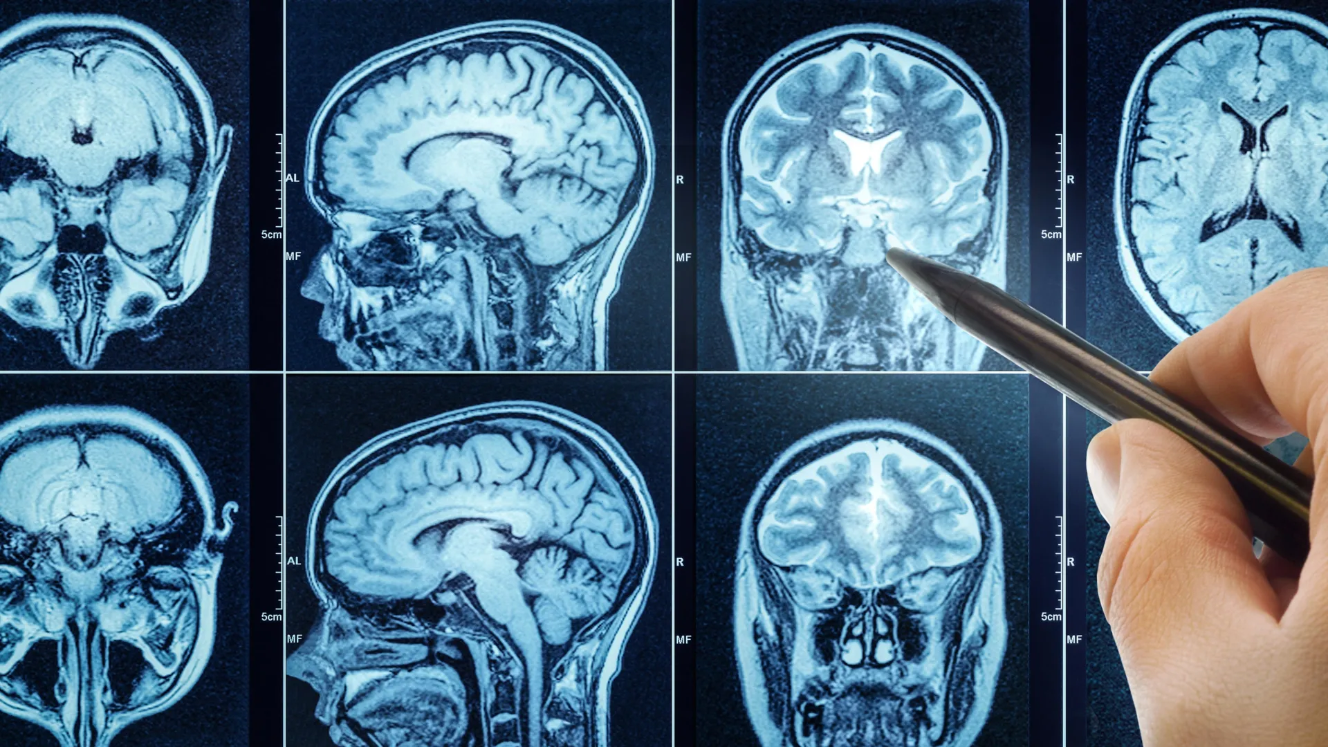
Two areas of the brain may work in combination to tell the brain when it’s “feeling” tired. People with depression and post-traumatic stress disorder (PTSD) often experience cognitive fatigue. Results of the study may provide a way for physicians to better evaluate and treat people who experience such fatigue.In experiments with healthy volunteers undergoing functional MRI imaging, scientists have found increased activity in two areas of the brain that work together to react to, and possibly regulate, the brain when it’s “feeling” tired and either quits or continues exerting mental effort.The experiments, designed to help detect various aspects of brain fatigue, may provide a way for physicians to better evaluate and treat people who experience overwhelming mental exhaustion, including those with depression and post-traumatic stress disorder (PTSD), the scientists say.
A report on the NIH-funded study was published online June 11 in the Journal of Neuroscience, detailing results on 18 female and 10 male healthy adult volunteers given tasks to exercise their memory.
“Our lab focuses on how [our minds] generate value for effort,” says Vikram Chib, Ph.D., associate professor of biomedical engineering at the Johns Hopkins University School of Medicine and a research scientist at Kennedy Krieger Institute. “We understand less about the biology of cognitive tasks, including memory and recall, than we do about physical tasks, even though both involve a lot of effort.” Anecdotally, Chib says, scientists know cognitive tasks are tiring, and relatively less about why and how such fatigue develops and plays out in the brain.
The 28 study participants, who ranged in age from 21 to 29, were paid $50 to participate in the study, and were told they could receive additional payments based on their performance and choices. All participants received a baseline MRI scan before the experiments began.
The tests of their working memory, which took place while undergoing subsequent MRI scans of their brains, included looking at a series of letters, in sequence, on a screen and recalling the position of certain letters. The farther back a letter was in the series of letters, the harder it was to recall its position, increasing the cognitive effort expended. The participants were given feedback on their performance after each test and opportunities to receive increasing payments ($1-$8) with more difficult recall exercises. The participants also were asked before and after each test to self-rate their level of cognitive fatigue.

Overall, the test results found increased activity and connectivity in two brain areas when participants reported cognitive fatigue: the right insula, an area deep in the brain that has been associated with feelings of fatigue, and the dorsal lateral prefrontal cortex, areas on both sides of the brain that control working memory. For each participant, activity in both brain locations during cognitive fatigue increased by more than twice the level of baseline measurements taken before starting the tests.
“Our study was designed to induce cognitive fatigue and see how people’s choices to exert effort change when they feel fatigue, as well as identify locations in the brain where these decisions are made,” says Chib.
Notably, Chib and his research team members Grace Steward and Vivian Looi found that the financial incentives need to be high in order for participants to exert increased cognitive effort, suggesting that external incentives prompt such effort.
“That outcome wasn’t entirely surprising, given our previous work finding the same need for incentives in spurring physical effort,” says Chib.
“The two areas of the brain may be working together to decide to avoid more cognitive effort unless there are more incentives offered. However, there may be a discrepancy between perceptions in cognitive fatigue and what the human brain is actually capable of doing,” says Chib.
Fatigue is linked with many neurological conditions, including PTSD and depression, says Chib. “Now that we’ve likely identified some of the neural circuits for cognitive effort in healthy people, we need to look at how fatigue manifests in the brains of people with these conditions,” he adds.

Chib says it may be possible to use medication or cognitive behavior therapy to combat cognitive fatigue, and the current study using decision tasks and functional MRI could be a framework for objectively classifying cognitive fatigue.
Functional MRI uses blood flow to measure broad areas of activity in the brain; however, it does not directly measure neuron activation, nor more subtle nuances in brain activity.
“This study was performed in an MRI scanner and with very specific cognitive tasks. It will be important to see how these results generalize to other cognitive effort and real-world tasks,” says Chib.
Funding for the research was provided by the National Institutes of Health (R01HD097619, R01MH119086).

Read more →

New research shows Monday stress is etched into your biology

A research study led by Professor Tarani Chandola from the Department of Sociology, Faculty of Social Sciences at the University of Hong Kong (HKU) has revealed that Mondays uniquely drive long-term biological stress, regardless of working status, with implications for heart health.
The research has identified a striking biological phenomenon: older adults who feel anxious on Mondays exhibit significantly higher long-term stress hormone levels, up to two months later. This “Anxious Monday” effect, observed in both working and retirees, points to a deep-rooted link between the start of the week and dysregulation of the body’s stress response system, a known driver of cardiovascular disease (CVD). Published in the Journal of Affective Disorders, the study analyzed data from over 3,500 older adults participating in the English Longitudinal Study of Ageing (ELSA).
Key Findings 23% Higher Cortisol Levels: Older adults reporting Monday anxiety had 23% elevated cortisol levels in hair samples (reflecting cumulative exposure over two months) compared to peers anxious on other days. Non-Workers Not Spared: The effect persisted among retirees, challenging assumptions that workplace stress alone explains Monday’s toll. CVD Connection: Mondays are linked to a 19% spike in heart attacks — this study identifies HPA-axis dysregulation as a potential biological bridge. Not just higher levels of Monday anxiety: Only 25% of the Monday effect was due to greater feelings of anxiety on Mondays. The rest was because of the greater effect of feeling anxious on Mondays compared to other days.The hypothalamic-pituitary-adrenal (HPA) axis regulates stress hormones like cortisol, which, when chronically elevated, contribute to hypertension, insulin resistance, and immune dysfunction. While prior research noted higher cortisol on weekdays versus weekends, this is the first study to pinpoint Mondays as uniquely disruptive. The findings suggest societal rhythms — not just job demands — embed themselves in human physiology, with lasting health risks.
“Mondays act as a cultural ‘stress amplifier,'” said Professor Chandola. “For some older adults, the week’s transition triggers a biological cascade that lingers for months. This isn’t about work — it’s about how deeply ingrained Mondays are in our stress physiology, even after careers end.
The study underscores how the “Monday blues” can become biologically embedded, with chronic stress hormone dysregulation posing long-term cardiovascular risks. Addressing Monday-specific stress could unlock new strategies to combat heart disease in aging populations.

Read more →

Cough medicine turned brain protector? Ambroxol may slow Parkinson’s dementia

Dementia poses a major health challenge with no safe, affordable treatments to slow its progression.
Researchers at Lawson Research Institute (Lawson), the research arm of St. Joseph’s Health Care London, are investigating whether Ambroxol — a cough medicine used safely for decades in Europe — can slow dementia in people with Parkinson’s disease.
Published on June 30 in the prestigious JAMA Neurology, this 12-month clinical trial involving 55 participants with Parkinson’s disease dementia (PDD) monitored memory, psychiatric symptoms and GFAP, a blood marker linked to brain damage. Parkinson’s disease dementia causes memory loss, confusion, hallucinations and mood changes. About half of those diagnosed with Parkinson’s develop dementia within 10 years, profoundly affecting patients, families and the health care system.
Led by Cognitive Neurologist Dr. Stephen Pasternak, the study gave one group daily Ambroxol while the other group received a placebo. “Our goal was to change the course of Parkinson’s dementia,” says Pasternak. “This early trial offers hope and provides a strong foundation for larger studies.”
Key findings from the clinical trial include: Ambroxol was safe, well-tolerated and reached therapeutic levels in the brain Psychiatric symptoms worsened in the placebo group but remained stable in those taking Ambroxol. Participants with high-risk GBA1 gene variants showed improved cognitive performance on Ambroxol A marker of brain cell damage (GFAP) increased in the placebo group but stayed stable with Ambroxol, suggesting potential brain protection.Although Ambroxol is approved in Europe for treating respiratory conditions and has a long-standing safety record — including use at high doses and during pregnancy — it is not approved for any use in Canada or the U.S.
“Current therapies for Parkinson’s disease and dementia address symptoms but do not stop the underlying disease,” explains Pasternak. “These findings suggest Ambroxol may protect brain function, especially in those genetically at risk. It offers a promising new treatment avenue where few currently exist.”
Ambroxol supports a key enzyme called glucocerebrosidase (GCase), which is produced by the GBA1 gene. In people with Parkinson’s disease, GCase levels are often low. When this enzyme doesn’t work properly, waste builds up in brain cells, leading to damage. Pasternak learned about Ambroxol during a fellowship at The Hospital for Sick Children (SickKids) in Toronto, where it was identified as a treatment for Gaucher disease — a rare genetic disorder in children caused by a deficiency of GCase.
He is now applying that research to explore whether boosting GCase with Ambroxol could help protect the brain in Parkinson’s-related diseases. “This research is vital because Parkinson’s dementia profoundly affects patients and families,” says Pasternak. “If a drug like Ambroxol can help, it could offer real hope and improve lives.”
Funded by the Weston Foundation, this study is an important step toward developing new treatments for Parkinson’s disease and other cognitive disorders, including dementia with Lewy bodies. Pasternak and his team plan to start a follow-up clinical trial focused specifically on cognition later this year.

Read more →

Multisensory VR forest reboots your brain and lifts mood—study confirms

In Japan, Shinrin Yoku or forest bathing has already been used for therapeutic applications, for instance, to lower blood pressure and stress levels. For their study, the researchers wanted to find out whether forest bathing – consciously immersing oneself in nature – can also be effective when done virtually, and focused on whether the positive effect is stronger when several senses are addressed simultaneously.
For the project, a high-quality 360° VR video was produced in Europe’s largest Douglas fir forest, the Sonnenberg nature reserve near Parchim – complete with original sounds and the scent of essential oils from the Douglas fir. The participants experienced the virtual forest scenery either as a full sensory experience (with images, sound and scent) or in a reduced form whereby forest stimuli appealing to just a single sense – visual, auditory or olfactory – were used. In variants where only hearing or scent was activated, participants were placed in a neutral virtual environment to minimize visual stimuli and the influence of VR technology.
Significantly better effect with sensory combination
More than 130 participants were first put into an acute stress situation using stress-inducing images. Then, equipped with VR glasses, they experienced one of the four forest stimulation/ bathing variants. The results show that the combination of all three sensory stimuli led to a significantly greater improvement in mood and a stronger feeling of connection with nature compared to when individual sensory stimuli were presented. In addition to positive effects on mood, there were also limited improvements in working memory – the cognitive function that enables us to store, process and retrieve information in the short term.
However, the researchers point out that the effects are area-specific and cannot yet be considered universally valid. Further studies with larger samples are needed to confirm the results and provide a better understanding of the mechanisms behind the restorative effects of virtual nature experiences.
“We can already say that digital nature experiences can absolutely produce an emotional effect – even if they don’t replace actual nature,” reports Leonie Ascone, lead author of the study and researcher in the Neuronal Plasticity working group at the University Medical Center Hamburg-Eppendorf (UKE).
Potential for clinics, waiting rooms and urban spaces
Simone Kühn, head of the study and Director of the Center for Environmental Neuroscience at the Max Planck Institute for Human Development, adds: “Especially in places with limited access to nature – such as clinics, waiting areas or urban interiors – multisensory VR applications or targeted nature staging could support mental well-being. The images, sounds and scents of nature offer previously underestimated potential for improving mood and mental performance in everyday situations.” Kühn conducts intensive research into the effects of the environment on the human brain and, together with colleagues from universities in Vienna, Exeter and Birmingham, was recently able to prove that just from watching nature videos, patients perceive physical pain as less intense (Steininger et al., 2025).
In brief: Forest bathing in Virtual Reality improves emotional well-being and increases connectedness to nature, particularly when several senses (sight, hearing, smell) are simultaneously engaged The study used a 360° VR forest video complete with original sounds and the scent of Douglas fir essential oils There is potential for application especially in clinical, urban and other environments with limited access to nature

Read more →

Pregnancy’s 100-million-year secret: Inside the placenta’s evolutionary power play

An international research team led by scientists from the University of Vienna has uncovered new insights into how specialized cell types and communication networks at the interface between mother and fetus evolved over millions of years. These discoveries shed light on one of nature’s most remarkable innovations – the ability to sustain a successful pregnancy. The findings have just been published in Nature Ecology & Evolution.
Pregnancy that lasts long enough to support full fetal development is a hallmark evolutionary breakthrough of placental mammals – a group that includes humans. At the center of this is the fetal-maternal interface: the site in the womb where a baby’s placenta meets the mother’s uterus, and where two genetically distinct organisms – mother and fetus – are in intimate contact and constant interaction. This interface has to strike a delicate balance: intimate enough to exchange nutrients and signals, but protected enough to prevent the maternal immune system from rejecting the genetically “foreign” fetus.
To uncover the origins and mechanisms behind this intricate structure, the team analyzed single-cell transcriptomes – snapshots of active genes in individual cells – from six mammalian species representing key branches of the mammalian evolutionary tree. These included mice and guinea pigs (rodents), macaques and humans (primates), and two more unusual mammals: the tenrec (an early placental mammal) and the opossum (a marsupial that split off from placental mammals before they evolved complex placentas).
A Cellular “Atlas of Mammal Pregnancy”
By analyzing cells at the fetal-maternal interface, the researchers were able to trace the evolutionary origin and diversification of the key cell types involved. Their focus was on two main players: placenta cells, which originate from the fetus and invade maternal tissue, and uterine stromal cells, which are of maternal origin and respond to this invasion.
Using molecular biology tools, the team identified distinct genetic signatures – patterns of gene activity unique to specific cell types and their specialized functions. Notably, they discovered a genetic signature associated with the invasive behavior of fetal placenta cells that has been conserved in mammals for over 100 million years. This finding challenges the traditional view that invasive placenta cells are unique to humans, and reveals instead that they are a deeply conserved feature of mammalian evolution. During this time, the maternal cells weren’t static, either. Placental mammals, but not marsupials, were found to have acquired new forms of hormone production, a pivotal step toward prolonged pregnancies and complex gestation, and a sign that the fetus and the mother could be driving each other’s evolution.
Cellular Dialogue: Between Cooperation and Conflict
To better understand how the fetal-maternal interface functions, the study tested two influential theories about the evolution of cellular communication between mother and fetus.

The first, the “Disambiguation Hypothesis,” predicts that over evolutionary time, hormonal signals became clearly assigned to either the fetus or the mother – a possible safeguard to ensure clarity and prevent manipulation. The results confirmed this idea: certain signals, including WNT proteins, immune modulators, and steroid hormones, could be clearly traced back to one source tissue.
The second, the “Escalation Hypothesis” (or “genomic Conflict”), suggests an evolutionary arms race between maternal and fetal genes – with, for example, the fetus boosting growth signals while the maternal side tries to dampen them. This pattern was observed in a small number of genes, notably IGF2, which regulates growth. On the whole, evidence pointed to fine-tuned cooperative signaling.
“These findings suggest that evolution may have favored more coordination between mother and fetus than previously assumed,” says Daniel J. Stadtmauer, lead author of the study and now a researcher at the Department of Evolutionary Biology, University of Vienna. “The so-called mother-fetus power struggle appears to be limited to specific genetic regions. Rather than asking whether pregnancy as a whole is conflict or cooperation, a more useful question may be: where is the conflict?”
Single-Cell Analysis: A Key to Evolutionary Discovery
The team’s discoveries were made possible by combining two powerful tools: single-cell transcriptomics – which captures the activity of genes in individual cells – and evolutionary modeling techniques that help scientists reconstruct how traits might have looked in long-extinct ancestors. By applying these methods to cell types and their gene activity, the researchers could simulate how cells communicate in different species, and even glimpse how this dialogue has evolved over millions of years.
“Our approach opens a new window into the evolution of complex biological systems – from individual cells to entire tissues,” says Silvia Basanta, co-first author and researcher at the University of Vienna. The study not only sheds light on how pregnancy evolved, but also offers a new framework for tracking evolutionary innovations at the cellular level – insights that could one day improve how we understand, diagnose, or treat pregnancy-related complications.
The research was conducted in the labs of Mihaela Pavličev at the Department of Evolutionary Biology, University of Vienna, and Günter Wagner at Yale University. Wagner is Professor Emeritus at Yale and a Senior Research Fellow at the University of Vienna. The study was supported by the John Templeton Foundation and the Austrian Science Fund (FWF).

Read more →

North Carolina Braces For Medicaid Cuts Because of Trump’s Bill

President Trump’s domestic policy law jeopardizes plans to reopen one rural county’s hospital — and health coverage for hundreds of thousands of state residents.The only hospital in Martin County, N.C., closed in 2023, but the electricity is still on inside. Air conditioning continues to keep its empty patient rooms cool. And the county still pays the bills for the building’s medical gas system.That is because the people of Martin County, in rural eastern North Carolina, have been determined to keep the beige brick building from deteriorating — and to somehow reopen their hospital, which had been struggling financially for years.When North Carolina expanded Medicaid later in 2023, after the hospital shuttered, offering government health insurance to the state’s low-income adults, Martin County saw an opportunity. Plans materialized to partly reopen the hospital, largely because federal dollars were pouring into the state to cover patients’ care under Medicaid.But those plans are now in jeopardy, as is Medicaid coverage for hundreds of thousands of North Carolina residents, after Congress passed President Trump’s sweeping domestic policy bill. To help pay for tax cuts, the bill slashes federal spending on Medicaid, leaving states that expanded the program under Obamacare in a particularly difficult spot.If Medicaid expansion is eliminated in North Carolina, Martin General Hospital almost surely will not reopen — “a catastrophic and deadly consequence,” said Paul Roberson, a real estate agent and community leader in Williamston, where a sign in front of the hospital reads, “CLOSED. If you need immediate assistance, dial 911.”“Not having the hospital here is costing lives,” Mr. Roberson said, noting that the nearest hospital was about a 30-minute drive away. “This is the most important thing for us.”We are having trouble retrieving the article content.Please enable JavaScript in your browser settings.Thank you for your patience while we verify access. If you are in Reader mode please exit and log into your Times account, or subscribe for all of The Times.Thank you for your patience while we verify access.Already a subscriber? Log in.Want all of The Times? Subscribe.

Read more →

New tech tracks blood sodium without a single needle

In a new study, researchers demonstrated long-term, non-invasive monitoring of blood sodium levels using a system that combines optoacoustic detection with terahertz spectroscopy. Accurate measurement of blood sodium is essential for diagnosing and managing conditions such as dehydration, kidney disease and certain neurological and endocrine disorders.
Terahertz radiation, which falls between microwaves and the mid-infrared region of the electromagnetic spectrum, is ideal for biological applications because it is low-energy and non-harmful to tissues, scatters less than near-infrared and visible light and is sensitive to structural and functional biological changes.
“For biomedical applications, terahertz spectroscopy still faces two key challenges: detecting molecules other than water in complex biological samples and penetrating thick tissue layers to enable detection inside the body,” said research team leader Zhen Tian from Tianjin University in China. “By adding optoacoustic detection, we were able to overcome these challenges and demonstrate the first in vivo detection of ions using terahertz waves. This is an important step toward making terahertz-based techniques practical for clinical use.”
In Optica, Optica Publishing Group’s flagship journal for high-impact research, the researchers describe their new multispectral terahertz optoacoustic system and show that it can be used for noninvasive, long-term monitoring of sodium concentration in live mice without the need for any labels. Preliminary tests performed with human volunteers were also promising.
“With further development, this technology could be used to monitor sodium levels in patients without the need for blood draws,” said Tian. “The real-time sodium measurements could be used to safely correct imbalances in critical patients while avoiding dangerous neurological complications that can occur when sodium levels rapidly change.”
Using sound to cut the noise
The new work is part of a larger project aimed at advancing and implementing terahertz technology in the biomedical field using terahertz optoacoustic techniques. One key aim of the project is to reduce signal interference caused by water, which strongly absorbs terahertz radiation.

To overcome this interference, the researchers developed a modular system that irradiates the sample with terahertz waves. As the sample absorbs these waves, it vibrates the sodium ions connected to water molecules in the blood, creating ultrasound waves that are detected with an ultrasonic transducer. This technique, known as optoacoustic detection, converts the absorbed terahertz energy into sound waves for measurement.
“Terahertz optoacoustic technology represents a groundbreaking advancement for biomedical applications by effectively overcoming the water absorption barrier that has historically limited these applications,” said Tian. “The broader significance of this work extends far beyond blood sodium detection. This technology has the capability to identify various biomolecules — including sugars, proteins, and enzymes — by recognizing their unique terahertz absorption signatures.”
Tracking sodium without needles
To test their new system, the researchers showed that it could measure increases in blood sodium levels in blood vessels under the skin of living mice on the millisecond timescale for over 30 minutes. These measurements were taken from the ear, with the skin surface cooled to 8 °C to dampen the background optoacoustic signal from water.
The researchers also demonstrated that the terahertz optoacoustic system could quickly distinguish between high and low sodium levels in human blood samples. Finally, they noninvasively measured sodium ion levels in the blood vessels of the hands of healthy volunteers. They found that the detected optoacoustic signal from sodium was proportional to the amount of blood flow under the skin surface, even though measurements were collected without any skin cooling. While more work is needed, these results suggest that the system could be useful for non-invasive real-time monitoring.
The researchers say that adapting the system for human use will require identifying suitable detection sites on the human body — such as inside of the mouth — that can tolerate rapid cooling and allow strong signal detection with minimal water background noise. They are also exploring alternative signal processing methods that might make it possible to suppress water interference without the need for cooling, making the approach more practical for clinical diagnostics.

Read more →

Scientists reverse Parkinson’s symptoms in mice — Could humans be next?

Groundbreaking research by the University of Sydney has identified a new brain protein involved in the development of Parkinson’s disease and a way to modify it, paving the way for future treatments for the disease.
Parkinson’s disease is the second most common neurological condition after dementia, with over 150,000 people in Australia living with the condition.
The research team, led by Professor Kay Double from the Brain and Mind Centre, has spent more than a decade studying the biological mechanisms underpinning the condition, with the aim of finding new treatments to slow or stop its progression.
In 2017, the team published a paper in Acta Neuropathologica, identifying for the first time the presence of an abnormal form of a protein – called SOD1 – in the brains of patients diagnosed with Parkinson’s disease.
Normally, the SOD1 protein provides protective benefits to the brain but, in Parkinson’s patients, it becomes faulty, causing the protein to clump and damage brain cells.
The newest study by the same University of Sydney team, published in Acta Neuropathologica Communications,builds on this research. It found that targeting the faulty SOD1 protein with a drug treatment improved the motor function in mice bred to have Parkinson-like symptoms.
Professor Double said: “All the mice we treated saw a dramatic improvement in their motor skills which is a really promising sign it could be effective in treating people who have Parkinson disease too.

“We hoped that by treating this malfunctioning protein, we might be able to improve the Parkinson-like symptoms in the mice we were treating – but even we were astonished by the success of the intervention.”
Methodology
The study involved two groups of mice bred to have Parkinson-like symptoms. One group of mice was treated with a special copper supplement for three months, while the other received a placebo.
Throughout the study, the mice receiving the placebo saw a decline in their motor symptoms. The mice receiving the special copper supplement, however, did not develop movement problems.
Professor Double said: “The results were beyond our expectations and suggest, once further studies are carried out, this treatment approach could slow the progression of Parkinson’s disease in humans.”
What is Parkinson’s disease?
Parkinson’s disease is a degenerative neurological disorder in which dopamine-producing cells in the brain die, leading to a range of symptoms including tremors, muscle stiffness, slow movement and impaired balance.

At present there is no known cure and only limited treatments, although researchers hope understanding the causes of the disease will lead to improved treatments.
Professor Double said: “As our understanding of Parkinson’s disease grows, we are finding that there are many factors contributing to its development and progression in humans – and faulty forms of the SOD1 protein is likely one of them.
“Just as researchers found with HIV, Parkinson’s disease is a complex condition that likely requires multiple interventions. A single treatment may have a small effect when used alone but, when combined with other interventions, contributes to a significant overall improvement in health.”
The researchers’ next step is to identify the best approach to targeting the faulty SOD1 protein in a clinical trial, which could be the start of a new therapy to slow the development of Parkinson’s disease.

Read more →

Tiny twitches, big breakthrough: New clues to catch Parkinson’s sooner

Midbrain dopamine (DA) neurons, located in the substantia nigra pars compacta (SNc) and the ventral tegmental area (VTA), are essential for regulating movement, emotion, and reward processing. Dysfunction of DA neurons in the SNc has been linked to the Parkinson’s disease (PD). Previous research mostly focuses on the functions of DA neurons in mood regulation and reward mechanisms. Their role in more subtle and spontaneous behaviors remains poorly understood.
In a study published in Translational Psychiatry, a team led by Prof. Xuemei Liu and Prof. Pengfei Wei from the Shenzhen Institutes of Advanced Technology of the Chinese Academy of Sciences discovered that behaviors such as rearing, walking, and hunching are associated with the loss of SNc DA neurons, but not VTA DA neurons. These subtle features serve as key behavioral biomarkers of SNc DA neuron loss, enhancing the translational value of PD models.
Using a machine learning-enhanced three-dimensional spontaneous behavior analysis system, researchers examined the detailed motor behaviors in two mouse models of dopamine neuron depletion: a subacute MPTP-induced PD model and an AAV-mediated DA neuron loss model. This approach enabled the capture of detailed and nuanced behavioral features that traditional two-dimensional methods may overlook.
Researchers observed reductions in rearing and hunching behavior in the PD model, which correlates directly with the loss of DA neurons in the SNc but not the VTA. Although overall motor deficits were not significant, notable behavioral lateralization was revealed in the PD mice.
Moreover, using the AAV-induced ablation model, researchers confirmed that climbing, a behavior similar to rearing, was also strongly correlated with the loss of DA neurons in the SNc, and the damage to SNc neurons significantly reduced motor function and influenced the lateralization of movement.
These findings highlight the significance of rearing behavior and behavioral lateralization as potential behavioral markers for tracking the progression of PD. “Connecting behavioral changes with targeted neural damage advances the understanding of PD progression and offers valuable insights into improving treatment strategies,” said Prof. Liu.

Read more →