Your thoughts can harm your neck and back during lifting tasks

The mental distress of cognitive dissonance — encountering information that conflicts with how we act or what we believe — can lead to added pressure on the neck and low back during lifting and lowering tasks, new research suggests.
When study participants were told they were performing poorly in a precision lowering experiment in the lab, after initially being told they were doing well, their movements were linked to increased loads on vertebrae in their neck and low back.
Results showed that the higher the cognitive dissonance score, the greater the extent of loading on the upper and lower parts of the spine.
The finding suggests cognitive dissonance may be a previously unidentified risk factor for neck and low back pain, which could have implications for risk prevention in the workplace, according to researchers.
“This increased spine loading occurred under just one condition with a fairly light load — you can imagine what this would be like with more complex tasks or higher loads,” said senior author William Marras, executive director of the Spine Research Institute at The Ohio State University. “Basically, the study scratched the surface of showing there’s something to this.”
The research was published recently in the journal Ergonomics.

Marras’ lab has been studying daily living and occupational forces on the spine for decades. About 20 years ago, he found that psychological stress could influence spine biomechanics, using a study design that involved having a fake argument with a graduate student in front of research participants.
“We found that in certain personality types, the loads in the spine increased by up to 35%,” Marras said. “We ended up finding that when you’re under that kind of psychosocial stress, what you tend to do is what we call co-activate muscles in your torso. It creates this tug of war in the muscles because you’re always tense.
“In this study, to get at that mind-body connection, we decided to look at the way people think and, with cognitive dissonance, when people are disturbed by their thoughts.”
Seventeen research participants — nine men and eight women aged 19-44 — completed three phases of an experiment in which they placed a light-weight box within a square on a surface that was moved left and right, up and down. After a short practice run, researchers gave almost exclusively positive feedback during the first of two 45-minute trial blocks. During the second, the feedback increasingly suggested participants were performing in an unsatisfactory way.
To arrive at a cognitive dissonance score for each participant, changes during the experiment to blood pressure and heart rate variability were combined with responses to two questionnaires assessing discomfort levels as well as positive and negative affect — feeling strong and inspired versus distressed and ashamed.

Wearable sensors and motion-capture technology were used to detect peak spinal loads in the neck and low back: both compression of vertebrae and vertebral movement, or shear, from side to side (lateral) and forward and back (A/P).
Statistical modeling showed that, on average, peak spinal loads on cervical vertebrae in the neck were 11.1% higher in compression, 9.4% higher in A/P shear and 19.3% higher in lateral shear during the negative-feedback trial block compared to the baseline measures from the practice run. Peak loading in the lumbar region of the low back — an area that bears the brunt of any spinal loading — increased by 1.7% in compression and 2.2% in shear during the final trial block.
“Part of the motivation here was to see whether cognitive dissonance can manifest itself not only in the low back — we thought we’d find it there, but we didn’t know what we’d find in the neck. We did find a pretty strong response in the neck,” said Marras, a professor of integrated systems engineering with College of Medicine academic appointments in neurosurgery, orthopaedics and physical medicine and rehabilitation.
“Our tolerance to shear is much, much lower than it is to compression, so that’s why that’s important,” he said. “A small percentage of load is no big deal for one time. But think about when you’re working day in and day out, and you’re in a job where you’re doing this 40 hours a week — that could be significant, and be the difference between a disorder and not having a disorder.”
Marras is also principal investigator on a federally funded multi-institution clinical trial assessing different treatments for low back pain that range from medication to exercise to cognitive behavioral therapy.
“We’re trying to unravel this onion and understand all the different things that affect spine disorders because it’s really, really complex,” he said. “Just like the whole system has got to be right for a car to run correctly, we’re learning that that’s the way it is with the spine. You could be in physically great shape, but if you’re not thinking correctly or appropriately, or you have all these mental irregularities, like cognitive dissonance, that will affect the system. And until you get that right, you’re not going to be right.
“We’re looking for causal pathways. And now we can say cognitive dissonance plays a role and here’s how it works.”
This research was supported by internal Spine Research Institute funds. Co-authors included first author Eric Weston, a former integrated systems engineering graduate student at Ohio State; Afton Hassett of the University of Michigan; and Safdar Khan and Tristan Weaver of Ohio State.


Read more →

Running throughout middle age keeps 'old' adult-born neurons 'wired'

A new study provides novel insight into the benefits of exercise, which should motivate adults to keep moving throughout their lifetime, especially during middle age. Long-term exercise profoundly benefits the aging brain and may prevent aging-related memory function decline by increasing the survival and modifying the network of the adult-born neurons born during early adulthood, and thereby facilitating their participation in cognitive processes.
Aging often is accompanied by cognitive decline. Among the first structures of the brain affected are the hippocampus and adjacent cortices, areas essential for learning and memory. Deficits in cognitive ability are associated with reduced hippocampal volume and degradation of synaptic connectivity between the hippocampus and the (peri)-entorhinal cortex.
Increasing evidence indicates that physical activity can delay or prevent these structural and functional reductions in older adults. A new study by Florida Atlantic University and CINVESTAV, Mexico City, Mexico, provides novel insight into the benefits of exercise, which should motivate adults to keep moving throughout their lifetime, especially during middle age.
For the study, researchers focused on the effects of long-term running on a network of new hippocampal neurons that were generated in young adult mice, at middle age. These “mice on the run” demonstrate that running throughout middle age keeps old adult-born neurons wired, which may prevent or delay aging-related memory loss and neurodegeneration.
Adult-born neurons are thought to contribute to hippocampus-dependent memory function and are believed to be temporarily important, during the so-called ‘critical period’ at about three to six weeks of cell age, when they can fleetingly display increased synaptic plasticity. However, these new neurons do remain present for many months, but it was unclear whether those born in early adulthood remain integrated into neural networks and whether their circuitry is modifiable by physical activity in middle age.
To address these questions, researchers used a unique rabies virus-based circuit tracing approach with a long-time interval between the initial labeling of new neurons and subsequent analysis of their neural circuitry in rodents. More than six months after tagging of the adult-born neurons with a fluorescent reporter vector, they identified and quantified the direct afferent inputs to these adult-born neurons within the hippocampus and (sub)cortical areas, when the mice were middle-aged.

Results of the study, published in the journal eNeuro, show long-term running wires ‘old’ new neurons, born during early adulthood, into a network that is relevant to the maintenance of episodic memory encoding during aging.
“Long-term exercise profoundly benefits the aging brain and may prevent aging-related memory function decline by increasing the survival and modifying the network of the adult-born neurons born during early adulthood, and thereby facilitating their participation in cognitive processes,” said Henriette van Praag, Ph.D., corresponding author, an associate professor of biomedical science in FAU’s Schmidt College of Medicine and a member of the FAU Stiles-Nicholson Brain Institute.
Findings from the study showed long-term running significantly increased the number of adult-born neurons and enhanced the recruitment of presynaptic (sub)-cortical cells to their network.
“Long-term running may enhance pattern separation ability, our ability to distinguish between highly similar events and stimuli, a behavior closely linked to adult neurogenesis, which is among the first to display deficits indicative of age-related memory decline,” said Carmen Vivar, Ph.D., corresponding author, Department of Physiology, Biophysics and Neuroscience, Centro de Investigacion y de Estudios Avanzados del IPN in Mexico.
Aging-related memory function decline is associated with the degradation of synaptic inputs from the perirhinal and entorhinal cortex onto the hippocampus, brain areas that are essential for pattern separation, and contextual and spatial memory.

“We show that running also substantially increases the back-projection from the dorsal subiculum onto old adult-born granule cells,” said van Praag. “This connectivity may provide navigation-associated information and mediate the long-term running-induced improvement in spatial memory function.”
Results from the study show that running not only rescued perirhinal connectivity but also increased and altered the contribution of the entorhinal cortices to the network of old adult-born neurons.
“Our study provides insight as to how chronic exercise, beginning in young adulthood and continuing throughout middle age, helps maintain memory function during aging, emphasizing the relevance of including exercise in our daily lives,” said Vivar.
Study co-authors are Ben Peterson, Ph.D., currently a postdoc at UC Davis; Alejandro Pinto, FAU’s Schmidt College of Medicine and Stiles-Nicholson Brain Institute; and Emma Janke, a recent graduate of the University of Pennsylvania.
This research was supported in part by the FAU Stiles-Nicholson Brain Institute and the Jupiter Life Sciences Initiative (awarded to van Praag), and by the Fondo de Investigación Científica y Desarrollo Tecnológico del Cinvestav (Proyectos SEP-Cinvestav), (awarded to Vivar).

Read more →

Study finds brain connectivity, memory improves in older adults after walking

A new University of Maryland School of Public Health study reveals how walking strengthens connections within and between three of the brain’s networks, including one associated with Alzheimer’s disease, adding to the growing evidence that exercise improves brain health.
Published this month in the Journal for Alzheimer’s Disease Reports, the study examined the brains and story recollection abilities of older adults with normal brain function and those diagnosed with mild cognitive impairment, which is a slight decline in mental abilities like memory, reasoning and judgment and a risk factor for Alzheimer’s.
“Historically, the brain networks we studied in this research show deterioration over time in people with mild cognitive impairment and Alzheimer’s disease,” said J. Carson Smith, a kinesiology professor with the School of Public Health and principal investigator of the study. “They become disconnected, and as a result, people lose their ability to think clearly and remember things. We’re demonstrating that exercise training strengthens these connections.”
The study builds upon Smith’s previous research, which showed how walking may decrease cerebral blood flow and improve brain function in older adults with mild cognitive impairment.
Thirty-three participants, who ranged between 71 and 85 years old, walked while supervised on a treadmill four days a week for 12 weeks. Before and after this exercise regimen, researchers asked participants to read a short story and then repeat it out loud with as many details as possible.
Participants also underwent functional magnetic resonance imaging (fMRI) so researchers could measure changes in communication within and between the three brain networks that control cognitive function: Default mode network – Activates when a person isn’t doing a specific task (think daydreaming about the grocery list) and is connected to the hippocampus — one of the first brain regions affected by Alzheimer’s disease. It’s also where Alzheimer’s and amyloid plaques, a prime suspect for Alzheimer’s disease found around nerve cells, show up in tests. Frontoparietal network — Regulates decisions made when a person is completing a task. It also involves memory. Salience network — Monitors the external world and stimuli and then decides what deserves attention. It also facilitates switching between networks to optimize performance.After 12 weeks of exercise, researchers repeated the tests and saw significant improvements in participants’ story recall abilities.
“The brain activity was stronger and more synchronized, demonstrating exercise actually can induce the brain’s ability to change and adapt,” Smith said. “These results provide even more hope that exercise may be useful as a way to prevent or help stabilize people with mild cognitive impairment and maybe, over the long term, delay their conversion to Alzheimer’s dementia.”
Researchers also observed stronger activity within the default mode network, within the salience network and in the connections between the three networks.

Read more →

Ultrasound Pulses to Brain Send Mice Into a Hibernation-Like State

Experiments offer an intriguing hint at technology that could induce torpor in humans in the future.For many animals, life is a cycle of scarcity as well as plenty. Hibernating creatures curl up underground in winter, slowing their metabolism so they can make it to spring without food. Even laboratory mice, if deprived of food, can enter a state called torpor, a kind of standby mode that economizes energy.It’s something humans have long fantasized about for ourselves: If we ever leave this planet and travel through space, we will experience our own time of scarcity. Science fiction writers tend to imagine a mysterious technology that keeps humans in stasis, able to survive centuries of silence before emerging into a new life. For now, it’s a technology that’s out of reach.But as scientists work to understand states like torpor and hibernation, tantalizing details about how the brain controls metabolism have emerged. Researchers reported in the journal Nature Metabolism on Thursday that they’ve been able to send mice into a torpor-like state by targeting a specific part of the brain with short bursts of ultrasound. It’s unclear exactly why ultrasound has this effect, but the findings suggest that studying the neural circuits involved in torpor could reveal ways to manipulate metabolism beyond the lab.Ultrasound devices, which generate high-frequency sound waves, are best known for their imaging powers. But they have also been used by neuroscientists to stimulate neurons. Correctly tuned, the soundwaves can travel deep into the brain, said Hong Chen, a professor of biomedical engineering at Washington University in St. Louis and an author of the new paper. In 2014, William Tyler, now at the University of Alabama, Birmingham, and his colleagues applied ultrasound to a sensory region in the brain and found that it enhanced a subject’s sense of touch. A growing body of work is exploring ultrasound as a treatment for disorders like depression and anxiety.Curious about a brain region that regulates body temperature in rodents, Dr. Chen and her colleagues constructed tiny ultrasound mouse caps. The devices trained six bursts, each consisting of 10 seconds of ultrasound, on the selected area of the rodent’s brain (Researchers who study the brain with ultrasound must tune their devices carefully to avoid heat that can damage tissues).The mice, the researchers noticed, stopped moving. Measurements of their body temperature, heart rate and metabolism showed a pronounced dip. The mice stayed in this state for about an hour after the ultrasound bursts, and then returned to normal.Looking closer at neurons involved in this response, the researchers identified a protein in their brain membranes, TRPM2, that appears to be sensitive to ultrasound; when the researchers reduced levels of the protein in mice, the mice became resistant to ultrasound’s effects.That’s an important step toward understanding how ultrasound affects neurons, said Davide Folloni, a researcher at the Icahn School of Medicine at Mount Sinai in New York City, who studies the brain using ultrasound; the details have largely been elusive.But it’s also possible that heat generated by the ultrasound, and not just the ultrasound itself, is affecting TRPM2 in the brains of mice, a point that was raised by Masashi Yanagisawa and Takeshi Sakurai of the University of Tsukuba in Japan, in separate interviews. The two have studied neurons in this brain area, and their connection to states of torpor. Both may be in play, Dr. Chen said.In one of the most tantalizing parts of the study, the researchers checked to see whether animals that don’t typically experience torpor — rats — behaved differently when the brain region was stimulated with ultrasound. Indeed, they seemed to slow down, and their body temperatures dropped.“We have to be careful with the rat data,” Dr. Chen cautions. So far, they only have information about temperature, not metabolic rate and other factors.Could ultrasound be a way to change the metabolism of larger animals with no history of torpor, like humans? It’s an intriguing idea, Dr. Sakurai said.“At this stage,” he said, “it remains an unanswered question.”

Read more →

New superbug-killing antibiotic discovered using AI

Published25 minutes agoShareclose panelShare pageCopy linkAbout sharingImage source, McMaster UniversityBy James GallagherHealth and science correspondentScientists have used artificial intelligence (AI) to discover a new antibiotic that can kill a deadly species of superbug.The AI helped narrow down thousands of potential chemicals to a handful that could be tested in the laboratory.The result was a potent, experimental antibiotic called abaucin, which will need further tests before being used.The researchers in Canada and the US say AI has the power to massively accelerate the discovery of new drugs.It is the latest example of how the tools of artificial intelligence can be a revolutionary force in science and medicine. AI breakthrough could spark medical revolutionStopping the superbugsAntibiotics kill bacteria. However, there has been a lack of new drugs for decades and bacteria are becoming harder to treat, as they evolve resistance to the ones we have. More than a million people a year are estimated to die from infections that resist treatment with antibiotics. The researchers focused on one of the most problematic species of bacteria – Acinetobacter baumannii, which can infect wounds and cause pneumonia.You may not have heard of it, but it is one of the three superbugs the World Health Organization has identified as a “critical” threat. It is often able to shrug off multiple antibiotics and is a problem in hospitals and care homes, where it can survive on surfaces and medical equipment.Dr Jonathan Stokes, from McMaster University, describes the bug as “public enemy number one” as it’s “really common” to find cases where it is “resistant to nearly every antibiotic”.Image source, McMaster University Artificial intelligenceTo find a new antibiotic, the researchers first had to train the AI. They took thousands of drugs where the precise chemical structure was known, and manually tested them on Acinetobacter baumannii to see which could slow it down or kill it. This information was fed into the AI so it could learn the chemical features of drugs that could attack the problematic bacterium. The AI was then unleashed on a list of 6,680 compounds whose effectiveness was unknown. The results – published in Nature Chemical Biology – showed it took the AI an hour and a half to produce a shortlist. The researchers tested 240 in the laboratory, and found nine potential antibiotics. One of them was the incredibly potent antibiotic abaucin.Laboratory experiments showed it could treat infected wounds in mice and was able to kill A. baumannii samples from patients. However, Dr Stokes told me: “This is when the work starts.” The next step is to perfect the drug in the laboratory and then perform clinical trials. He expects the first AI antibiotics could take until 2030 until they are available to be prescribed. Curiously, this experimental antibiotic had no effect on other species of bacteria, and works only on A. baumannii. Many antibiotics kill bacteria indiscriminately. The researchers believe the precision of abaucin will make it harder for drug-resistance to emerge, and could lead to fewer side-effects. Image source, Getty ImagesIn principle, the AI could screen tens of millions of potential compounds – something that would be impractical to do manually.”AI enhances the rate, and in a perfect world decreases the cost, with which we can discover these new classes of antibiotic that we desperately need,” Dr Stokes told me.The researchers tested the principles of AI-aided antibiotic discovery in E. coli in 2020, but have now used that knowledge to focus on the big nasties. They plan to look at Staphylococcus aureus and Pseudomonas aeruginosa next.”This finding further supports the premise that AI can significantly accelerate and expand our search for novel antibiotics,” said Prof James Collins, from the Massachusetts Institute of Technology. He added: “I’m excited that this work shows that we can use AI to help combat problematic pathogens such as A. baumannii.”Follow James on Twitter.

Read more →

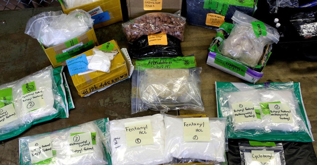
House Passes Bill to Make Penalties Permanent for Fentanyl-Related Drugs

The legislation earned quiet endorsement from the Biden administration and a solid group of Democratic votes, but exposed divisions over how to tackle the country’s most pressing drug crisis.The House of Representatives passed legislation on Thursday that would make permanent harsh criminal penalties and strict controls on fentanyl-related drugs, with scores of Democrats joining nearly all Republicans in a vote that reflected the political challenges of tackling what both parties consider America’s most pressing drug crisis.The bill, approved by a vote of 289 to 133, would permanently list fentanyl-related drugs as Schedule I controlled substances, a designation that mandates severe prison sentences for highly addictive, nonmedicinal chemicals, and which is now set to expire at the end of 2024.The bipartisan vote reflected agreement among Republicans and a solid bloc of Democrats that stiffening penalties for fentanyl-related drugs is a necessary component of the federal response to the crisis. According to the Centers for Disease Control and Prevention, fentanyl-related drugs were the cause of most of the roughly 75,000 synthetic opioid overdose deaths that occurred in 2022.“We should vote to advance this bill that we agree on and that does help stop the bad guys,” Representative Morgan Griffith, Republican of Virginia and an author of the bill, said on the House floor. “Once fentanyl analogues are permanently made Schedule I, Congress can build off this and deal with the illicit crisis.”But there are deep divisions over the ramifications of doing so, making the fate of the legislation unclear in the Democratic-led Senate.Many Democrats, along with public health and civil rights groups, note that harsh sentences for fentanyl-related drugs have driven up incarceration rates and disproportionately affected people of color. They argue that further criminalizing them will only worsen the crisis and have called for a public health response including better public education, more addiction treatment and recovery services, as well as overdose prevention.The White House last week came out in support of the House bill, while urging that Congress consider its other recommendations, including narrower mandatory minimum sentences that would apply only to cases in which the substance could be linked to death or serious bodily injury.But on the House floor on Thursday, Representative Frank Pallone Jr. of New Jersey, the top Democrat on the Energy and Commerce Committee, broadly denounced the G.O.P. bill, calling it “one-sided” and a futile attempt “to incarcerate our way out of a public health crisis.”“This war on drugs — mandatory sentencing, incarcerate everybody — has not worked,” Mr. Pallone said. “It didn’t work on other drugs.”Still, a large group of Democrats, some of them from competitive districts, lined up in support of the measure, eager to show they were working to tackle the synthetic opioid crisis at a time when Republicans have attempted to portray their party as weak on the issue.Representative Angie Craig of Minnesota, one of the 74 Democrats to cross party lines and support the bill, said she was “not going to let perfect be the enemy of good here.”“We’ve got an American crisis here at hand, and I think what you saw from the White House is that they recognize this is a crisis,” Ms. Craig said, noting Thursday’s bill “is what can pass the House, and we’ll see what happens in the Senate.”The debate was just the latest and most focused fight to play out over fentanyl in Congress, where the synthetic opioid crisis has featured prominently in other politically charged policy battles, such as how to address rising threats from China, and a bitter standoff over border security and immigration. Republicans in particular have frequently cited the surge of fentanyl-related deaths across the country as a reason to clamp down on immigration and impeach Alejandro N. Mayorkas, the homeland security secretary, even though the bulk of such drugs are brought in through ports of entry by U.S. citizens.Under Schedule I, a person caught trafficking 10 grams of fentanyl would receive a minimum prison sentence of five years, while a person carrying 100 grams would receive a minimum sentence of 10 years. According to the Drug Enforcement Administration, with some fentanyl analogues, a few milligrams can be a deadly dose.The legislation makes exceptions for drugs already listed elsewhere — such as fentanyl itself, which, as an ingredient in various federally approved medicines, appears on Schedule II — and for institutions researching fentanyl analogues for potential beneficial use.But Democrats raised concern that the bill contains no instructions for delisting fentanyl-related drugs later found to be beneficial, or reducing or vacating the sentences of people convicted of related offenses.A companion bill in the Senate so far has only Republican backing, and Democratic leaders were unsure how many of their members might back the effort — particularly after the White House statement supporting it.The administration has proposed coupling the permanent Schedule I designation of fentanyl-related drugs with the narrower application of the mandatory minimum sentences, as well as a mechanism for delisting fentanyl-related drugs discovered to have medicinal properties and for reducing or vacating any related criminal sentences. It has also called for a study of how the permanent scheduling would affect research, civil rights and the illicit production and trafficking of fentanyl analogues.Many of those proposals have been included in bipartisan bills still pending in Congress.

Read more →

Deadly virus structures point toward new avenues for vaccine design

By comparing the structures of protein complexes from different lineages of the dangerous Lassa virus, a Scripps Research team identified new antibodies and vaccine targets.
Every year, hundreds of thousands of people in West Africa become infected with Lassa virus, which can cause Lassa fever and lead to severe illness, long-term side effects or death. There are currently no widely approved treatments or vaccines for the disease. Now, scientists at Scripps Research have determined the structure of the critical protein complex that lets Lassa virus infect human cells. The research, published online in Cell Reports, also identified new antibodies that bind to these proteins and neutralize the virus, paving the way toward more effective vaccines and treatments for Lassa virus.
“This work is a big step forward in our ability to isolate new antibodies to relevant sites of vulnerability on the virus, and it provides a basis to conduct rational vaccine design to broadly protect people against many lineages of the Lassa virus,” says senior author Andrew Ward, PhD, professor of Integrative Structural and Computational Biology at Scripps Research. “These new reagents described in the paper are already being put to good use and yielding exciting new results.”
Like many viruses, Lassa virus exists in a variety of lineages, each with slight variations in its genes. This diversity has made it challenging to pinpoint antibodies that recognize all versions of Lassa virus. Scientists have also struggled to isolate Lassa glycoproteins — the spike-like proteins that surround the virus and are the target of most antibodies. In the infectious virus, these glycoproteins exist in complexes of three, called trimers. For decades, however, scientists were only able to isolate glycoproteins in the lab as single proteins and not in their trimer complexes.
In 2022, Ward and colleagues discovered how to use nanoparticles to hold the glycoproteins together into trimers. In the new work, they used that technique to isolate and structurally characterize trimers of the glycoproteins from four different Lassa virus lineages. Surprisingly, the glycoprotein structures from the distinct lineages were extremely similar.
“We were hoping to see more obvious differences that would explain why antibodies didn’t recognize all the lineages,” says Hailee Perrett, a Scripps Research graduate student and first author of the work. “Instead, we found a very high level of conservation across the peptide and sugar components of the protein.”
Using the same stable glycoproteins, Ward, Perrett and their colleagues next used blood samples from patients who had recovered from Lassa virus to isolate antibodies against the glycoprotein trimers. They found new antibodies and characterized previously discovered antibodies that recognize different lineages of the Lassa virus glycoprotein, which may be useful in developing a treatment or preventive vaccine against the virus.
The team is already planning future experiments to pinpoint more antibodies against the Lassa virus glycoproteins, as well as further analyzing the protein structures to identify places on the glycoproteins that are ideal for targeting with drugs.
“Our goals were to not only try and define some of the structural details of these different Lassa viruses, but to provide foundational protocols and resources for the field,” says Perrett. “We hope our approaches and initial findings help push the science in this field forward.”
This work was supported by a David C. Fairchild Endowed Fellowship, the Achievement Rewards for College Scientists Foundation, the National Institutes of Health (1F31Al172358, R01 AI165692, R01 AI171438), the Netherlands Organisation for Scientific Research, the amfAR Mathilde Krim Fellowship in Biomedical Research (#110182-69-RKVA), a Vici fellowship from the Netherlands Organisation for Scientific Research, the Fondation Dormeur in Vaduz, the Deutsche Forschungsgemeinschaft (197785619/SFB1021), the International AIDS Vaccine Initiative (INV008352/OPP1153692) and the Bill and Melinda Gates Foundation (OPP1170236).

Read more →

Plant-based diets good for the heart

Published8 hours agoShareclose panelShare pageCopy linkAbout sharingImage source, Getty ImagesBy James GallagherHealth and science correspondentGiving plants the starring role in your diet is good for heart health, a review of four decades of data shows. Researchers in Denmark showed vegetarian and vegan diets cut levels of cholesterol and fats in the blood that increase heart attacks. The effect – equivalent to about a third that of taking daily drugs – was “really substantial”, they said.But experts said meat and dairy had their own health benefits – and not all meat-free diets were actually healthy. The research pulled together the 30 trials since 1982 in which scientists gave volunteers a set diet and tracked its impact on heart health. In total, nearly 2,400 people from around the world were involved.High levels of bad cholesterol lead to fatty deposits building up in blood vessels, which can eventually cause heart attacks or strokes. The results, published in the European Heart Journal, showed vegetarian and vegan diets:cut bad cholesterol by 10%cut total cholesterol by 7%cut apolipoprotein B (the main protein in bad cholesterol) by 14%”That corresponds to a third of the effect of a cholesterol-lowering statin [pill] – so that’s really substantial,” Prof Ruth Frikke-Schmidt, who conducted the work, at Rigshospitalet, in Denmark, told BBC News. The studies would have needed to have controlled people’s diets for years or decades to see how that change in the blood played out.But Prof Frikke-Schmidt used data from trials of statins to estimate maintaining such a diet for 15 years could cut the risk of cardiovascular disease by 20%. The World Health Organization estimates cardiovascular disease kills nearly 18 million people every year. Despite the health benefits of following a more plant-based diet, Prof Frikke-Schmidt warned that anyone following such a diet should not come off drugs they have been prescribed because they are at risk of heart disease. Sugary drinksShe choses to eat a mostly plant-based diet, with some chicken and white fish for “my health, the environment and because I like it”.Other diets that incorporate meat, such as the Mediterranean diet, have also been shown to be healthy. Prof Frikke-Schmidt said meat did not have to be excluded but “the important message is ‘plant-based'”, as this was good for both health and the environment.But it is worth noting people on the trials were given “healthy” vegetarian and vegan meals. Vegetables, fruits, nuts, pulses such as chickpeas and wholegrains are very different to sweets, crisps and sugary drinks despite both being meat-free.Podcast: Are ultra-processed vegan meals actually healthy?How healthy are vegan ready meals?”Not all plant-based diets are equal,” Prof Aedin Cassidy, from Queen’s University Belfast, said. And diets such as “those including refined carbohydrates, processed foods high in fat/salt” would still be unhealthy.There have also been questions about the current wave of highly processed vegan foods, which are markedly different to a vegan diet from the 1980s.Quadram Institute chief scientific officer Prof Martin Warren said: “Animal-based products such as meat do represent nutrient-dense foods that have other benefits. “Similarly, crop-based diets can be low in certain micronutrients – so in general, reducing meat consumption but maintaining a broad and varied diet is good for health.”Follow James on Twitter. More on this storyHow healthy are vegan ready meals?Published2 October 2021

Read more →

People who live to be 90+ with superior thinking skills are resilient to Alzheimer's pathology in their brains

A University of California, Irvine-led team of researchers have discovered that the oldest-old, those who live to be 90+ and have superior cognitive skills, have similar levels of brain pathology as Alzheimer’s patients, however, they also have less brain pathology of other neurodegenerative diseases that cause memory and thinking problems.
The study, “Superior Global Cognition in Oldest-Old is Associated with Resistance to Neurodegenerative Pathologies: Results from the 90+ Study,” was published in the Journal of Alzheimer’s Disease.
“People who are 90+ and still have good memory and thinking abilities tend to have similar levels of Alzheimer’s pathology in their brains,” Roshni Biswas, post-doctoral scholar with The 90+ Study. “Our findings indicate that while Alzheimer’s Disease neuropathological changes and vascular changes are common in their brains, these individuals are less susceptible to other types of neurodegenerative changes such as Lewy body disease.”
Age is the primary risk factor for cognitive issues, such as Alzheimer’s, Lewy body disease and other related dementias. Over the past 30 years, the number of people aged 90 and older in the U.S. has nearly tripled, and this number is projected to quadruple in the next four decades.
With this rise in age, many people see increased problems with memory and brain function. However, little data is available on the changes in the brains of 90+ people who maintain superior cognitive abilities, despite their age.
The objective of the study was to examine the brain features of people without cognitive impairment and their relation to superior cognitive skills and reasoning in those that are 90+.
“There are some individuals who can maintain high levels of cognitive function well into advanced ages,” said María M. Corrada, ScD, co-principal investigator of the study and professor in the Department of Neurology at UCI School of Medicine. “Further research into the factors that enable these individuals to maintain their cognitive function could provide insights into how to preserve cognitive health despite advanced age.”
The study results were derived by analyzing autopsy data from 102 cognitively normal individuals who died at a mean age of 97.6 years. They also used cognitive test scores from people taken between two to twelve months before death. The average age of study participants at the time of their last visit was 97.1 years of age.
“In our future research, we will examine how lifestyle habits and health conditions are associated with superior cognition in individuals who are 90+ and the factors that contribute to maintaining stable cognitive function over time,” said Biswas.
The 90+ Study is a longitudinal study on aging and dementia that was initiated in 2003 to study the oldest-old population, which is the fastest growing age group in the United States.
With more than 2000 participants enrolled, it is now one of the largest studies of its kind in the world. The project has produced several significant findings regarding cognitive function, health and lifestyle habits in the oldest-old population information obtained during life.
This work was supported by the National Institutes of Health.

Read more →

Epigenetic landscape modulates pioneer transcription factor binding

Like thread tightly wrapped around a spool, DNA is wrapped around histones and packaged into structures called nucleosomes. Scientists at St. Jude Children’s Research Hospital are exploring how a type of transcription factor called a pioneer transcription factor accesses DNA even when it is tightly wound. Their work revealed how the epigenetic landscape influences transcription factor binding. Problems with transcription have been implicated in numerous cancers, so this more detailed understanding of the process may aid in developing future therapeutics. The study was published today in Nature.
The nucleosome packaging of DNA can physically block transcription factors that regulate gene expression from accessing their binding sites. Restricting access to DNA is an integral part of how transcription is regulated. However, pioneer transcription factors can bind to their target piece of DNA even within compacted chromatin and are also known to promote the binding of other transcription factors.
Among pioneer transcription factors are the so-called Yamanaka factors which include Oct4 and are used to induce pluripotency (the ability to give rise to different cell types). How pioneer transcription factors access tightly wound DNA was unclear. To better understand the process, scientists at St. Jude used cryo-electron microscopy (cryo-EM) and biochemistry to investigate how Oct4 interacts with nucleosomes.
“Building on prior work to understand the dynamic behavior of nucleosomes, we wanted to understand how other factors might utilize those dynamic changes to access chromatin,” said corresponding author Mario Halic, Ph.D., St. Jude?Department of Structural Biology. “Oct4 did not bind where we anticipated it might — rather than binding inside the nucleosome, we found that it bound a little bit outside.”
“One of the main findings is that epigenetic modifications can affect transcription factor binding and cooperativity,” Halic added. “The existing epigenetic state of chromatin can determine how transcription factors will cooperatively bind to chromatin.”
The epigenetic impact
Results show that the first Oct4 molecule binding “fixes” the nucleosome in a position that increases the exposure of other binding sites, thus promoting the binding of additional transcription factors and explaining transcription factor cooperativity.?They also found that Oct4 contacts histones, and these interactions promote chromatin opening and influence cooperativity. Their work also showed that modifications at histone H3K27 affect the positioning of DNA by Oct4. These findings explain how the epigenetic landscape can regulate Oct4 activity to ensure proper cell programming.
Notably, the researchers used endogenous human DNA sequences instead of artificial sequences to assemble their nucleosomes. This allowed them to study the dynamic nature of the nucleosome, despite it being more challenging to work with.
“In this work, we used real genomic DNA sequences to study transcription factors in the context of where they function,” said first author Kalyan Sinha, Ph.D., St. Jude Department of Structural Biology. “This strategy allowed us to discover that the first binding event of Oct4 positions the nucleosomal DNA in a manner that allows cooperative binding of additional Oct4 molecules to internal sites. In addition, we observed exciting interactions with histone tails and have seen that histone modifications can alter those interactions. Together, these findings provide new insights into the pioneering activity of Oct4.”
“Histone modifications affect how DNA is positioned and how transcription factors can bind cooperatively,” Sinha added, “which means in cells, if you have the same DNA sequence, different epigenetic modifications can result in different, combinatorial effects on transcription factor binding.”

Read more →