Biodegradable ultrasound opens the blood-brain barrier

A new, biodegradable ultrasound far more powerful than previous devices could make brain cancers more treatable, University of Connecticut researchers report in the June 14 issue of Science Advances.
Brain cancer affects more than 24,000 people in the US every year, and more than 18,000 Americans will die of one in 2023, according to the American Cancer Society.
When someone is diagnosed with a cancerous brain tumor, it is usually removed surgically and then chemotherapy is used to mop up the remaining cancer cells left behind. But brain cancers are particularly resistant to chemotherapy because the lining of the blood vessels prevents large molecules that could potentially harm the brain from passing through. These also prevent useful chemo-drugs and other therapeutics from killing brain cancer cells and treating other brain diseases. One safe and effective way to get past the blood-brain barrier, as it’s known, is to use ultrasound to jiggle cells enough to open pores large enough to allow the medicine to pass through.
But getting ultrasound through the thick human skull is not easy. Generally, multiple powerful ultrasound devices must be strategically placed around the skull and carefully focused on the site of the tumor with an MRI machine immediately after chemotherapy is administered in the hospital. The process takes five or six hours and the powerful ultrasound can be damaging to tissue. It is rarely done more than once, even though most patients with aggressive brain cancers receive chemotherapy for months. Applying ultrasound every time the patient received chemotherapy would be much more effective. But because the MRI-ultrasound process is so cumbersome, it is rarely performed.
“We can avoid all of that by using an implanted device” within the brain itself, says biomedical engineer Thanh Nguyen. “We can repeatedly use it, allowing chemo to penetrate the brain and kill off tumor cells.” There is already an implantable ultrasound device commercially available, but it is made of ceramic materials that are potentially toxic and must be surgically removed after treatment is finished.
Nguyen’s lab specializes in biodegradable, piezoelectric polymers. Piezoelectric means that a material vibrates when a small electrical current runs through it. They had constructed a safe, biodegradable piezoelectric ultrasound brain implant before, but it wasn’t as powerful as the traditional piezoelectric ceramics. So the Nguyen lab with graduate students Thinh T. Le and Meysam Chorsi, who is co-advised by Engineering Professor Horea Ilies and Engineering Dean Kazem Kazerounian, along with postdoc Feng Lin, used a totally new technique to produce a biodegradable polymer ultrasound just as powerful as those made of ceramics.
The team wanted to use crystals of glycine, an amino acid that is a common protein in the body and has been recently found to be strongly piezo-electric. Glycine is safe and biodegradable, but too much so; it quickly dissolves in water. Glycine piezoelectric crystals are also brittle and easily shatter, making handling the material and fabricating it into a useful ultrasound device extremely challenging.
The researchers came up with a novel solution. They grew glycine crystals and then intentionally shattered them into pieces just a few hundred nanometers in size. They then spun them (under high voltage in a process called electrospinning) with polycaprolactone (PCL), a biodegradable polymer, to make piezoelectric films composed of nanofibers of glycine and PCL. Under a small driven voltage (~ 0.15 Vrms), the film can generate ultrasound at 334 kilo-Pascals, about the same as a ceramic ultrasound brain implant. The team coats the glycine-PCL film in other biodegradable polymers to protect it. Poly-L-Lactide (PLLA), one possible coating, takes approximately six weeks to break down.
The researchers tested the device in mice with brain cancer. They treated the mice with PTX (paclitaxel), a potent chemotherapy chemical that is effective against brain cancer but difficult to get past the blood-brain barrier. The glycine-PCL ultrasound successfully enabled PTX to bypass the blood-brain-barrier — the tumors shrank and the treatment doubled the lifetime of mice with brain cancer compared to those mice who received no treatment. The combined glycine-PCL ultrasound + PTX treatment was also much more effective for the mice than treating with PTX alone, or PTX and ultrasound from the original, less powerful version of the Nguyen lab’s biodegradable ultrasound device, based on PLLA.
In addition to the aforementioned therapeutic efficacy, the team has already done a six-month safety look at the device implanted inside the brain, and found it had no adverse effects on the health of the mice. They will now begin testing safety and efficacy in large animals.
This research was funded by National Institutes of Health (NIH) grants R21NS116095 and RO1NS131310 to the Nguyen lab.

Read more →

New images capture unseen details of the synapse

Scientists have created one of the most detailed 3D images of the synapse, the important juncture where neurons communicate with each other through an exchange of chemical signals. These nanometer scale models will help scientists better understand and study neurodegenerative diseases such as Huntington’s disease and schizophrenia.
The new study appears in the journal PNAS and was authored by a team led by Steve Goldman, MD, PhD, co-director of the Center for Translational Neuromedicine at the University of Rochester and the University of Copenhagen. The findings represent a significant technical achievement that allows researchers to study the different cells that converge at individual synapses at a level of detail not previously achievable.
“It is one thing to understand the structure of the synapse from the literature, but it is another to see the precise geometry of interactions between individual cells with your own eyes,” said Abdellatif Benraiss, PhD, a research associate professor in the Center for Translational Neuromedicine and co-author of the study. “The ability to measure these extremely small environments is a young field, and holds the potential to advance our understanding of a number of neurodegenerative and neuropsychiatric diseases in which synaptic function is disturbed.”
The researchers used the new technique to compare the brains of healthy mice to mice carrying the mutant gene that causes Huntington’s disease. Prior research in Goldman’s lab has shown that dysfunctional astrocytes play a key role in the disease. Astrocytes are members of a family of support cells in the brain called glia, and help maintain the proper chemical environment at the synapse.
The researchers focused on synapses that involve medium spiny motor neurons, the progressive loss of these cells is a hallmark of Huntington’s disease. The researchers first had to identify synapses hidden within the tangle of the three different cells that converge at the site: the pre-synaptic axon from a distant neuron; its target, the post-synaptic medium spiny motor neuron; and the fiber processes of a neighboring astrocyte.
To do so, the investigators employed viruses to assign separate fluorescent tags to the axons, motor neurons, and astrocytes. They then removed the brains, imaged the areas of interest by multiphoton microscopy, and used a technique called infrared branding that employs lasers to create reference points in the brain tissue, which allowed the researchers to later relocate the cells of interest.
The team then examined the brain tissue using a serial block-face scanning electron microscope located at the University of Copenhagen, a research tool created to study the smallest structures of the brain. The device uses a diamond knife to serially remove and image ultrathin slices of brain tissue, creating 3D, nanometer scale models of the labeled cells and their interactions at the synapse.
“The models reveal the geometry and structural relationships between astrocytes and their partnered synapses, which is important because these cells must interact in a specific manner at the synapse,” said Carlos Benitez Villanueva, PhD, senior associate in Center for Translational Neuromedicine and first author of the study. “This approach gives us the ability to measure and describe the geometry of the synaptic environment, and to do so as a function of glial disease.”
In the brains of healthy mice, the team observed that astrocytic processes engaged with and completely enveloped the space around the disk-shaped synapse, creating a tight bond. In contrast, the astrocytes in Huntington’s mice were not as effective in investing or sequestering the synapse, leaving large gaps. This structural flaw allows potassium and glutamate — chemicals that regulate communication between cells — to leak from the synapse, potentially disrupting normal cell-cell communication.
Astrocyte dysfunction been linked with other conditions, including schizophrenia, amyotrophic lateral sclerosis, and frontotemporal dementias. The researchers believe this technique could greatly improve our understanding of the precise structural basis for those diseases. In particular, they point out that this technique might be used to evaluate the effectiveness of cell replacement strategies, which replace sick glial cells with healthy ones, for treating these diseases.
Additional co-authors include Hans Stephensen and Jon Sporring with the University of Copenhagen, and Rajmund Mokso with Lund University in Sweden. The study was supported with funding from the Novo Nordisk Foundation and the Lundbeck Foundation.

Read more →

The Viking disease can be due to gene variants inherited from Neanderthals

Many men in northern Europe over the age of 60 suffer from the so-called Viking disease, which means that the fingers lock in a bent position. Now researchers at Karolinska Institutet, together with colleagues, have used data from over 7,000 affected individuals to look for genetic risk factors for the disease. The findings, which have been published in Molecular Biology and Evolution, show that three of the strongest risk factors are inherited from Neanderthals.
Up to 30 percent of men in northern Europe over 60 suffer from a condition called Dupuytren’s contracture. The condition is sometimes called the Viking disease because it mainly affects individuals with northern European ancestry. The disease is significantly more common in men than women and usually begins as a lump in the palm of the hand that grows and causes one or more fingers to lock in a bent position. The condition is usually not painful, but the nodules may sometimes be tender to pressure.
The researchers in the study, led by Hugo Zeberg from Karolinska Institutet and Svante Pääbo from Max Planck Institute for Evolutionary Anthropology, set out to investigate whether genetic variants inherited from Neanderthals are involved in the disease.
Neanderthals lived in Europe and western Asia until about 40,000 years ago, when they were replaced by modern humans. However before Neanderthals disappeared, they mixed with modern humans. As a result, between one and two percent of the genomes of people with roots outside of Africa come from Neanderthals.
“Since Dupuytren’s contracture is rarely seen in individuals of African descent, we wondered whether gene variants from Neanderthals can partly explain why people outside of Africa are affected,” says Hugo Zeberg, assistant professor at the department of Physiology and Pharmacology, Karolinska Institutet.
The researchers used data from three large clinical cohorts in the US, UK, and Finland, which allowed them to compare the genomes of 7,871 sufferers and 645,880 healthy controls. They identified 61 genetic risk factors for Dupuytren’s contracture. The researchers found that three of these were inherited from Neanderthals, and these included the second and third most important risk factors.
The study is further evidence that the intermingling between Neanderthals and our ancestors has important consequences for the prevalence of some diseases, particularly among certain groups.
“This is a case where the meeting with Neanderthals has affected who suffers from illness, although we should not exaggerate the connection between Neanderthals and Vikings,” says Hugo Zeberg.
The study was financed by The Swedish Research Council, The Swedish Brain Foundation, The Erik Philip-Sörensen Foundation, Petrus och Augusta Hedlunds Stiftelse, and Emil och Wera Cornells Stiftelse.

Read more →

Further hope for base-edited T-cell therapy to treat resistant leukemia

Three young patients with relapsed T-cell leukemia have now been treated with base-edited T-cells, as part of a ‘bench-to-bedside’ collaboration between UCL and Great Ormond Street Hospital for Children (GOSH).
The data from the NHS clinical trial, published in The New England Journal of Medicine and funded by the MRC, shows how donor CAR T cells were engineered using cutting edge gene editing technology to change single letters of their DNA code so they could fight leukemia.
The experience of using the cells in three patients is shared, and includes 13-year-old Alyssa from Leicester, who last year was the first person in the world to be treated on the trial for T-cell acute lymphoblastic leukemia (T-ALL)*. This is a cancer of white blood cells and is usually treated with chemotherapy, but if it comes back, can be hard to clear.
Within four weeks of receiving the cells Alyssa’s leukemia was undetectable and she went on to have a successful bone marrow transplant, and is still well and at home almost a year later.
A second teenager cleared their leukemia within a similar time period and is now recovering at home after their transplant. Sadly, while a third child responded to the CAR T cell therapy, their course was complicated by serious infections and their family agreed with the clinical team to move to palliative care.
This first human application of base-editing technology was designed and developed by a team of researchers at UCL, led by Professor Waseem Qasim (UCL Great Ormond Street Institute of Child Health and Honorary Consultant at GOSH), working with Dr Robert Chiesa and the Bone Marrow Transplant/CART/Haematology teams at GOSH.

The project is supported by the Wellcome Trust and National Institute of Health and Care Research (NIHR).
Professor Waseem Qasim, Professor of Cell and Gene Therapy at UCL, said: “It’s nice to be able to see the fruits of a long period of work coming together from multiple teams and being brought into play for new treatments. It’s still early, and we need more follow up and to treat more patients to know how it might impact treatments long term.”
Dr Robert Chiesa said: “It is really crucial that children affected by cancer who failed standard of care have access to innovative strategies in the context of clinical trials such as this. A research hospital such as GOSH offers the ideal setting for developing experimental approaches that might offer hope to children with otherwise very poor prognosis. This is possible due to the dedication of scientists, doctors, nurses and allied professionals working for these children and their families.”
To generate banks of ‘universal’ anti T-cell CAR T-cells for the study, the researchers used healthy donor T-cells, arranged by the Anthony Nolan registry. They then made changes to the cells using ‘base editing’, which works by chemically converting single nucleotide bases (letters of the DNA code) which carry instructions for a specific protein, in order to prevent them being produced.
The steps were: Removing existing receptors so that T-cells from a donor can be banked and used without matching — making them ‘universal’. Removing a ‘flag’ called CD7 that identifies them as T-cells (CD7 T-cell marker). Without this step the T-cells — which are designed to recognise and attack cancerous cells — could also kill each other. Removing a second ‘flag’ called CD52. This makes the edited cells invisible to some of the strong drugs given to the patient during the treatment process. Adding a Chimeric Antigen Receptor (CAR) which recognises the CD7 T-cell receptor on leukemic T-cells. The cells become armed against CD7 and recognise and fight T-cell leukemia.Professor Qasim said: “Base editing involves making changes to single letters of DNA code to change signals and stop genes being expressed, without having to make a cut to the chromosomes. It works really well for engineering T cells.”

The clinical trial for this treatment is still open and aims to recruit up to 10 NHS patients with T-cell leukemia, who have exhausted all conventional treatment options, referred by NHS children’s leukemia specialists. Patients are treated in the Bone Marrow Transplant Department at GOSH under the care of the BMT/CART/Haematology teams. Any patients eligible to receive treatment under the NHS and interested in this trial should approach their specialist healthcare provider.
If shown to be widely successful, the teams hope that it can be offered to more children and earlier in their treatment journey when they are less sick. With additional funding, they also hope to make it available for adults in the future.
The researchers also believe the base editing technique could be used for multiple other conditions, where changes in single letters of DNA cause illness such as sickle cell disease.
Professor Qasim explained: “The technology itself could also have wide reaching applications for corrections of certain inherited conditions such as sickle cell disease. As the technology matures and is shown to be safe, it could be applied quite widely, although there will need to be careful testing and longer-term studies.”
The cells were manufactured as part of a long-standing research programme led by Professor Qasim at UCL Great Ormond Street Institute of Child Health. Thanks to early funding from Great Ormond Street Hospital Children’s Charity (GOSH Charity), Professor Qasim has been a pioneer in developing new CAR T-cell treatments using innovative gene editing techniques.

Read more →

Building a new vaccine arsenal to eradicate polio

Despite some of the most successful international vaccination campaigns in history, the poliovirus continues to circulate around the world, posing a threat of neurological damage and even paralysis to anyone who is not vaccinated.
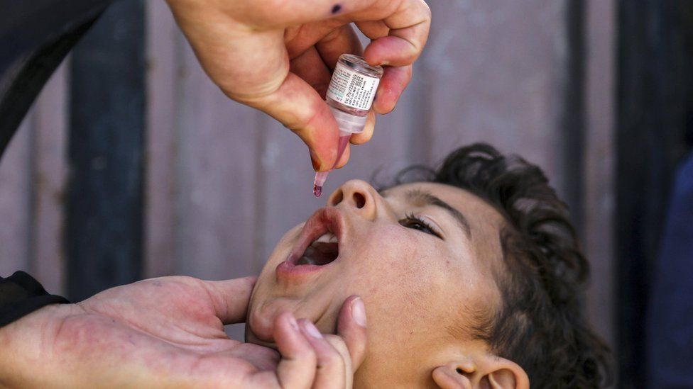
While the original polio strains, called wildtype, have largely been eliminated, new strains can develop from the oral polio vaccine (OPV), which is the one most used in the developing world. Oral vaccines use live, weakened virus that occasionally mutates to an active form, leading to outbreaks even in countries believed to have eliminated polio.
Scientists at UCSF and the UK’s National Institute of Biological Standards and Control (NIBSC) have developed two novel oral polio vaccines (nOPVs) to bolster the World Health Organization’s most recent push to finally eradicate polio, which began two years ago using the first nOPV developed by the same team. These are the first new polio vaccines in 50 years.
Like the first nOPV, the two newest nOPVs, which were described in Nature on June 14, are made from weakened poliovirus that has been genetically engineered to reduce reversion to dangerous forms of the virus. The development of these new vaccines was led jointly by Raul Andino, PhD, UCSF professor of microbiology and immunology, and Andrew Macadam, PhD, a virologist at NIBSC.
“With such variation in vaccination within and between countries, poliovirus has persisted into the 21st century, with sometimes tragic consequences,” said Andino, co-senior author of the paper along with Macadam. “We’ve designed these new vaccines using lessons learned from many years of fighting polio and believe they will help eliminate the disease once and for all.”
The evolving battle against polio
Polio is insidious: it is usually asymptomatic, but can cause severe disability, paralysis or death in about one in every hundred children. It spreads via fecal or oral particles, so it is particularly problematic in regions with poor sanitation. In the first half of the 20th century, polio outbreaks routinely rolled through the US, leading to a race to develop vaccines.

The first effective polio vaccines emerged in the 1950s, kicking off massive campaigns to immunize every person, with an emphasis on children. The inactivated polio vaccine (IPV), made of dead poliovirus, was given via injection, while the oral polio vaccine (OPV), made of weakened poliovirus, was given on a sugar cube or in a candy. Today, IPV is the vaccine of choice in countries with robust healthcare, and OPV — the cheaper, easier-to-administer option — is used otherwise.
In populations where everyone is immunized early in life, it doesn’t matter whether they receive IPV or OPV, although these vaccines act in different ways in the environment. People vaccinated with IPV can still get infected with any polio that happens to be circulating. They will not get sick, but they can silently transmit the virus to the unvaccinated. People vaccinated with OPV can’t silently transmit circulating polio in this way, but they can shed the weakened virus they were inoculated with and spread it to the unvaccinated. If the weakened virus mutates, it can become pathogenic polio once more.
In populations with unvaccinated children — whether due to refusal to vaccinate, natural disaster, or war — such vaccine-derived polio can spread widely, causing severe disease in the unlucky few.
While the original, or “wildtype,” poliovirus has only been recently detected in Afghanistan and Pakistan, vaccine-derived polio has been detected in countries as far flung as Syria, the Democratic Republic of Congo, and the U.S. In fact, there have been more cases of vaccine-derived polio than wildtype in recent years, creating an urgency to counter this new source of polio.
In 2017, Andino and his colleagues discovered how OPV reverts to its harmful form: a single mutation restores the virus’s capacity to migrate from the human gut and into the nervous system. Within a few years, the group had devised a trio of mutations that make such genetic reversion much less likely and packaged it into a new vaccine.

That vaccine, nOPV2, earned the WHO’s first-ever emergency use listing for a vaccine in 2020 and was quickly manufactured and distributed.
“Over 600 million doses were delivered to more than 28 countries, and in ten instances it stopped ongoing outbreaks of vaccine-derived polio,” said Andino. “It gave us a lot more confidence that this actually was working as anticipated.”
Covering all the bases with polio eradication
Despite its effectiveness, nOPV2 only protects against one of three strains of polio, and cases of polio have recently emerged in Israel, which is heavily vaccinated, as well as in pockets of the US where people refuse to vaccinate their kids.
Even where there are no polio cases in hospitals, polio continues to be detected in wastewater in major cities. There may be 99% fewer polio cases today than there were 30 years ago, but the last 1% has proven hard to snuff out.
“If there’s polio anywhere, it will come back where there are gaps in vaccination,” Andino said.
The latest work from Andino’s group takes the solution they crafted for nOPV2 — the three mutations that usually prevent the vaccine from becoming dangerous over time — and engineers it into the other two types of OPV. The resulting vaccines, nOPV1 and nOPV3, effectively prevented polio in animal models. All three are much safer than the original OPVs, which can occasionally cause paralysis in those who get the vaccine, although this is rare (on the order of one case per two million children vaccinated).
The two new vaccines are currently being tested in clinical trials to ensure that they are both effective and do not revert to dangerous forms in humans. Andino is hopeful they will be incorporated into bivalent or trivalent combinations with nOPV2. Children of the future will be equally protected from polio for life, and perhaps the world will someday experience decades in which zero polio is detected.
“The perception that polio is gone is a dangerous one,” said Andino. “For instance, just in India, 500,000 children are born each week, an enormous number of susceptible people. We now have what we need to protect them.”

Read more →

Pandemic akin to war, lawyer tells Covid inquiry

Published14 JuneShareclose panelShare pageCopy linkAbout sharingImage source, ReutersBy Philippa RoxbyHealth reporterThe pandemic was a “confused period akin to war”, the lawyer for the Department of Health and Social Care has told the Covid inquiry.Fiona Scolding KC said the department had been faced with a series of “hugely unpalatable options” and decisions had been “finely balanced”.With the benefit of hindsight, some of those decisions might have been different, she added.The public inquiry on the UK’s response to Covid has finished its second day.The first part of the inquiry – called Resilience and preparedness – will hear from key politicians, civil servants, scientists, unions, health and care organisations, as well as groups representing victims and their relatives.Over the next six weeks, about 70 people are expected to take the stand on this subject.On Tuesday, the first day, Baroness Hallett, who is chairing the inquiry, said there were three key questions:Was the UK properly prepared?Was the response appropriate?What lessons can be learned for the future?The lead lawyer for the inquiry, Hugo Keith KC, described the “death, misery and incalculable loss” caused by the virus, saying the UK may not have been well prepared “at all”.On Wednesday, in her opening statement, Ms Scolding said the government would not argue it had got everything right in its response to Covid.”Contrary decisions could rationally have been made resulting in a different set of outcomes,” she said.But the pandemic had been the “greatest challenge ever faced by the NHS and care sector”.And it was necessary “to recognise the context of the time was very different to what we know now”.Ms Scolding asked the inquiry “not to impose a retroscope on decision making” but described testing and diagnostics as a “significant weakness” in the early part of the pandemic, compared with that in some other countries.Matthew Hill, from the Government Office for Science, which also represents current and former chief scientific advisers, told the inquiry the UK had had other areas of weakness when the pandemic started, including the:absence of a major diagnostic industryunderlying health inequalities and conditions within the UK population lack of excess capacity in the NHS – even in normal timesHe said there were questions over the investment in public-health infrastructure before Covid and “whether it had responded effectively to previous pandemic threats”.Mr Hill confirmed that the role of the Scientific Advisory Group for Emergencies (Sage), who met regularly during the pandemic, was to provide independent scientific advice to the UK government, not to decide policy.That was the role of politicians and decision-makers, he said.”It is for them, and not the experts, to weigh the competing factors against one another, make the trade-offs and come to decision on policy,” Mr Hill added.’Impending wave’The first two experts questioned on Wednesday afternoon – both epidemiologists – said a coronavirus had already jumped to humans on a significant scale twice in the 21st Century, in the form of severe acute respiratory syndrome (Sars) and Middle East respiratory syndrome (Mers).And it had been “a reasonable bet”, before 2020, another one might follow, infectious-diseases expert and former professor of international public health Jimmy Whitworth said.By mid-January 2020, people in the international public-health community had been aware the Covid-19 outbreak had been “out of the ordinary”, he said.”It wasn’t going to die away – it was something bigger than that,” Mr Whitworth said.And by end of January, people in public health had been very clear it had been “an impending wave that was coming to UK”.”For those of us who had memories of Sars, the parallels were something that gave us shivers”, Mr Whitworth added.Asked about the dangers of a virus being leaked, deliberately or accidentally, from a laboratory, Cambridge University fellow in emerging infectious diseases Dr Charlotte Hammer said she was aware of four incidents involving the Sars virus. But when it came to controlling an outbreak once it had started to infect thousands of people, the virus’s origins mattered “very little”.What is the UK Covid-19 inquiry?It is about going through what happened and learning lessonsNo-one will be found guilty or innocentAny recommendations made do not have to be adopted by governmentsThe inquiry has no formal deadline but is due to hold public hearings until 2026Scotland is holding a separate inquiry in addition to the wider UK oneThis video can not be playedTo play this video you need to enable JavaScript in your browser.How many Covid deaths have there been?The UK saw one of the worst first waves of Covid in Europe, in spring 2020. In April and May that year, about 160,000 deaths were registered – 60,000 more than expected, based on the years just before the pandemic. But by that winter, the UK had been overtaken by many countries in Eastern Europe that had seemed to escape the first wave. The UK’s Office for National Statistics has continued to analyse these figures for Europe and, as of July last year, put the UK in the middle of the pack. According to Department of Health figures, 227,321 people across the UK died with Covid recorded on their death certificate.Covid vaccines have prevented many deaths and serious illness from the virus – more than 151 million doses have been given in the UK. More on this story’Little thought’ about lockdown impact pre-pandemic, inquiry hearsPublished13 JuneThe questions we want the Covid inquiry to answerPublished13 JuneWhat is the UK Covid inquiry and how does it work?Published1 hour agoNew judge appointed to lead Scottish Covid inquiryPublished27 October 2022Related Internet LinksUK Covid-19 InquiryThe BBC is not responsible for the content of external sites.

Read more →

Super-engineered vaccines created to help end polio

Published33 minutes agoShareclose panelShare pageCopy linkAbout sharingImage source, SAMIULLAH POPAL/EPA-EFE/REX/ShutterstockBy James GallagherHealth and science correspondentScientists have “super-engineered” polio vaccines to prevent them mutating into a dangerous form that can cause outbreaks and paralysis.The oral vaccines contain weakened live polio viruses and the genetic redesign locks them into that weakened state. The US and UK teams have now created upgraded vaccines against all three types of polio. However, better vaccines still need to reach every child in order to stop the disease. Polio can spread into the nervous system, causing paralysis. Cases have fallen by more than 99% since the late 1980s and about 20 million people who would have been paralysed can walk thanks to vaccines. The original or “wild” poliovirus is now contained to small pockets of Afghanistan and Pakistan and the oral vaccines play a pivotal role in the attempt to rid the world of polio. “The issue is they’re genetically unstable,” Dr Andrew Macadam, from the UK’s National Institute for Biological Standards and Control, told BBC News.It takes only one mutation to turn the safe polio vaccine back into a virus that can move out of a child’s stomach, invade their nervous system and cause paralysis. And if those viruses spread from an immunised child – through their contaminated faeces – there is a risk of infecting the unvaccinated and triggering an outbreak. There are now more cases of “vaccine-derived polio” than of the wild poliovirus and the polio detected in London’s sewers last year was connected to the oral vaccine. Polio: Do we have to worry about it once again?So the researchers have genetically altered the weakened virus even further to make it much harder for it to start causing paralysis again. “By genetically modifying this part of the virus, we could modify this region so it couldn’t revert and this I think has been remarkably successful,” Dr Macadam said.Prof Raul Andino, from University of California San Francisco, said he was “super-proud” of the scientific effort showing the vaccine was “50 to 100 times more stable”.Image source, Getty ImagesIn March 2021, the World Health Organization made the researchers’ vaccine against type-two polio available for emergency use.Since then, it has been used more than 650 million times – and, Dr Macadam said, “we’ve never seen a reversion” back to dangerous poliovirus. Now, in the journal Nature, the researchers have detailed the creation of stable vaccines against polio types one and three. The first-stage human trials of the upgraded vaccines have already been conducted – and, the researchers say, the data, which is still being analysed, is “very promising”. The trio represent the first new polio vaccines in 50 years. “I don’t think there’s any question that they’re helpful, the new vaccines address the instability question, but it doesn’t address the coverage issue,” Dr Macadam said.’Impressive science’Tackling the last 1% of polio cases has proven stubborn. The original goal was to completely eradicate polio by the year 2000 – but delivering vaccines to some of the poorest and most conflict-ridden parts of the world has been a challenge. Prof Alan Barrett, from the Sealy Institute for Vaccine Sciences, at the University of Texas, called the “super-engineered” vaccines a feat of “impressive science”.”[But] will it lead the endgame to the finish line? That is a big question,” he added.Joseph Swan, from the World Health Organization and the Global Polio Eradication Initiative, said more stable vaccines were a “significant part” of the plan for a polio-free world.But, he said: “Simply having these new and better tools will not get us over the finish line – vaccination, not just vaccines, is what will end polio.”There was now a “unique opportunity” to eradicate wild poliovirus – but vaccine-derived polio outbreaks were causing problems in places facing “complex humanitarian emergencies”, in the Democratic Republic of Congo and Yemen. Follow Jameson Twitter.

Read more →

New Obesity Drugs Come With a Side Effect of Shaming

Wegovy and other drugs expose a social tension between a quest to medicate illness and a stigmatizing belief that obese people lack sufficient willpower to lose weight.Eileen Isotalo was always able to lose weight, but always gained it back. Now 66, her first diet was with Weight Watchers at age 14. She went on to try one diet after another and bought so many books on weight loss that she thinks she has more than the public library.In desperation, she finally went to a weight management clinic at the University of Michigan. She had sleep apnea and aching knees, but could not curb her appetite.“It’s just this drive to eat,” said Ms. Isoltalo, a retired interior design coordinator. “It’s almost like this panic feeling when you start craving food.”“My mental shame was profound,” she said.Now, though, since she started taking Wegovy, one of a new class of drugs for obesity that was prescribed by her doctor at the clinic, those cravings are gone. She has lost 50 pounds and jettisoned the dark clothes she wore to hide her body. Her obesity-related medical problems have vanished along with much of the stigma that caused her to retreat from family and friends.But like others at the clinic, she still struggles with the fear others will judge her for receiving injections to treat her obesity rather than finding the willpower to lose weight and keep it off.Yet the drug, she said, “changed my life.”Wegovy and drugs like it make this “a very exciting time in the field,” said Dr. Susan Yanovski, co-director of the office of obesity research at the National Institute of Diabetes and Digestive and Kidney Diseases.About 100 million Americans, or 42 percent of the adult population, have obesity, according to the Centers for Disease Control and Prevention. For the first time, people with obesity, who faced a lifetime of medical jeopardy, can escape the ruthless trap of fruitless dieting and see their obesity-related health problems mitigated, along with the weight loss.But there is still the taint.“There’s a moral component to it,” Dr. Yanovski said. “People really believe that people with obesity just need to summon their willpower and they think that taking a medicine is the easy way out.”Unlike other chronic diseases, obesity is on full public display, Dr. Yankovski said. “No one looks at you and knows you have high cholesterol of high blood pressure,” she said.Obesity, she added, “is one of the most stigmatized conditions out there. “Wegovy’s maker, Novo Nordisk, reports that doctors in the United States have written about 110,000 prescriptions for the drug.Cydni Elledge for The New York TimesWegovy and a similar but less effective medication, Saxenda, are the only ones in their class of drugs so far to be approved for the treatment of obesity — others like Ozempic and Mounjaro are diabetes drugs but also spur weight loss.Novo Nordisk, Wegovy’s maker, reports that doctors in the United States have written about 110,000 prescriptions for the drug. Citing a huge demand, the company recently put its advertising for Wegovy on hold.“We can’t make enough,” said Ambre James-Brown, a Novo Nordisk spokeswoman. Supplies are so limited that the company is only selling the drug in the United States, Norway and Denmark, the company’s corporate headquarters. Its high list price of $13,492 a month puts it out of reach for most whose insurance will not cover it. But increasingly many insurers do.The drugs have arrived at a time when researchers have documented the risks of obesity and the futility of prescribing only diet and exercise as a treatment. Decades of studies have consistently shown that very few people can lose excess weight and keep it off with lifestyle changes alone.People with obesity are at risk for a variety of serious medical conditions, including diabetes, hypertension, high cholesterol, sleep apnea and nonalcoholic fatty liver disease, a leading reason for liver transplants in the United States.Losing weight can make some of these complications vanish.Katarra Ewing of Detroit will readily tell anyone who asks that she takes Wegovy, which helped her lose 90 pounds. But she said some longtime friends fell away after she lost weight.Cydni Elledge for The New York TimesYet the belief persists — fed by diet gurus, influencers and an industry selling supplements and diet plans — that if people really really tried, they could shed pounds.So those who take a drug like Wegovy often end up in uncomfortable situations that are influenced by the common view that obesity is a lifestyle choice.At the University of Michigan clinic there are those like Ms. Isotalo whose reluctance to admit to taking Wegovy stems from her conviction that those who take it are often thought to be cheating.Another patient, though, Katarra Ewing of Detroit, readily tells anyone who asks that she takes the drug. She tried diets, but it was Wegovy that allowed her to lose 90 pounds.She came to the weight management clinic after her all-night shift at a Ford factory, ebullient and vibrant, wearing a vivid green sweater. She has more energy now that she lost the weight, her mood is brighter, her high blood pressure gone.But she discovered an unintended social consequence to weight loss, as many longtime friends fell away.“Only my genuine friends are left and that’s a very small number,” Ms. Ewing said.Obesity medicine specialists say they are not surprised — they see the same thing after people lose weight with bariatric surgery.Relationships shift because obesity is such a defining condition. People of normal weight may feel superior to a friend with obesity and that helps define a relationship — until the friend loses weight. Other friends who themselves have obesity may use the condition as a bonding factor in the relationship. Now that is gone.Another issue is the drugs’ reputation as vanity medications, which has been amplified by comedians’ punchlines at the Oscars and in other high-profile settings.But when Samuel Simpson came to the weight management clinic, he considered losing weight to be a matter of life or death.Mr. Simpson was terrified he’d face the fate of his mother, brother and sister, all of whom had obesity and diabetes. They all developed kidney failure that ultimately killed them, each dying at the age of 59.Amy Rothberg, the medical director of Rewind, a company that counsels diabetic patients, and a professor of medicine at the University of Michigan. “I don’t think it’s a matter of willpower,” she told one patient.Cydni Elledge for The New York TimesHis first appointment with Dr. Amy Rothberg at the clinic was nearly two years ago, when he was 58. He had obesity and diabetes. Although he was taking high doses of insulin to lower his blood sugar, his kidneys were starting to fail.“I was so afraid,” he said. “Was I going to end up on dialysis like everyone else? I’d be history.”He began with a diet and then Dr. Rothberg added Mounjaro, a drug by Eli Lilly that appears to be even more powerful than Wegovy in eliciting weight loss, but is, so far, only approved for people with diabetes.Now he’s lost 44 pounds, 20 percent of his original weight, and his diabetes is in remission. The weight loss, he said, “turned my life around.”He will tell those who ask how he lost the weight,“I’m not like the roadside preacher but when someone asks me how I did this I will tell them,” he said.Art Regner had a different issue. A garrulous color commentator for the Detroit Red Wings hockey team, he said he was not ready to resort to medication. But when he came to Dr. Rothberg’s clinic he was chagrined. He’d regained 22 of the 76 pounds he lost by dieting.Dr. Rothberg, who is also the medical director of Rewind, a company that counsels diabetic patients, suggested Wegovy or Mounjaro. But Mr. Regner felt he should have enough willpower to do it on his own. He knows his blood sugar is high and is aware of the consequences of diabetes.Dr. Rothberg gently explained to him that it was not his fault he kept regaining weight every time he lost some.“I think biology is conspiring against you,” she said. “I don’t think it’s a matter of willpower.”Mr. Regner was not swayed. “I believe in myself,” he said. “I wake up in the morning and look in the mirror and say, ‘Are you going to do it or aren’t you?’”

Read more →

First illustration of the molecular machinery that makes cilia beat

The first image of the structures that power human cilia — the tiny, hairlike projections that line our airways — has been produced by a team involving UCL researchers and could lead to much-needed treatments for people with rare cilial diseases.
The study, published in Nature, combined advanced microscopy and artificial intelligence techniques to create a detailed snapshot of the structure of human cilia. These are the microscopic projections on the cells that line our lungs, ears and sinuses and beat rhythmically to keep the lungs clear from mucus and bacteria. People who inherit the rare condition primary ciliary dyskinesia (PCD) have defective cilia that can’t effectively remove debris from the airways, and so suffer from breathing difficulties and chronic lung infections.
For the first time, the scientists visualised the molecular ‘nano-machinery’ that causes cilia to beat, visible as identical structures dotted every 96 nanometres along the cilia length. These structures come together to form the axoneme. In healthy airways, this complex structure is tightly controlled, with molecules precisely arranged to make cilia beat in a rhythmic, wave-like motion, around a million times a day.
In people with PCD, the team found that cilia don’t beat correctly because key elements of the axoneme structure are missing, caused by genetic mutations. This new information could lead to new medicines that target these defects, making cilia beat properly.
Study co-author, Professor Hannah Mitchison (UCL Great Ormond Street Institute of Child Health), said: “Treatments for PCD currently work to clear people’s airways and prevent infection. Our findings offer the possibility of molecular medicines to precisely target tiny defects in the axoneme and make cilia beat as they should.
“Molecular medicines are showing promise for other rare diseases, and COVID-19 research has unlocked new ways to deliver these drugs directly to the lung. If we can combine these advances with our new findings, my hope is that we’ll bring molecular medicines to people with PCD within the next 5 to 10 years.”
The team’s research could also prove useful for infertility, as sperm cells rely on a similar axoneme structure in their tails to propel themselves forward.
The research team was a global collaboration, with scientists based across the UK, US, Netherlands, China and Egypt. “It can be difficult to study rare diseases like PCD, because patients are spread thinly across the world. In the UK, we think around 9,000 families may be affected by PCD,” said Professor Mitchison. “Our study was made possible by a fantastic international collaboration between clinical scientists, biologists and members of the rare disease community willing to take part in our research.”
In addition to human cilia, the team examined the axoneme structure of a single-celled alga called Chlamydomonas reinhardtii, which uses two tail-like projections on its surface to swim. Despite being separated by more than 1 billion years of evolution, the alga’s tails shared structural similarities with the human airway cilia, highlighting the importance of the axoneme throughout evolution.
This study involved collaborators at Harvard Medical School, Alexandria University, University of Leicester, Amsterdam University Medical Centers, Guy’s and St Thomas’ NHS Foundation Trust and Imperial College London.
At UCL, the study was supported by NIHR Great Ormond Street Hospital Biomedical Research Centre, the Ministry of Higher Education in Egypt and a MRC UCL Confidence in Concept grant.

Read more →

Microplastics stick around in human airways

Research shows humans might inhale about 16.2 bits of microplastic every hour, which is equivalent to a credit card over an entire week. And these microplastics — tiny debris in the environment generated from the degradation of plastic products — usually contain toxic pollutants and chemicals.
Inhaled microplastics can pose serious health risks, so understanding how they travel in the respiratory system is essential for prevention and treatment of respiratory diseases. In Physics of Fluids, by AIP Publishing, researchers from the University of Technology Sydney, Western Sydney University, Urmia University, Islamic Azad University, the University of Comilla, and Queensland University of Technology developed a computational fluid dynamics model to analyze microplastic transport and deposition in the upper airway.
“Millions of tons of these microplastic particles have been found in water, air, and soil. Global microplastic production is surging, and the density of microplastics in the air is increasing significantly,” said author Mohammad S. Islam. “For the first time, in 2022, studies found microplastics deep in human airways, which raises the concern of serious respiratory health hazards.”
The team explored the movement of microplastics with different shapes (spherical, tetrahedral, and cylindrical) and sizes (1.6, 2.56, and 5.56 microns) and under slow and fast breathing conditions.
Microplastics tended to collect in hot spots in the nasal cavity and oropharynx, or back of the throat.
“The complicated and highly asymmetric anatomical shape of the airway and complex flow behavior in the nasal cavity and oropharynx causes the microplastics to deviate from the flow pathline and deposit in those areas,” said Islam. “The flow speed, particle inertia, and asymmetric anatomy influence the overall deposition and increase the deposition concentration in nasal cavities and the oropharynx area.”
Breathing conditions and microplastic size influenced the overall microplastic deposition rate in airways. An increased flow rate led to less deposition, and the largest (5.56 micron) microplastics were deposited in the airways more often than their smaller counterparts.
The authors believe their study highlights the real concern of exposure to and inhalation of microplastics, particularly in areas with high levels of plastic pollution or industrial activity. They hope the results can help inform targeted drug delivery devices and improve health risk assessment.
“This study emphasizes the need for greater awareness of the presence and potential health impacts of microplastics in the air we breathe,” said author YuanTong Gu.
In the future, the researchers plan to analyze microplastic transport in a large scale, patient-specific whole lung model that includes environmental parameters such as humidity and temperature.

Read more →