In rats, loss of immune cells leads non-moms to care for newborns

Immune system changes in the pregnant body that protect the fetus appear to extend to the brain, where a decrease in immune cells late in gestation may factor into the onset of maternal behavior, new research in rats suggests.
In adult female rats that had never given birth — which typically don’t like being around babies — depletion of these cells sped up their care for rat newborns that were placed in their cage.
The loss of these cells, called microglia, and the related uptick in motherly attentiveness were also associated with changes to neuron activity in several regions of the rat brain, suggesting shifts in immune function have a role in regulating maternal behavior.
Microglia are well-known for their link to brain injury and disease because of their protective activities under those circumstances, and for helping enable brain development. But these findings suggest they have another job entirely in the adult brain.
“Our data shows that microglia are probably also really important for plasticity in the adult brain — its ability to adapt to all kinds of changes — and contribute to normal behavioral function,” said co-senior author Kathryn Lenz, associate professor of psychology at The Ohio State University.
The findings also suggest that feeling maternal is much more than just a hormonal response.
“The standard way of thinking up to now about mothers is that hormones are the primary drivers of maternal care. But this really draws attention to the importance of immune changes to maternal behavior,” said co-senior author Benedetta Leuner, associate professor of psychology at Ohio State. “These data are novel and potentially paradigm-shifting.”
The research was published recently in the journal Neuropsychopharmacology.

Read more →

New scanning methods can detect deadly heart condition before symptoms appear

Combining two types of heart scan techniques could help doctors to detect the deadly heart condition hypertrophic cardiomyopathy (HCM) before symptoms and signs on conventional tests appear, according to a new study led by UCL researchers.
The research, funded by the British Heart Foundation and published in the journal Circulation, opens the prospect of treating the condition at the earliest stages.
Being able to detect HCM earlier than ever before will also assist trials investigating gene therapies and drug treatments aimed at stopping the disease developing in those at risk.
HCM is an inherited condition that affects around 1 in 500 people in the UK. It causes the muscular walls of the heart to become thicker than normal, affecting how well the heart can pump blood around the body. It is a leading cause of heart failure and sudden cardiac death.
Researchers from UCL, Barts Heart Centre and University of Leeds studied the hearts of three groups: healthy people, people who already had HCM, and people with an HCM-causing genetic mutation but no overt signs of disease (no heart muscle thickening).
To do this, they used two cutting-edge heart scanning techniques: cardiac diffusion tensor imaging (cDTI), a type of MRI scan that shows how individual heart muscle cells are organised and packed together (the heart’s microstructure), and cardiac MRI perfusion (perfusion CMR), which detects problems with the small blood vessels supplying the heart muscle (microvascular disease).
The scans showed that people with overt signs of HCM have very abnormal organisation of their heart muscle cells, and a high rate and severity of microvascular disease compared to healthy volunteers.

Read more →

Why does skin get 'leathery' after too much sun? Bioengineers examine cellular breakdown

Received wisdom says that staying out in the sun too long can make your skin tougher over time. Think about the “leathery” complexions of farmers, road crews and others who work long hours outdoors, or someone who spends too much time in tanning booths, or too much time on the beach during the summer.
Surprisingly, though, very little research has been done to explain why this happens on a biological level — until now.
A study from Binghamton University researchers recently published in the Journal of the Mechanical Behavior of Biomedical Materials explores how ultraviolet radiation can alter the microstructure of human skin. Particularly affected is collagen, the fibrous protein that binds together tissue, tendon, cartilage and bone throughout our bodies.
“We don’t want to put a fear factor in here saying ‘don’t go out in the sun,'” said Binghamton University Associate Professor of Biomedical Engineering Guy German. “But extended periods of time under UV light can toughen up your skin as well as lead to a higher risk of carcinogenic problems.”
Leading the research with German at the Thomas J. Watson College of Engineering and Applied Science’s Department of Biomedical Engineering are PhD student Abraham Ittycheri, Zachary Lipsky, PhD ’21, and Assistant Professor Tracy Hookway.
The new study builds on previous research from German and Lipsky that focused on the outer stratum corneum, which is the top layer of skin. This time, the Binghamton team compared full-thickness skin samples before and after various levels of UV exposure.
“One way to characterize the material characteristics of skin is by conducting a mechanical stretch test on it,” Ittycheri said. “If it stretches very easily, it’s relatively compliant, but if it’s much harder to stretch it, you can characterize it as much stiffer. My experiment was to see what the isolated effects of UV light would be and compare it with a scenario where a skin is not exposed to UV light.”
The researchers found that as the skin absorbed more UV radiation, the collagen fibers in it became more tightly packed together, leading to increased stiffness and tissue that is harder to break. German sees correlations with the cross?linkage theory of aging, which proposes that the accumulation of undesirable molecular bonds over time can cause cellular dysfunction.

Read more →

Study optimizes patient-specific stem cell-based therapy for Parkinson's disease

Scientists from Scripps Research and Cardiff University made key discoveries in support of a new stem cell-based therapy for Parkinson’s disease. The approach, called an autologous therapy, uses induced pluripotent stem cells (iPSCs) — made from a patient’s own skin or blood cells — to replace the neurons in the brain that are lost in Parkinson’s. Transplants of a person’s own cells eliminates the need for immunosuppression.
In a new study, the researchers used iPSCs made from the skin cells of two people with Parkinson’s disease to make young neurons that were successfully transplanted into a rat model with the disease. They used the animal model to pinpoint exactly at what stage of development the iPSC-derived neurons should be transplanted to become mature neurons that can reverse signs of disease in the rat brain.
The research, published in Stem Cells and Development on June 22, 2023, is a crucial step forward for clinical trials of autologous iPSC-derived neurons in human patients with Parkinson’s disease.
“This paper reports important progress toward development of an autologous cell replacement therapy for Parkinson’s disease,” says senior author Jeanne Loring, PhD, professor emeritus and director of the Center for Regenerative Medicine at Scripps Research and co-founder of Aspen Neuroscience, Inc. “These results give us confidence that personalized therapy is feasible for Parkinson’s disease.
For over a decade, researchers have been developing methods for using iPSCs — which can form any cell type in the body — to treat a variety of diseases characterized by defective or missing cells. People with Parkinson’s disease have lost neurons in the brain that produce a chemical messenger called dopamine. The resulting low levels of dopamine lead to the symptoms of Parkinson’s, which are typically tremors and slow movement. There is currently no cure for the disease, and there are only limited options for controlling symptoms. Nearly 90,000 people are diagnosed with Parkinson’s each year, and 10 million people worldwide are living with the disease.
Today, clinical trials are already underway using stem cells to replace dopamine-producing neurons in the brains of people with Parkinson’s disease, but the trials all use unmatched cells from the lab or a donor, rather than cells from a patient’s own body.
“When you transplant neurons derived from someone else’s cells, those cells will be rejected by the immune system, requiring the use of immunosuppressive drugs that are often not well tolerated” says Loring.

Read more →

New therapeutic target for Parkinson's disease discovered

Northwestern Medicine scientists have uncovered a new mechanism by which mutations in a gene parkin contribute to familial forms of Parkinson’s disease. The discovery opens a new avenue for Parkinson’s therapeutics, scientists report in a new study.
The Northwestern scientists discovered that mutations in parkin result in a breakdown of contacts between two key workers in the cell — lysosomes and mitochondria.
Mitochondria are the main producers of energy in cells, and lysosomes recycle cellular debris that accumulates during normal function of our cells. These organelles are especially important in our brains because neurons are highly dependent on energy production by mitochondria, and because of their activity, neurons produce an abundance of cellular debris that must be cleared by lysosomes.
In a prior study, published in Nature, Dr. Dimitri Krainc, chair of neurology and director of Simpson Querrey Center for Neurogenetics at Northwestern University Feinberg School of Medicine, and his group discovered that lysosomes and mitochondria form contacts with each other. After the initial discovery, Northwestern scientists tried to understand the function of these contacts in Parkinson’s disease.
In the new study published in Science Advances, the investigators report that lysosomes help mitochondria by providing key metabolites for their function. Mitochondria must import many of their essential ingredients, but it has not been well known where some of these metabolites come from. On the other hand, lysosomes serve as recycling factories in cells and, therefore, produce many breakdown products that could be used by other organelles such as mitochondria.
In this work, scientists found that lysosomes provide important amino acids that support the function of mitochondria. However, they also found that in some forms of Parkinson’s disease, lysosomes cannot serve as a “helping hand” to mitochondria because the contacts between the two organelles are disrupted. This results in dysfunctional mitochondria and ultimately degeneration of vulnerable neurons in Parkinson’s disease.
“Findings from this study suggest that dysregulation of mitochondria-lysosome contacts contributes to the Parkinson’s disease pathophysiology,” said Krainc, the study’s corresponding author. “We propose that restoring such mitochondria-lysosome contacts represents an important new therapeutic opportunity for Parkinson’s disease.”
From a broader perspective, this study opens a new avenue of research in neurodegenerative disorders, by highlighting the importance of direct communication and collaboration between cellular organelles in the pathogenesis of these disorders.
The first author of the study is Dr. Wesley Peng who recently completed the medical scientist training program (MD-PhD) at Northwestern and currently serves as a neurology resident at Mass General Brigham and Harvard Medical School. Other contributors to the study include Leonie Schroder, Pingping Song and Yvette Wong.
The study was supported by the following National Institute on Aging grant AG066333, National Institute of Neurological Disorders and Stroke (NINDS) grants NS109252 and NS122257, all from the National Institutes of Health.

Read more →

More than just lifestyle and genes: New factor influencing excess body weight discovered

What determines whether we become overweight? Aside from lifestyle, predisposition plays a role, but genes cannot fully explain the inherited propensity to accumulate excess weight. A new study by Charité — Universitätsmedizin Berlin in Science Translational Medicine* shows that a kind of formatting of the DNA code in one gene that is associated with satiety is implicated in a slightly elevated risk of excess body weight — at least in women. This “epigenetic marking” is established early on during the embryonic stage.
People who are overweight, especially those who are severely overweight, are at increased risk of a number of serious diseases such as cardiovascular disease, diabetes, and cancer. It is a growing health issue. All over the world, the number of overweight people is increasing. In the European Region, two out of three adults (59 percent) are overweight or obese, according to the World Health Organization.
But what determines whether people will become overweight? Genetic predisposition plays a major role, alongside lifestyle. The similarity of the body mass index (BMI) in identical twins ranges from 40 to 70 percent. Even identical twins raised in different families still show the same significant similarity. Scientists have identified several genetic variants that influence a person’s body weight — and with it, the risk of developing obesity. But even taken all together, they cannot explain the heritability that has been observed. Researchers began to suspect there must be additional non-genetic factors that affect a person’s propensity to gain excess weight.
Satiety gene is not altered, but formatted
Researchers led by Prof. Peter Kühnen, Director of the Department of Pediatric Endocrinology at Charité, have now identified one such factor in their recent study. According to their findings, women’s risk of being overweight increases by about 44 percent if there are an especially large number of methyl groups adhering to the POMC (pro-opiomelanocortin) gene, which is responsible for the feeling of satiety. Methyl groups are tiny chemical units the body uses to mark the letters in the DNA code to activate or deactivate genes without modifying the sequence of letters in the DNA. In other words, the effect is much like highlighting a section of a text without rewriting the text itself. This type of “DNA formatting” is known as epigenetic marking.
For their study, the team of researchers analyzed the “formatting” of the POMC gene in more than 1,100 people. They found more methyl groups attached to the satiety gene in obese women with a BMI of over 35 than in women with normal body weight. “A 44 percent increase in the risk of obesity is about the same as the effect that has been observed for individual gene variants as well,” says Kühnen. “By comparison, socioeconomic factors have a much stronger effect. They can increase the risk by a factor of two to three. As for why the methylation effect only shows up in women, we don’t know yet.”
The POMC gene is “formatted” very early on during embryonic development, as the researchers showed by comparing methylation patterns in more than 15 sets each of identical and fraternal twins. While the “formatting” of the satiety gene was the same in most of the identical twins, there was hardly any correlation in the fraternal twins. “This indicates that the epigenetic marking of the POMC gene is established shortly after the egg and sperm cells merge, before the fertilized egg divides into two twin embryos,” explains Lara Lechner, the study’s first author, who works at the Department of Pediatric Endocrinology. This means the very early stage of pregnancy is crucial.

Read more →

Stanford President Resigns After Report Finds Flaws in his Research

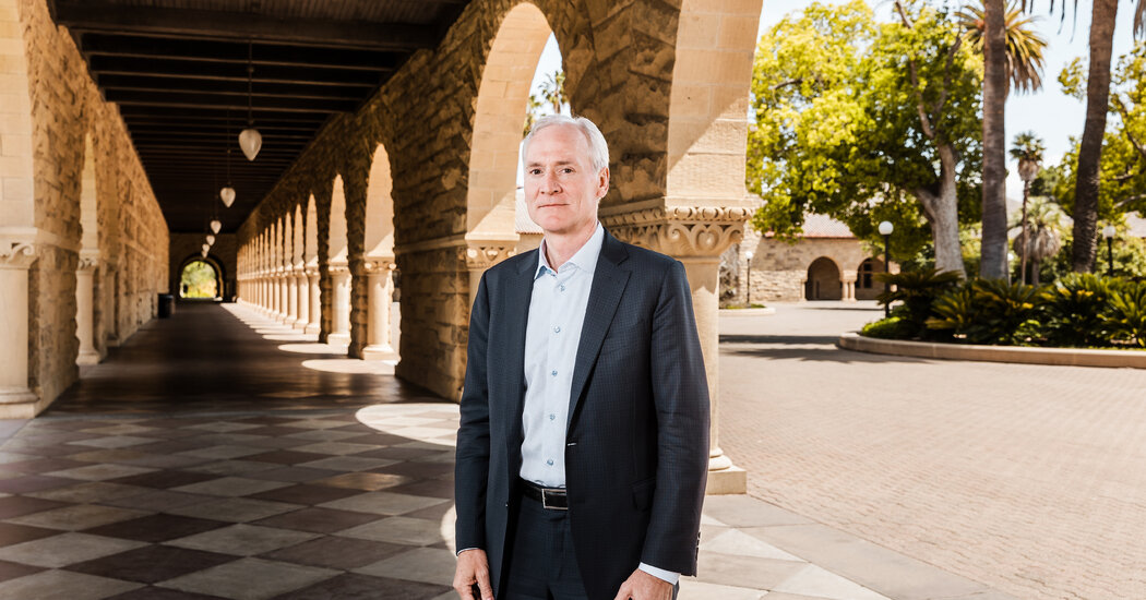
Marc Tessier-Lavigne was cleared of accusations of scientific fraud and misconduct. But the review said his work had “multiple problems” and “fell below customary standards of scientific rigor.”Following months of intense scrutiny of his scientific work, Marc Tessier-Lavigne announced Wednesday that he would resign as president of Stanford University after an independent review of his research found significant flaws in studies he supervised going back decades.The review, conducted by an outside panel of scientists, refuted the most serious claim involving Dr. Tessier-Lavigne’s work — that an important 2009 Alzheimer’s study was the subject of an investigation that found falsified data and that Dr. Tessier-Lavigne had covered it up.The panel concluded that the claims, published in February by The Stanford Daily, the campus newspaper, “appear to be mistaken” and that there was no evidence of falsified data or that Dr. Tessier-Lavigne had otherwise engaged in fraud.But the review also stated that the 2009 study, conducted while he was an executive at the biotech company Genentech, had “multiple problems” and “fell below customary standards of scientific rigor and process,” especially for a paper of such potential consequences.As a result of the review, Dr. Tessier-Lavigne said he would retract a 1999 paper that appeared in the journal Cell and two others that appeared in Science in 2001. Two other papers published in Nature, including the 2009 Alzheimer’s study, would also undergo what was described as comprehensive correction.Stanford is known for its leadership in scientific research, and even though the claims involved work published before Dr. Tessier-Lavigne’s arrival at the university in 2016, the allegations reflected poorly on the university’s integrity.In a statement describing his reasons for resigning, Dr. Tessier-Lavigne said, “I expect there may be ongoing discussion about the report and its conclusions, at least in the near term, which could lead to debate about my ability to lead the university into the new academic year.”Dr. Tessier-Lavigne, 63, will relinquish the presidency at the end of August but remain at the university as a professor of biology.The university named Richard Saller, a professor of European studies, as interim president, effective Sept. 1.As president of Stanford, Dr. Tessier-Lavigne is known for starting the university’s first new school in 70 years, the Doerr School of Sustainability. Opened last year, the school’s stated mission is to seek a solution to climate change.The panel’s 89-page report, based on more than 50 interviews and a review of more than 50,000 documents, concluded that members of Dr. Tessier-Lavigne’s labs engaged in inappropriate manipulation of research data or deficient scientific practices, resulting in significant flaws in five papers that listed Dr. Tessier-Lavigne as the principle author.In several instances, the panel found, Dr. Tessier-Lavigne took insufficient steps to correct mistakes, and it questioned his decision not to seek a correction in the 2009 paper after follow-up studies revealed that its key finding was incorrect.The flaws cited by the panel involved a total of 12 papers, in which Dr. Tessier-Lavigne was listed either as principle author or co-author. As a noted neuroscientist, he has published more than 200 papers, focusing primarily on the cause and treatment of degenerative brain diseases. Beginning in the 1990s, he has worked at multiple institutions, including Stanford, Rockefeller University, the University of California, San Francisco, and Genentech, a biotechnology company.The accusations had first surfaced years ago on PubPeer, an online crowdsourcing site for publishing and discussing scientific work. But they resurfaced after the student newspaper, The Stanford Daily, published a series of articles questioning the accuracy and honesty of work produced in laboratories overseen by Dr. Tessier-Lavigne.The newspaper first reported claims last November that images were manipulated in published papers listing Dr. Tessier-Lavigne as either lead author or co-author.In February, the campus newspaper published an article with more serious claims of fraud involving the 2009 paper that Dr. Tessier-Lavigne published while a senior scientist at Genentech. The Stanford Daily report said an investigation by Genentech found that the 2009 study contained falsified data, and that Dr. Tessier-Lavigne tried to keep its findings hidden.It also reported that a postdoctoral researcher who worked on the study had been caught by Genentech falsifying data.Both Dr. Tessier-Lavigne and the former researcher, now a medical doctor practicing in Florida, strongly denied the claims, which relied heavily on unnamed sources.Noting that, in some cases, it was unable to identify the unnamed sources cited in The Stanford Daily story, the review panel said that The Daily’s claim that “Genentech had conducted a fraud investigation and made a finding of fraud” in the study “appear to be mistaken.” No such investigation had been conducted, the report said.Following the newspaper’s initial report about manipulated studies in November, Stanford’s board formed a special committee to review the claims, headed by Carol Lam, a Stanford trustee and former federal prosecutor. The special committee then engaged Mark Filip, a former federal judge in Illinois, and his law firm, Kirkland & Ellis, to run the review.In January, it was announced that Mr. Filip also had enlisted the five-member scientific panel — which included a Nobel laureate and a former Princeton president — to examine the claims from a scientific perspective.Genentech had touted the 2009 study as a breakthrough, with Dr. Tessier-Lavigne characterizing the findings during a presentation to Genentech investors as a completely new and different way of looking at the Alzheimer’s disease process.The study focused on what it said was the previously unknown role of a brain protein — Death Receptor 6 — in the development of Alzheimer’s.As has been the case with many new theories in Alzheimer’s, a central finding of the study was found to be incorrect. Following several years of attempts to duplicate the results, Genentech ultimately abandoned the line of inquiry.Dr. Tessier-Lavigne left Genentech in 2011 to head Rockefeller University, but, along with the company, published subsequent work acknowledging the failure to confirm key parts of the research.More recently, Dr. Tessier-Lavigne told the publication STAT NEWS that there had been inconsistencies in the results of experiments, which he blamed on impure protein samples.The failure of Dr. Tessier-Lavigne’s Genentech laboratory to assure the samples’ purity was one of the scientific process problems cited by the panel, which also criticized Dr. Tessier-Lavigne’s decision to not correct the original paper as “suboptimal” but within the bounds of scientific practice.In his statement, Dr. Tessier-Lavigne said that he had earlier attempted to issue corrections to the Cell and Science papers, but that Cell had declined to publish a correction and Science failed to publish one after agreeing to do so.The panel’s findings confirmed a report released in April by Genentech, which said its own internal review of The Stanford Daily’s claims did not find any evidence of “fraud, fabrication, or other intentional wrongdoing.”Most of the panel’s report, about 60 pages, is a detailed appendix of analysis of images in 12 published scientific papers in which Dr. Tessier-Lavigne served either as author or co-author, some dating back 20 years.The panel found multiple instances of images in the papers that had been duplicated or spliced but concluded that Dr. Tessier-Lavigne had not participated in the manipulation, was not aware of them at the time, and had not been reckless in failing to detect them.Oliver Whang

Read more →

Astronomers find new type of stellar object

An international team led by astronomers from the Curtin University node of the International Centre for Radio Astronomy Research (ICRAR) has discovered a new type of stellar object that challenges our understanding of the physics of neutron stars.
The object could be an ultra-long period magnetar, a rare type of star with extremely strong magnetic fields that can produce powerful bursts of energy.
Until recently, all known magnetars released energy at intervals ranging from a few seconds to a few minutes. The newly discovered object emits radio waves every 22 minutes, making it the longest period magnetar ever detected.
The research was published today in the journal Nature.
Astronomers discovered the object using the Murchison Widefield Array (MWA), a radio telescope on Wajarri Yamaji Country in outback Western Australia.
Lead author Dr Natasha Hurley-Walker said the magnetar, named GPM J1839-10, is 15,000 light-years away from Earth in the Scutum constellation.
“This remarkable object challenges our understanding of neutron stars and magnetars, which are some of the most exotic and extreme objects in the Universe,” she said.

Read more →

Understanding brain network connecting temperature sensation and emotion may help prevent heatstroke

A research group from the Nagoya University Graduate School of Medicine in Japan has identified the neural pathways in the brain that influence thermoregulatory behaviors in rats. Thermoregulatory behaviors are the behavioral processes by which all animals regulate body temperature in response to changes in the environment. These findings, published in the Journal of Neuroscience, provide a better understanding of the neural network of the brain, and also suggest new medical strategies for the prevention of heatstroke.
All animals, including humans, use thermoregulatory behaviors to maintain body temperature within certain limits, even when the surrounding temperature is very different. Strategies include finding warmer or cooler spaces and adjusting body posture. More specific examples include a turtle basking in the sun or a person who uses air conditioning and wears a t-shirt and short pants in the summer.
In a previous study, a research group at Nagoya University reported that thermoregulatory behavior requires a region of the brain called the lateral parabrachial nucleus (LPB). The group included Nagoya University graduate student Takaki Yahiro, Lecturer Naoya Kataoka, and Professor Kazuhiro Nakamura. In a new study, the same team identified two different groups of neurons in the LPB that transmit thermosensory information from skin thermoreceptors to different areas of the forebrain.
The two groups of neurons were found to form distinct thermosensory pathways. The first group transmits a warm and cold sensation to a region called the median preoptic nucleus (MnPO). Meanwhile, the other group transmits only a cold sensation to the central nucleus of the amygdala (CeA). The amygdala is found deep within the temporal lobe of the brain and is essential for different aspects of emotional processing and behavior, including anxiety, fear, and threat response. But researchers did not know that it was also involved in thermoregulatory behavior.
The Nagoya University researchers suspect that the neural pathways they identified form unpleasant emotions to drive thermoregulatory behaviors. Their findings may contribute to a better understanding of the causes of heat stroke and hypothermia. For some people, the neural pathways may not form unpleasant emotions in response to sensations of heat and cold. As a result, they may not act even when the surrounding temperatures change.
To test this possibility, the researchers blocked one of the thermosensory pathways from the LPB in rats. When the pathway to MnPO was blocked, rats did not avoid heat, leading some to experience increases in body temperature above the normal range. In contrast, blocking the pathway to the CeA abolished the rat’s behavior to avoid cold.
A similar process may occur in humans, especially among older adults. According to Nakamura, “As people age, the generation of heat and cold discomfort through the LPB may be weakened due to the reduced temperature sensitivity of the skin’s thermosensors. This may cause them to fail to perform their thermoregulatory behavior.”
Therefore, older people should avoid basing their thermoregulatory behavior on subjective factors such as emotions. Instead, they should respond to objective changes in air temperature and humidity. For example, a person should move somewhere cooler based on thermometer and hygrometer readings in the summer, even when not experiencing discomfort.

Read more →

New prostate cancer biomarkers provide hope to millions of men

University of South Australia researchers have identified three new biomarkers for prostate cancer to help identify and differentiate potentially aggressive cases of the disease which kills more than 300,000 men each year.
An international team of scientists led by UniSA Professor of Molecular Medicine Doug Brooks has made the breakthrough, which assists pathologists when visualising prostate cancer in patient tissue samples.
The new biomarkers, when used together, will assist clinicians in determining which patients require immediate, radical treatment compared to those who need close monitoring.
With more than one million men diagnosed with prostate cancer worldwide each year, the research breakthrough is significant.
The UniSA-based team has collaborated with the Australian company Envision Sciences on the technology to improve patient management and treatment outcomes.
“It is anticipated this will lead to long-term improvements in the way prostate cancer is diagnosed and graded,” Prof Brooks says.
“The biomarkers are remarkably sensitive and specific in accurately visualising the progress of the cancer and confirming its grade. This discovery has led to the commercial development of a test designed to determine how advanced and aggressive the cancer is and whether immediate treatment is needed.”
Envision Sciences, which funded the development and translation of the technology at UniSA, has signed a commercialisation agreement with the largest tissue diagnostic pathology company in the US, Quest Diagnostics, to take the technology into clinical practice.

Read more →