Red meat consumption associated with increased type 2 diabetes risk

People who eat just two servings of red meat per week may have an increased risk of developing type 2 diabetes compared to people who eat fewer servings, and the risk increases with greater consumption, according to a new study led by researchers from Harvard T.H. Chan School of Public Health. They also found that replacing red meat with healthy plant-based protein sources, such as nuts and legumes, or modest amounts of dairy foods, was associated with reduced risk of type 2 diabetes.
The study will be published on Thursday, October 19, in The American Journal of Clinical Nutrition.
“Our findings strongly support dietary guidelines that recommend limiting the consumption of red meat, and this applies to both processed and unprocessed red meat,” said first author Xiao Gu, postdoctoral research fellow in the Department of Nutrition.
While previous studies have found a link between red meat consumption and type 2 diabetes risk, this study, which analyzed a large number of type 2 diabetes cases among participants being followed for an extended period of years, adds a greater level of certainty about the association.
Type 2 diabetes rates are increasing rapidly in the U.S. and worldwide. This is concerning not only because the disease is a serious burden, but it also is a major risk factor for cardiovascular and kidney disease, cancer, and dementia.
For this study, the researchers analyzed health data from 216,695 participants from the Nurses’ Health Study (NHS), NHS II, and Health Professionals Follow-up Study (HPFS). Diet was assessed with food frequency questionnaires every two to four years, for up to 36 years. During this time, more than 22,000 participants developed type 2 diabetes.
The researchers found that consumption of red meat, including processed and unprocessed red meat, was strongly associated with increased risk of type 2 diabetes. Participants who ate the most red meat had a 62% higher risk of developing type 2 diabetes compared to those who ate the least. Every additional daily serving of processed red meat was associated with a 46% greater risk of developing type 2 diabetes and every additional daily serving of unprocessed red meat was associated with a 24% greater risk.

Read more →

Physical theory improves protein folding prediction

Proteins are important molecules that perform a variety of functions essential to life. To function properly, many proteins must fold into specific structures. However, the way proteins fold into specific structures is still largely unknown. Researchers from the University of Tokyo developed a novel physical theory that can accurately predict how proteins fold. Their model can predict things previous models cannot. Improved knowledge of protein folding could offer huge benefits to medical research, as well as to various industrial processes.
You are literally made of proteins. These chainlike molecules, made from tens to thousands of smaller molecules called amino acids, form things like hair, bones, muscles, enzymes for digestion, antibodies to fight diseases, and more. Proteins make these things by folding into various structures that in turn build up these larger tissues and biological components. And by knowing more about this folding process, researchers can better understand more about the processes that constitute life itself. Such knowledge is also essential to medicine, not only for the development of new treatments and industrial processes to produce medicines, but also for knowledge of how certain diseases work, as some are examples of protein folding gone wrong. So, to say proteins are important is putting it mildly. Proteins are the stuff of life.
Encouraged by the importance of protein folding, Project Assistant Professor Koji Ooka from the College of Arts and Sciences and Professor Munehito Arai from the Department of Life Sciences and Department of Physics embarked on the hard task of improving upon the prediction methods of protein folding. This task is formidable for many reasons. In particular, the computational requirements to simulate the dynamics of molecules necessitate a powerful supercomputer. Recently, the artificial intelligence-based program AlphaFold 2 accurately predicts structures resulting from a given amino acid sequence; but it cannot give details of the way proteins fold, making it a black box. This is problematic, as the forms and behaviors of proteins vary such that two similar ones may fold in radically different ways. So, instead of AI, the duo needed a different approach: statistical mechanics, a branch of physical theory.
“For over 20 years, a theory called the Wako-Saitô-Muñoz-Eaton (WSME) model has successfully predicted the folding processes for proteins comprising around 100 amino acids or fewer, based on the native protein structures,” said Arai. “WSME can only evaluate small sections of proteins at a time, missing potential connections between sections farther apart. To overcome this issue, we produced a new model, WSME-L, where the L stands for ‘linker.’ Our linkers correspond to these nonlocal interactions and allow WSME-L to elucidate the folding process without the limitations of protein size and shape, which AlphaFold 2 cannot.”
But it doesn’t end there. There are other limitations of existing protein folding models that Ooka and Arai set their sights on. Proteins can exist inside or outside of living cells; those within are in some ways protected by the cell, but those outside cells, such as antibodies, require additional bonds during folding, called disulfide bonds, which help to stabilize them. Conventional models cannot factor in these bonds, but an extension to WSME-L called WSME-L(SS), where each S stands for sulfide, can. To further complicate things, some proteins have disulfide bonds before folding starts, so the researchers made a further enhancement called WSME-L(SSintact), which factors in that situation at the expense of extra computation time.
“Our theory allows us to draw a kind of map of protein folding pathways in a relatively short time; mere seconds on a desktop computer for short proteins, and about an hour on a supercomputer for large proteins, assuming the native protein structure is available by experiments or AlphaFold 2 prediction,” said Arai. “The resulting landscape allows a comprehensive understanding of multiple potential folding pathways a long protein might take. And crucially, we can scrutinize structures of transient states. This might be helpful for those researching diseases like Alzheimer’s and Parkinson’s — both are caused by proteins which fail to fold correctly. Also, our method may be useful for designing novel proteins and enzymes which can efficiently fold into stable functional structures, for medical and industrial use.”
While the models produced here accurately reflect experimental observations, Ooka and Arai hope they can be used to elucidate the folding processes of many proteins that have not yet been studied experimentally. Humans have about 20,000 different proteins, but only around 100 have had their folding processes thoroughly studied.

Read more →

NUS scientists develop innovative magnetic gel that heals diabetic wounds three times faster

Diabetic patients, whose natural wound-healing capabilities are compromised, often develop chronic wounds that are slow to heal. Such non-healing wounds could cause serious infections resulting in painful outcomes such as limb amputation. To address this global healthcare challenge, a team of researchers from the National University of Singapore (NUS) engineered an innovative magnetic wound-healing gel that promises to accelerate the healing of diabetic wounds, reduce the rates of recurrence, and in turn, lower the incidents of limb amputations.
Each treatment involves the application of a bandage pre-loaded with a hydrogel containing skin cells for healing and magnetic particles. To maximise therapeutic results, a wireless external magnetic device is used to activate skin cells and accelerate the wound healing process. The ideal duration of magnetic stimulation is about one to two hours.
Lab tests showed the treatment coupled with magnetic stimulation healed diabetic wounds about three times faster than current conventional approaches. Furthermore, while the research has focussed on healing diabetic foot ulcers, the technology has potential for treating a wide range of complex wounds such as burns.
“Conventional dressings do not play an active role in healing wounds,” said Assistant Professor Andy Tay, who leads the team comprising researchers from the Department of Biomedical Engineering at NUS College of Design and Engineering as well as the NUS Institute for Health Innovation & Technology. “They merely prevent the wound from worsening and patients need to be scheduled for dressing change every two or three days. It is a huge cost to our healthcare system and an inconvenience to patients.”
In contrast, the unique NUS invention takes a comprehensive ‘all-in-one’ approach to wound healing, accelerating the process on several fronts.
“Our technology addresses multiple critical factors associated with diabetic wounds, simultaneously managing elevated glucose levels in the wound area, activating dormant skin cells near the wound, restoring damaged blood vessels, and repairing the disrupted vascular network within the wound,” explained Asst Prof Tay.
The NUS team described their innovation in a paper published in the scientific journal, Advanced Materials, on 8 September 2023. The research was conducted in collaboration with scientists from the Agency for Science, Technology and Research, Nanyang Technological University, Sun Yat-sen University and Wuhan University of Technology.

Read more →

A miniature magnetic resonance imager made of diamond

The development of tumors begins with miniscule changes within the body’s cells; ion diffusion at the smallest scales is decisive in the performance of batteries. Until now the resolution of conventional imaging methods has not been high enough to represent these processes in detail. A research team lead by the Technical University of Munich (TUM) has developed diamond quantum sensors which can be used to improve resolution in magnetic imaging.
Nuclear magnetic resonance (NMR) is an important imaging method in research which can be used to visualize tissue and structures without damaging them. The technique is better known from the medical field as Magnetic Resonance Imaging (MRI), where the patient is moved into a bore of a large magnet on a table. The MRI device creates a very strong magnetic field which interacts with the tiny magnetic fields of the hydrogen nuclei in the body. Since the hydrogen atoms are distributed in a particular way amongst different types of tissues, it becomes possible to differentiate organs, joints, muscles and blood vessels.
NMR methods can also be used to visualize the diffusion of water and other elements. Research for example often involves observing the behavior of carbon or lithium in order to explore the structures of enzymes or processes in batteries. “Existing NMR methods provide good results, for example when it comes to recognizing abnormal processes in cell colonies,” says Dominik Bucher, Professor for Quantum Sensing at TUM. “But we need new approaches if we want to explain what happens in the microstructures within the single cells.”
Sensors made of diamond
The research team produced a quantum sensor made of synthetic diamond for this purpose. “We enrich the diamond layer, which we provide for the new NMR method, with special nitrogen and carbon atoms already during growth,” explains Dr. Peter Knittel of the Fraunhofer Institute for Applied Solid State Physics (IAF). After growth, electron irradiation detaches individual carbon atoms from the diamond’s perfect crystal lattice. The resulting defects arrange themselves next to the nitrogen atoms — a so-called nitrogen-vacancy center has been created. Such vacancies have special quantum mechanical properties needed for sensing. “Our processing of the material optimizes the duration of the quantum states, which allows the sensors to measure for longer,” adds Knittel.
Quantum sensors pass the first test
The quantum state of the nitrogen-vacancy centers interacts with magnetic fields. “The MRI signal from the sample is then converted into an optical signal which we can detect with a high degree of spatial resolution,” Bucher explains.
In order to test the method, the TUM scientists placed a microchip with microscopic water-filled channels on the diamond quantum sensor. “This allows us to simulate microstructures of a cell,” says Bucher. The researchers were able to successfully analyze the diffusion of water molecules within the microstructure.
In the next step the researchers want to develop the method further to enable the investigation of microstructures in single living cells, tissue sections or the ion mobility of thin-film materials for battery applications. “The ability of NMR and MRI techniques to directly detect the mobility of atoms and molecules makes them absolutely unique compared to other imaging methods,” says Prof. Maxim Zaitsev of the University of Freiburg. “We now have found a way, how their spatial resolution, which is currently often deemed insufficient, can be significantly improved in future.”

Read more →

Two probiotics identified as promising hypertension treatments

An estimated 40% of the global adult population have high blood pressure, or hypertension, which puts people at risk of cardiovascular disease and other dangerous health conditions. Recent studies suggest that probiotics may offer a protective effect, but researchers have a limited understanding of why shaping the gut microbiota can regulate blood pressure.
A study published this week in mSystems adds 2 new strains to the list of potential antihypertensive probiotics. In experiments on hypertensive mice, treatment with the 2 probiotics, Bifidobacterium lactis and Lactobacillus rhamnosus, returned blood pressure to normal levels. The researchers also tracked how those probiotics altered the animals’ gut microbial mix over 16 weeks, identifying specific microbes and metabolic pathways that may help explain the protective effect.
“Accumulated evidence supports an antihypertensive effect of probiotics and probiotic fermented foods in both in vitro and in vivo experiments,” said computational biologist Jun Li, Ph.D., at the City University of Hong Kong. Her team worked with that of microbiologist Zhihong Sun, Ph.D., at Inner Mongolia Agricultural University, on the study. “So we believed that the dietary intake of probiotic foods would well supplement traditional hypertension treatment.”
Previous studies have connected the rising rates of hypertension worldwide to increasing consumption of sugar. It likely boosts blood pressure through many mechanisms — increased insulin resistance or salt retention, for example — but in recent years researchers have investigated sugar’s effect on the gut microbiome, as well.
In the new study, the researchers tested the 2 probiotic strains on mice that developed high blood pressure after consuming water mixed with fructose. Over 16 weeks, they measured the animals’ blood pressures every 4 weeks. They found that fructose-fed mice that received either probiotic showed significantly lower blood pressures than those fed a high fructose diet and not treated with probiotics.
In addition, the researchers found no difference between the blood pressure readings of fructose-fed mice that received probiotics and a control group of mice that only drank water. That suggests probiotic interventions would maintain blood pressure at normal levels, Li said.
The researchers used shotgun metagenomic sequencing to probe connections between the altered gut microbiota and the change in blood pressure. They found that a high-fructose diet in the mice led to an increase in Bacteroidetes and a decrease in Firmicutes bacteria; however, treatment with probiotics returned those populations to those found in the control group. In addition, the analysis identified new microbial signatures associated with blood pressure: Increased levels of Lawsonia and Pyrolobus bacteria, and reduced levels of Alistipes and Alloprevotella, were associated with lower blood pressure.
The researchers are now planning a large clinical trial to see if the protective effect of probiotics extend to people with hypertension. “Probiotics present a promising avenue in preventive medicine,” Sun said, “offering potential in regulating hypertension and reshaping our approach to cardiovascular health.”

Read more →

Molecular age of the eye determined

A team of researchers have mapped almost 6,000 proteins from different cell types within the eye by analyzing tiny drops of eye fluid that are routinely removed during surgery. Reporting October 19 in the journal Cell, the researchers used an AI model to create a “proteomic clock” from this data that can predict a healthy person’s age based on their protein profile. The clock revealed that diseases such as diabetic retinopathy and uveitis cause accelerated aging within specific cell types. Surprisingly, the researchers also detected proteins associated with Parkinson’s disease within eye fluid, which they say could offer a pathway to earlier Parkinson’s diagnoses.
“What’s amazing about the eye is we can look inside and see diseases happening in real time,” says senior author Vinit Mahajan, a surgeon and professor of ophthalmology at Stanford University. “Our primary focus was to connect those anatomical changes to what’s happening at the molecular level inside the eyes of our patients.”
The eye is a difficult organ to sample in living patients because, like the brain, it is non-regenerative and taking a tissue biopsy would cause irreparable damage. An alternative method is to use liquid biopsies — samples of fluid taken from near the cells or tissues of interest. Though liquid biopsies can provide a snapshot of what proteins are present in the region of interest, they have thus far been limited in their ability to measure large numbers of proteins within the small volumes of fluid, and they are also unable to provide information on which cells produced which proteins, which is important for diagnosing and treating diseases.
To map protein production by different types of cells within the eye, Mahajan’s team used a high-resolution method to characterize proteins in 120 liquid biopsies taken from the aqueous or vitreous humor of patients undergoing eye surgery. Altogether, they identified 5,953 proteins — ten times the number of proteins previously characterized in similar studies. Using a software tool they created called TEMPO, the researchers were able to trace each protein back to specific cell types.
To investigate the relationship between disease and molecular aging, the researchers built an AI machine learning model that can predict the molecular age of the eye based on a subset of 26 proteins. The model was able to accurately predict the age of healthy eyes but showed that diseases were associated with significant molecular aging. For diabetic retinopathy, the degree of aging increased with disease progression and this aging was accelerated by as much as 30 years for individuals with severe (proliferative) diabetic retinopathy. These signs of aging were sometimes observable before the patient displayed clinical symptoms of the underlying disease and lingered in patients who had been successfully treated.
The researchers also detected several proteins that are associated with Parkinson’s disease. These proteins are usually identified postmortem and current diagnostic methods aren’t capable of testing for them, which is one reason Parkinson’s diagnoses are so difficult. Screening for these markers in eye fluid could enable earlier diagnosis of Parkinson’s disease and later therapeutic monitoring.
The authors say that these results suggest that aging may be organ- or even cell-specific, which could yield advances in precision medicine and clinical trial design. “These findings demonstrate that our organs are aging at different rates,” says first author and ophthalmologist Julian Wolf of Stanford University. “The use of targeted anti-aging drugs could be the next step in preventative, precision medicine.”
“If we’re going to use molecular therapies, we should be characterizing the molecules in our patients,” says Mahajan. “I think reclassifying patients based on their molecular patterns and which cells are being affected can really improve clinical trials, drug selection, and drug outcomes.”
Next, the researchers plan to characterize samples from a larger number of patients and a broader range of eye diseases. They also say that their method could be used to characterize other difficult-to-sample tissues. For example, liquid biopsies of cerebrospinal fluid could be used to study or diagnose the brain, synovial fluid could be used to study joints, and urine could be used to study the kidneys.

Read more →

Stolen genes allow parasitic control of behavior

A team led by Tappei Mishina at the RIKEN Center for Biosystems Dynamics Research (BDR) has discovered that parasites manipulate their hosts using stolen genes that they likely acquired through a phenomenon called horizontal gene transfer. The study was published in the scientific journal Current Biology on October 19.
Many parasites manipulate the behavior of their hosts to ensure their survival and ability to reproduce. Horsehair worms display one of the most sophisticated examples of this type of control of behavior. Horsehair worms are born in water and use aquatic insects like mayflies to hitchhike to dry land, where they sit tight until they are eaten by terrestrial insects such as crickets or mantises. Once a horsehair worm reaches these hosts, it starts growing and manipulates the host’s behavior. The matured horsehair worm finally induces the host to jump into water, often to the host’s ultimate demise, so it can complete its life mission and reproduce.
Previous studies have suggested that horsehair worms hijack their hosts’ biological pathways and increase movement towards light, which leads the hosts to approach water. Scientists believe this is accomplished with molecules that mimic those of the hosts’ central nervous systems, but exactly how these parasites developed this kind of molecular mimicry has remained a mystery.
To answer this question, the researchers analyzed whole-body gene expression in a Chordodes horsehair worm before, during, and after manipulating its mantis host. They found over 3,000 hairworm genes that were expressed more when hosts were being manipulated, and 1,500 hairworm genes that were expressed less. On the other hand, gene expression in the mantis brains did not change, and in fact could not be distinguished from that found in uninfected mantises. These results indicate that horsehair worms produce their own proteins for manipulating their hosts’ nervous systems.
The researchers next searched a protein database to explore the origins of the genes that Chordodes horsehair worms use to manipulate mantises. “Strikingly, many of the horsehair worm genes that could play important roles in manipulating their hosts were very similar to mantid genes, suggesting that they were acquired through horizontal gene transfer,” says Mishina. Horizontal gene transfer is a biological process in which genes are transferred from one organism to another, but not through reproduction. It can have significant evolutionary consequences, allowing organisms to acquire new genes or functions rapidly, potentially helping them adapt to new environments or lifestyles.
Further analysis supported the idea that the molecular mimicry seen in the Chordodes horsehair worms is likely the result of horizontal gene transfer from mantises. In particular, over 1,400 Chordodes horsehair worm genes were found to match those in mantises, but were absent or very different from species of horsehair worms that do not use mantis hosts. The authors conclude that the numerous mimicry genes that they identified are likely the result of multiple horizontal gene-transfer events from various mantid species during the evolution of hairworms. These genes, particularly those associated with neuromodulation, attraction to light, and circadian rhythms, appear to play a role in host manipulation.
Horizontal gene transfer is one of the primary ways that bacteria evolve to resist antibiotics. Mishina believes that as we find more examples of horizontal gene transfer between multicellular organisms, we will gain insight into this phenomenon as well as evolution in general. “The many cases of horizontal gene transfer that we have found in the hairworm can be a good model for study,” Mishina says. “Using this model, we hope to identify the mechanisms underlying horizontal gene transfer and advance our understanding of evolutionary adaptation.”

Read more →

New 'subway map' of Lyme disease pathways identifies potential new treatment targets

Scientists at Tufts University School of Medicine have developed a genome-scale metabolic model or “subway map” of key metabolic activities of the bacterium that causes Lyme disease. Using this map, they have successfully identified two compounds that selectively target routes only used by Lyme disease to infect a host. Their research was published October 19 in the journal mSystems.
While neither medication is a viable treatment for Lyme because they have numerous side effects, the successful use of the computational “subway map” to predict drug targets and possible existing treatments demonstrates that it may be possible to develop micro-substances that only block Lyme disease while leaving other helpful bacteria untouched.
Genome-scale metabolic models (GEMs) collect all known metabolic information on a biological system, including the genes, enzymes, metabolites, and other information. These models use big data and machine learning to help scientists understand molecular mechanisms, make predictions, and identify new processes that might be previously unknown and even counter-intuitive to known biological processes.
Micro-Substances to Target Bacterium
Currently, Lyme disease is treated with broad-spectrum antibiotics that kill the Lyme bacterium Borrelia burgdorferi, but simultaneously also kill a wide range of the other bacteria that inhabit a host’s microbiome and perform many helpful functions. Some people with chronic Lyme symptoms or recurring Lyme disease take antibiotics for years, although it is against medical guidelines and there is no proof that it works.
“Most of the antibiotics we still use are based on discoveries that are decades old, and antibiotic resistance is an increasing problem across many bacterial diseases,” says Peter Gwynne, first author on the paper and research assistant professor of molecular biology and microbiology at Tufts University School of Medicine and the Tufts Lyme Disease Initiative. “There is a growing movement to find micro-substances that target a specific pathway in a single bacterium, rather than treating patients with broad spectrum antibiotics that wipe out the microbiome and cause antibiotic resistance.”
The two compounds identified using the “subway map” computational model are an anticancer drug with significant side effects that make it impractical to use in treating Lyme, and an asthma medication taken off the market because of its side effects. Both drugs identified by the model were tested in the lab and found to successfully kill Lyme bacteria — and only Lyme — in culture.

Read more →

Breakthrough in treatment for world's leading cause of kidney failure in children

A potential treatment for the world’s leading cause of kidney failure in children needing dialysis has been discovered by an international team of scientists. The University of Bristol-led breakthrough is published today [19 October] in Med.
The commonest cause of kidney failure in children is due to toxin producing bacteria that enters the circulation through the gut resulting in a disease called Haemolytic Uraemic Syndrome (HUS). There are different types of HUS — the most common is called Shiga toxin-associated haemolytic uraemic syndrome (STEC-HUS). As one of the most common causes of kidney problems in people of all ages, it can be particularly devastating in young children, often requiring kidney dialysis, with around one in 20 children developing life-long kidney failure or dying.
STEC-HUS commonly happens after a gut infection, associated with bloody diarrhea. Exactly why the kidney is so susceptible to injury in STEC-HUS has until now, remained unclear. The research, funded by the Medical Research Council and Kidney Research UK and led by scientists from Bristol Renal, wanted to identify the mechanism underpinning the disease pathway.
Using laboratory models, the team found a specific cell in the kidney called the podocyte — which plays a crucial role in renal function — is targeted by the Shiga toxin and then ‘talks’ to local blood vessels causing small blood clots to form. This is due to the activation of the ‘complement’ pathway, and can lead to an eventual loss of kidney function.
Critically, the team demonstrated in both mouse models and in human kidney cells that STEC-HUS can be successfully treated by inhibiting the complement pathway early in the disease process with a drug called Eculizumab.
Richard Coward, Professor of Renal Medicine at the University of Bristol and Consultant Paediatric Nephrologist at Bristol Royal hospital for Sick Children, and one of the study’s lead authors said: “As a children’s kidney doctor one of the most difficult and devastating diseases we treat is STEC-HUS, which causes kidney failure and death in some children. This is normally caused by a bacteria that enters the circulation via the gut causing bloody diarrhea.
“We have now discovered that a cell in the kidney called the podocyte is a key target cell of Shiga toxin and that it can be treated if the ‘complement’ pathway is blocked in the blood early in the disease.”
Dr Aisling McMahon, Executive director of research and policy at Kidney Research UK added: “This research has not only shown exactly how Shiga toxin is able to target the kidney and cause such devastating damage but has also discovered a way in which HUS could be stopped in its tracks using a drug that is already in clinical use. This is another great example of the importance of research in identifying new treatment options for patients, and we look forward to the next steps in this project.”

Read more →

How an ‘Effort-Reward Imbalance’ Can Make Work Miserable

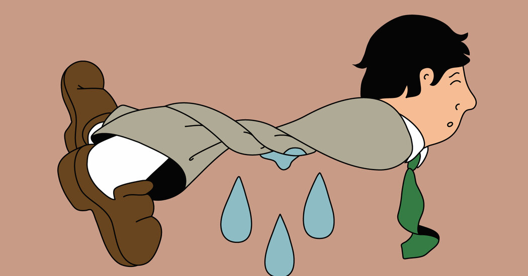
Experts offer tips on what to do if you’re not getting the respect or compensation you deserve.Life isn’t fair.It’s a phrase so often repeated that it has become a cliché. But studies have shown that humans are hard-wired to want their fair share, as are other animals that have cooperative relationships, like monkeys, birds and wolves.In one famous experiment, researchers trained two capuchin monkeys to hand them tokens in exchange for a cucumber snack. At first, the animals were happy with this arrangement — that is, until one of the monkeys received grapes instead, which are considered far more delicious. The other monkey, who continued to receive cucumbers, looked enraged, shook the walls of her enclosure and hurled the cucumbers out of reach.She would rather have nothing, it seemed, than receive an inferior reward.In the workplace, psychologists refer to this as effort-reward imbalance. The effort is the time, energy and emotional labor devoted to completing a task — and the rewards are what you get back from your workplace, such as compensation, benefits, recognition and opportunities.In humans, the perception that you are getting less than others for the same amount of work can contribute to symptoms associated with burnout and lead to a higher risk of depression. The need for fairness is most likely a biological predisposition to avoid exploitation, explained Sarah Brosnan, a professor of psychology, philosophy and neuroscience at Georgia State University who co-led the capuchin study.“We should care what we get relative to others,” she said. “We do best if we can work well with others, but it only benefits us if we’re working with someone who isn’t taking advantage of us.”If you feel that your efforts in the workplace are not in line with your rewards, here are some steps that you can take to examine the situation and, hopefully, find more balance.Get a reality check.Equity theory, which was developed in the 1960s by a behavioral psychologist, says that in order to feel motivated, employees need to be convinced that the rewards they receive are fair and similar to those that their counterparts are getting.But first ask yourself: Am I really being undervalued by my company, team or manager?“You might have a different view of your skills and your marketability than others do,” said Ben Dattner, an organizational psychologist and executive coach in New York City.If you are represented by a labor union, have a conversation with one of the leaders to get a better sense of how your compensation or other types of rewards compare to that of union members in similar roles. Consider also consulting with a career coach who can help you think through how to address potential inequitiesThink of it like a scale where effort is balanced with rewards, said Dennis Stolle, the senior director of applied psychology at the American Psychological Association.Ponder the intangible rewards too — are you learning a lot, deriving meaning from your work or making useful connections? Do you have a great boss or flexible hours? Do you receive recognition for your efforts?Sometimes the grass isn’t greener when you tally up those benefits.Take action.Once you identify your priorities, think: “What can I constructively do about this?” Dr. Dattner said.Have a direct conversation with your manager about your goals, Dr. Stolle said. Are they realistic? Are they in line with what the company needs and wants? During the conversation, be as concrete as possible about what you want, he advised.If you are looking for more compensation,take an objective look at the value you are adding to the company. This type of information will help your manager advocate on your behalf, Dr. Dattner said. Dr. Stolle noted that when it comes to the less tangible rewards, like the opportunity to advance, “there’s more room for miscommunication and hurt feelings.”He gave the hypothetical example of a young woman starting out in the marketing profession who wants to eventually be asked to attend client meetings. Her supervisor, however, is not aware she expects this to happen in her first year on the job. Having a conversation about her goals allows them to set a more concrete and realistic timeline.Stop overcommitting.Some people have difficulty stepping away from work, especially when technology enables us to stay continuously connected.But this can lead to what psychologists call overcommitment, where people pour themselves into their work, even when the rewards do not justify that level of effort.Overcommitment paired with low rewards can make workers especially vulnerable to emotional exhaustion, which is one of the signs of burnout, Dr. Stolle said.“When you reach emotional exhaustion, you’re just too tired to control your emotions anymore,” which can lead to tears or outbursts, Dr. Stolle said.If you have made your best effort and are still not being treated fairly, “you don’t have to live like that,” he added. “There are surely other opportunities. It may not happen tomorrow, but you can start looking.”

Read more →