Human mini guts reveal new insights into the process leading to Cronkhite-Canada syndrome and potential new therapies

Researchers at Baylor College of Medicine and collaborating institutions working with human intestinal organoids, also called mini guts, have shed new light on the potential causes of Cronkhite-Canada syndrome, a rare condition characterized by abundant non-cancerous growths or polyps in the intestine and other symptoms such as hair and nail loss and changes in skin pigmentation. Published in The Journal of Clinical Investigation, the study is the first to show a connection between high polyp proliferation and increased levels of serotonin produced by the intestinal epithelium.
The findings suggest a potential new approach to treat this disease with serotonin inhibitors already approved for other conditions, offering the possibility of improving the outcome of this disease for which the current prognosis is poor. The work also highlights not only the value of human organoid technology to help understand any disease but also to identify potential clinical applications.
“During my clinical rounds, I met a patient with Cronkhite-Canada syndrome and became very interested in better understanding this little known condition,” said first author Victoria Poplaski, a graduate student in Dr. Sarah Blutt’s lab at Baylor. “Patients with Cronkhite-Canada syndrome have diarrhea, weight loss, abdominal pain, anorexia and nausea. About 500 people have been diagnosed with this disease worldwide so there have not been many opportunities to investigate it to identify its causes and potential therapies.”
Poplaski and her colleagues applied their expertise in developing human intestinal organoids to take a closer look at what could be triggering so much proliferation in the patient’s intestine. Human intestinal organoids, or mini guts grown in the lab, model human intestinal cells, closely representing actual small intestine tissue and its functions.
Poplaski received a biopsy sample or tissue from the patient’s intestine and developed mini guts from it in the lab. “We discovered that, compared to mini guts from people without Cronkhite-Canada syndrome, the patient-derived mini guts were highly proliferative. They also had many more enteroendocrine cells, the cell type in the intestine that specializes in producing hormones, and the enteroendocrine cells abundantly present in the patient’s mini guts produced serotonin,” Poplaski said. “We confirmed these features in patient tissue biopsies.”
More than 90% of serotonin in the body is produced by the enteroendocrine cells in the intestinal tract. Although serotonin is best known for its role as a neurotransmitter in the central nervous system, it also affects a variety of functions in other organs, including some functions of the intestinal epithelium.
“Supporting the novel role of serotonin in intestinal epithelial proliferation, we found that treating non-Cronkhite-Canada syndrome organoids with serotonin increased their proliferation and inhibiting serotonin production in the Cronkhite-Canada syndrome organoids decreased proliferation, suggesting a link between local serotonin production and control of epithelial intestinal cell proliferation,” said Blutt, associate professor of virology and microbiology at Baylor and corresponding author of the work. “The findings suggest the potential benefit of serotonin inhibitors to treat this condition.”
This research is a perfect example of how organoid cultures can lead to new discoveries about the biology underlying rare diseases.
“When we started this research, some people said it was not worthwhile because initially we only had a single patient to study. I disagreed and thought we might find something worthwhile,” said co-author Dr. Mary Estes, Distinguished Service Professor of Virology and Microbiology and Cullen Foundation Endowed Chair at Baylor. She also is a member of Baylor’s Dan L Duncan Comprehensive Cancer Center. “The results from the organoids derived from that single patient yielded completely unexpected and exciting data that might lead to new therapies. Then we were able to confirm these results with organoids from a second patient. I am hoping this study will lead to contacts with more physicians caring for patients with Cronkhite-Canada syndrome and a clinical trial to test the new basis for a process that leads to this condition that we discovered in this work.”
Other contributors to this work include Carolyn Bomidi, Amal Kambal, Hoa Nguyen-Phuc, Sara C. Di Rienzi, Heather A. Danhof, Xi-Lei Zeng, Linda A. Feagins, Nan Deng, Eduardo Vilar, Florencia McAllister, Cristian Coarfa, Soyoun Min, Hyun Jung Kim, Richa Shukla and Robert Britton. The authors are affiliated with one of the following institutions: Baylor College of Medicine, the University of Texas at Austin Dell Medical School, the University of Texas MD Anderson Cancer Center-Houston or the Lerner Research Institute at Cleveland Clinic.

Read more →

Infant Deaths Have Risen for the First Time in 20 Years

The increases were particularly stark among babies born to Native American, Alaska Native and white mothers in 2022. Rates among Black infants remained highest of all.The number of American babies who died before their first birthdays rose last year, significantly increasing the nation’s infant mortality rate for the first time in two decades, according to provisional figures released Wednesday by the National Center for Health Statistics.The spike is a somber manifestation of the state of maternal and child health in the United States. Infant and maternal mortality, inextricably linked, are widely considered to be markers of a society’s overall health, and America’s rates are higher than those in other industrialized countries.The rates are particularly poor among Black and Native American mothers, who are roughly three times as likely to die during and after pregnancy, compared with white and Hispanic mothers. Their infants face up to double the risk of dying, compared with white and Hispanic babies.Overall life expectancy has declined in the United States in recent years, too, affecting white Americans as well as people of color. The declines were driven in part by the Covid-19 pandemic.The increase in infant mortality comes after a century of public health improvements, in which rates consistently and gradually declined almost every year with few exceptions, said Danielle M. Ely, a health statistician with the N.C.H.S. and the report’s lead author.The report did not delve into the cause of the increase, but most of the babies born in 2022 were conceived in 2021, when maternal deaths rose by 40 percent because of the pandemic and many pregnant women were taken ill.“Seeing an increase that hits the statistical significance mark indicates that this was a bigger jump than we’ve had in the last 20 years, and that is something we need to keep an eye on to see if it’s just a one-year anomaly or the start of increasing rates,” Dr. Ely said.One of the more disturbing findings was an increase in infant mortality among babies born to women ages 25 to 29. The rate increased to 5.37 per 1,000 live births last year, up from 5.15 deaths per 1,000 live births in 2021.Rates did not change for women in other age groups, even those who generally experience higher infant mortality rates, such as women younger than 20, those 20 to 24 and women 40 and older.Some 20,538 infants died in 2022, representing a 3 percent increase over the 19,928 babies who died in 2021. The infant mortality rate — defined as the number of babies who die before they are a year old for every 1,000 live births — also increased by a statistically significant 3 percent last year, to 5.6 infant deaths per 1,000 live births, up from 5.44 deaths per 1,000 live births in 2021, according to the new report.The mortality rate of babies who were between 4 weeks and a year old increased by 4 percent, while neonatal mortality rates — that of babies less than a month old — increased by 3 percent.Rates increased significantly among both premature babies born before 37 weeks of gestation and those born extremely early, at less than 34 weeks of gestation.Overall, the statistically significant increases in mortality rates were seen only among male infants, whose survival rates have always been slightly lower than those among females.Black infants have the highest mortality rate in the United States, rising slightly last year to 10.86 deaths per 1,000 live births, from 10.55 deaths per 1,000 live births in 2021, an increase that was not statistically significant.By contrast, the infant mortality rates of both white and Native American and Alaska Native babies increased by statistically significant amounts last year.Among white infants, the figure rose to 4.52 deaths per 1,000 live births from 4.36 deaths per 1,000 live births in 2021. Among Native American and Alaska Native babies, the figure increased to 9.06 deaths per 1,000 live births from 7.46 deaths per 1,000 live births in 2021.The two leading causes of infant deaths that were more prevalent last year were bacterial sepsis, caused by the body’s overwhelming reaction to an infection, and maternal health complications.Since infant deaths are relatively rare events involving small numbers of babies, statistically significant changes cannot easily be seen from year to year at the state level. Nevada was the only state that had a statistically significant decline in infant mortality, while four states — Georgia, Iowa, Missouri and Texas — experienced statistically significant increases in infant mortality last year.Texas banned abortions after six weeks of pregnancy in 2021, which may have played a role in the larger number of infant deaths the following year by preventing terminations of acutely ill fetuses.

Read more →

Scientists excited by AI tool that grades severity of rare cancer

Published1 day agoShareclose panelShare pageCopy linkAbout sharingBy Fergus WalshMedical editorArtificial intelligence is nearly twice as good at grading the aggressiveness of a rare form of cancer from scans as the current method, a study suggests.By recognising details invisible to the naked eye, AI was 82% accurate, compared with 44% for lab analysis.Researchers from the Royal Marsden Hospital and Institute of Cancer Research say it could improve treatment and benefit thousands every year.They are also excited by its potential for spotting other cancers early.AI is already showing huge promise for diagnosing breast cancers and reducing treatment times.Computers can be fed huge amounts of information and trained to identify the patterns in it to make predictions, solve problems and even learn from their own mistakes.What is AI and is it dangerous?AI named word of the year by Collins DictionaryDemis Hassabis: AI must not ‘move fast and break things'”We’re incredibly excited by the potential of this state-of-the-art technology,” said Professor Christina Messiou, consultant radiologist at The Royal Marsden NHS Foundation Trust and professor in imaging for personalised oncology at The Institute of Cancer Research, London.”It could lead to patients having better outcomes, through faster diagnosis and more effectively personalised treatment.” LISTEN to 5 Minutes On – AI and Cancer Diagnosis: “An absolute game changer” on BBC SoundsThe researchers, writing in Lancet Oncology, used a technique called radiomics to identify signs, invisible to the naked eye, of retroperitoneal sarcoma – which develops in the connective tissue of the back of the abdomen – in scans of 170 patients.With this data, the AI algorithm was able to grade the aggressiveness of 89 other European and US hospital patients’ tumours, from scans, much more accurately than biopsies, in which a small part of the cancerous tissue is analysed under a microscope.’Quicker diagnosis’When dental nurse Tina McLaughlan was diagnosed – in June last year, after stomach pain – with a sarcoma at the back of her abdomen, doctors relied on computerised-tomography (CT) scan images to find the problem. They decided it was too risky to give her a needle biopsy.The 65-year-old, from Bedfordshire, had the tumour removed and now returns to the Royal Marsden for scans every three months. She was not part of the AI trial but told BBC News it would help other patients.”You go in for the first scan and they can’t tell you what it is – they didn’t tell me through all my treatment, until the histology, post-op, so it would be really useful to know that straight away,” Ms McLaughlan said. “Hopefully, it would lead to a quicker diagnosis.”‘Personalised treatment’About 4,300 people in England are diagnosed with this type of cancer each year.Prof Messiou hopes the technology can eventually be used around the world, with high-risk patients given specific treatment while those at low risk are spared unnecessary treatments and follow-up scans.Dr Paul Huang, from the Institute of Cancer Research, London, said: “This kind of technology has the potential to transform the lives of people with sarcoma – enabling personalised treatment plans tailored to the specific biology of their cancer. “It’s great to see such promising findings.”Sign up for our morning newsletter and get BBC News in your inbox.More on this storySafer brain surgery using AI possible in two yearsPublished28 SeptemberGoogle boss: AI too important not to get rightPublished17 OctoberAI offers huge promise on breast cancer screeningPublished2 AugustHow AI is helping doctors spot breast cancersPublished9 JuneAI cuts treatment time for cancer radiotherapyPublished27 JuneAI ‘outperforms’ doctors diagnosing breast cancerPublished2 January 2020Around the BBC5 Minutes On – AI and Cancer Diagnosis – -An absolute game changer- – BBC SoundsRelated Internet LinksBiopsy – NHSThe BBC is not responsible for the content of external sites.

Read more →

Zimbabwe's cholera crisis fuelled by chronic water shortages

Published16 minutes agoShareclose panelShare pageCopy linkAbout sharingImage source, EPABy Farai SevenzoHarareFor months now, Zimbabwe has been battling to stem the spread of deadly cholera in its cities and villages because the country simply lacks clean water.”If the water comes at all it’s often dirty,” Regai Chibanda, a 46-year-old father of five from the sprawling township of Chitungwiza, told me.Cholera, an acute diarrhoeal infection caused by consuming food or water contaminated with the bacterium Vibrio cholerae, can spread quickly in cramped and dirty conditions. It has become a kind of grim reaper to this southern African nation – back in 2008-2009 more than 4,000 went to their graves when the water-borne disease struck in what was already a frenzied and turbulent time.It reflected the imploding political and economic crisis when hyper-inflation peaked at 80 billion per cent and heralded a historic power-sharing government that eventually got to grips with the situation.Today inflation is again rearing its head and cholera has spread across all of the country’s 10 provinces, mainly affecting children, often left unsupervised in the stifling heat as their parents try to work.This outbreak first struck back in February and as October ended official figures from the Health and Childcare Department are listing nearly 6,000 cases and some 123 suspected deaths. President Emmerson Mnangagwa, who won disputed polls in August for a second term in office, has promised a nationwide borehole drilling programme.This is to be supported by solar-powered water points, mainly to serve some 35,000 villages which do not have access to clean drinking water.In the capital, Harare, residents can go for weeks, or even months, without a regular supply of water from the Harare City Council. In Harare’s satellite township of Chitungwiza, more than 50 deaths were reported as October ended – all from cholera.Image source, EPAChitungwiza is a city all of its own given its size and population, but the infrastructure of its water works and civil planning have barely caught up with an ever-expanding population and a massive exodus from the villages to the city in the constant search for work.”In Chitungwiza things are not good as far as water is concerned. There have been many people affected by cholera and every year it’s the same,” said Mr Chibanda, who commutes by car daily to central Harare for his job as a printer. He said he had heard of several deaths in his neighbourhood.”Our water supply is not good, residents are resorting to buying mineral water from the supermarkets to save their lives but of course it’s hitting their pockets.”Out in Mutare, the main city in Manicaland’s eastern highlands, it is the same story – more infections from cholera and a city struggling to serve their residents with the most basic of needs – clean water.Social media is full of cholera information alerts, though a comment earlier this month on the health ministry’s Facebook page from a resident in the southern city of Bulawayo summed up the predicament for most: “How can we wash our hands? We don’t have running water in Bulawayo – for almost two weeks now.”Cholera is cheap to treat with rehydration salts – and easy to avoid altogether if people have access to clean water and decent toilet facilities.As recently as last week, Harare pharmacist Panashe Chawana, 26, told me he was seeing between two and three patients in the chemist for cholera medication – ranging from children to adults, all showing the classic symptoms of the runs and a desperate lack of energy”If it wasn’t for the public announcements, Harare would have seen much more. It’s only because people became aware of the dangers of un-purified water matching their symptoms that’s when they sought medical help,” says Mr Chawana. “And so we tell them to look out for white substance in their stool and prescribe things like [the antibiotic] Azithromycin. On the whole there’re less people coming in now.”But aid organisation Mercy Corps, in an appeal for borehole funding, has warned that the situation is far from improving.”Despite a significant decline in cases from July to August, we now witness a worrying spike of cholera cases, particularly among women and children. In Manicaland, many people have to use crowded water facilities, while others must rely on unsafe wells and rivers for drinking water, putting them at further risk,” Mildred Makore, the group’s country director, said in a statement.A few days ago the World Health Organization’s emergencies director Mike Ryan called cholera “a poster child of poverty, social injustice, climate change and conflict”. It is not straightforward to see which of these can be pinned on President Mnangagwa’s Zimbabwe, but the reported cholera cases point to a lack of will or ability or both to stem the occurrences by providing fresh water.In Harare’s southern suburbs of Budiriro and Mbare, stretching to informal settlements like Hopley, the search for water is a visual reality.Image source, AFPWheelbarrows are carted across many roads to community centres and churches with boreholes willing to open their taps and share their water.The government’s investment in fresh water supplies has been underwhelming, and here critics point to the disparities in wealth between those who can afford to sink boreholes in their backyards and those who cannot.In the towns, city councils – often run by the opposition – blame a desperate lack of investment by the government in providing new kit and cleaning chemicals to purify the water.That the government is always caught unawares shows a depressing underinvestment in the waterworks of its cities and its rural areas.Precious Shumba, director of the Harare Residents’ Trust, an NGO that says water shortages in the capital are worsening, urged the government to do more to help councils.”Local authorities cannot sustain service delivery from ratepayers alone,” he told Zimbabwe’s Independent newspaper – pointing to the cost of replacing broken pipes and chemicals.The paper reported that water-treating chemicals were costing the City of Harare up to US$3m (£2.5m) a month.Sewage management is also to blame, with Mr Shumba noting that industrial waste and effluent are continually being discharged into the tributaries and streams feeding Lake Chivero, which provides the main water supply for Harare.In more affluent areas of the capital, folks organise their own rubbish collections through community initiatives – but elsewhere the streets have turned into rubbish dumps because the authorities no longer organise collections.With the heavens set to open for the seasonal rains, many fear the dirt and filth accumulated over months mean cholera, which lurks in shallow pools of water, will prove difficult to defeat.They continue the battle of keeping their toddlers away from the water taps and puddles and running the daily gauntlet of what is or not safe to drink. Farai Sevenzo is a freelance journalist and film-maker based in Harare.More on Zimbabwe:Zimbabwe struggling after nurse exodus to UKHow Zimbabwe is still haunted by Robert Mugabe A quick guide to ZimbabweAround the BBCFocus on Africa podcasts

Read more →

Second Maryland Man to Receive an Altered Pig’s Heart Has Died

Researchers said they found signs of organ rejection, which the genetically modified tissue was supposed to prevent.A 58-year-old man with heart failure who received a new heart from a genetically modified pig died on Monday, nearly six weeks after receiving the pig organ, University of Maryland Medical Center officials announced on Tuesday.Lawrence Faucette, of Frederick, Md., was the second patient at the medical center to have had an ailing heart replaced with one from a pig that had been genetically modified so its organs would be more compatible with a human recipient and would not be rejected by the human immune system.The first patient, 57-year-old David Bennett, died last year, two months after his transplant. He had developed multiple complications, and traces of a virus that infects pigs were found in his new heart.Both of the patients had terminal heart disease when they received the transplanted organs, and neither managed to recover sufficiently to leave the hospital. But while doctors said that Mr. Bennett did not show any signs of acute rejection of the new heart, which is the most significant risk in organ transplants, they said that Mr. Faucette’s transplanted heart had started to display some initial signs of rejection.“We mourn the loss of Mr. Faucette, a remarkable patient, scientist, Navy veteran and family man, who just wanted a little more time to spend with his loving wife, sons and family,” said Dr. Bartley P. Griffith, the surgeon who performed the transplant at the University of Maryland Medical Center in Baltimore.Mr. Faucette was very engaged in his own care, reading and interpreting his own biopsies, said Dr. Muhammad M. Mohiuddin, a professor of surgery and the scientific program director of the cardiac xenotransplantation program at the University of Maryland School of Medicine.“Mr. Faucette’s last wish was for us to make the most of what we have learned from our experience, so others may be guaranteed a chance for a new heart when a human organ is unavailable,” Dr. Griffith said.After the surgery, the transplanted heart performed well, with no signs of rejection during the first month, and Mr. Faucette was able to do physical therapy with the aim of regaining his ability to walk, according to a statement from the University of Maryland.Like Mr. Bennett, the first patient to receive a pig’s heart, Mr. Faucette was rejected from transplant programs that use a traditional organ from a deceased human donor. He was too sick, suffering from advanced heart failure as well as peripheral vascular disease and other complications.He was in end-stage heart failure on Sept. 14 when he was admitted to the University of Maryland Medical Center, and shortly before the surgery, his heart stopped and he had to be resuscitated.His wife, Ann Faucette, said at that time that the couple was keeping expectations low, and hoping only for some more time to sit “on the front porch” and have coffee together.After his death, Ms. Faucette said that her husband was a kind and selfless man who hoped his experience would help save lives by advancing the field of xenotransplantation, or the transplantation of organs or tissues from an animal source into a human recipient.“He knew his time with us was short, and this was his last chance to do for others,” she said in a statement.Transplant surgeons at a number of medical centers have been working fervently to advance the field of xenotransplantation. Most of the work so far has involved transplanting kidneys from genetically modified pigs into brain-dead patients being maintained on ventilators, in order to demonstrate that the kidneys can make urine and perform other essential biological functions without being rejected.More than 100,000 Americans are living with end-stage organ disease, and there is an acute shortage of human donor organs. Most of those waiting for an organ need a kidney, but fewer than 25,000 kidney transplants are done each year and thousands die while on the waiting list.

Read more →

Panel Tells F.D.A. That CRISPR Sickle Cell Cure Is Safe Enough for Patients

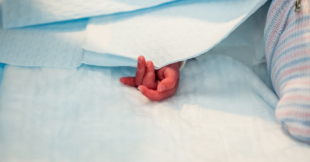
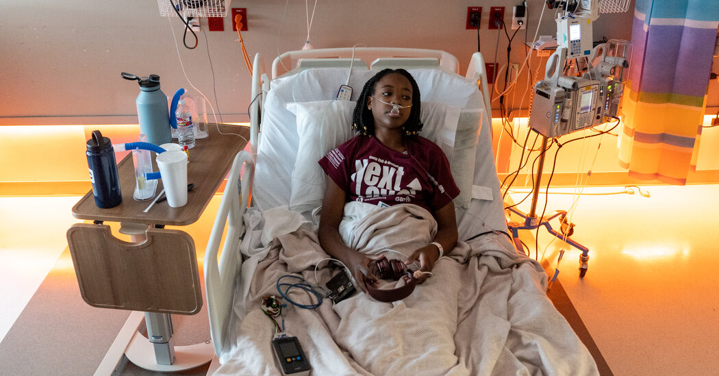
The decision by an advisory committee may lead to Food and Drug Administration approval of the first treatment for humans that uses the CRISPR gene-editing system.A panel of experts said on Tuesday that a groundbreaking treatment for sickle cell disease was safe enough for clinical use, setting the stage for likely federal approval by Dec. 8 of a powerful potential cure for an illness that afflicts more than 100,000 Americans.The Food and Drug Administration had previously found that the treatment, known as exa-cel and jointly developed by Vertex Pharmaceuticals of Boston and CRISPR Therapeutics of Switzerland, was effective. The panel’s conclusion on Tuesday about exa-cel’s safety sends it to the F.D.A. for a decision on greenlighting it for broad patient use.Exa-cel frees patients from the debilitating and painful effects of this chronic, deadly disease. If approved, the Vertex product would be the first medicine to treat a genetic disease with the CRISPR gene-editing technique.It could also be the first of a series of new options to cure the excruciating illness. By Dec. 20, the F.D.A. will decide on a second potential cure for sickle cell, a gene therapy devised by the company Bluebird Bio of Somerville, Mass.Sickle cell disease is caused by a gene mutation that makes blood cells misshapen, so that they resemble sickles or crescents. It affects millions of people worldwide, most of whom have African ancestry. The misshapen cells get stuck in blood vessels, causing strokes, organ damage and episodes of agonizing pain as muscles are starved of oxygen.Sickle cell’s toll starts early in life. Evelyn Islam of Milwaukee, now 8, had 22 blood transfusions and had to have her spleen removed before she was 3. “Gene therapy is our last hope for a cure,” said her mother, Melissa Nicole Allen.Ashley Valentine, a co-founder of the national advocacy group Sick Cells, at her parents’ home in Lisle, Ill. Her brother Marqus, pictured on the wall, died in 2020.Mustafa Hussain for The New York TimesBut the new gene therapies will come too late for many.Ashley Valentine, a co-founder of the national advocacy group Sick Cells, had to take three months off from work in 2016 to help her brother Marqus deal with symptoms of sickle cell. When he had a hip replacement in 2018, her father ended up accepting a layoff from his job to help care for him.“And that’s just us,” she said.Marqus died in 2020, at age 36, from a stroke caused by sickle cell.New treatments like the one that was endorsed on Tuesday are expected to cost millions of dollars per patient, though Vertex has not yet said what it will charge. But lifelong care for patients with the disease is also enormously expensive, costing the health care system an estimated $3 billion a year.It’s not yet clear how many people will seek the new therapy. The new therapies are also not easy to endure and come with hardships for patients, who will have to undergo chemotherapy and spend more than a month in the hospital. Family members are affected too — they may need to take time off work during the most intensive phase of the treatment.Additionally, most Americans with sickle cell are Black and may not trust a health care system that has often failed to provide the most basic preventive and therapeutic care for those with the disease. Some with sickle cell are anxious about undergoing a medical treatment that is on the cutting edge of biotechnology.But for doctors who have spent years watching patients suffer, and many parents who have seen their children endure years of agony, there is elation at what lies ahead.“We are finally at a spot where we can envision broadly available cures for sickle cell disease,” said Dr. John Tisdale, director of the cellular and molecular therapeutics branch at the National Heart, Lung and Blood Institute and a member of the advisory committee.Dana Jones of San Antonio wants her daughters Kyra, 18, and Kami, 20, to have a chance at one of the new therapies. Both had strokes that left them with learning disabilities — injuries that could probably have been avoided if they had been given a screening test and treatment long known to prevent nine out of 10 strokes in children with the disease. Kyra is now in intensive care as doctors try to control her pain.Ms. Jones is overwhelmed by the possibility that her daughters could be cured.“It is my prayer that Kami and Kyra can be cured of this awful disease and finally be able to truly live,” she said.A New Treatment and a New TechnologySickle cell disease is caused by a gene mutation that makes blood cells misshapen. The misshapen cells can get stuck in blood vessels, causing strokes, organ damage and episodes of agonizing pain.F. Gilbert/CDC, via Associated PressThe cause of sickle cell has been known for nearly 70 years, but research lagged, a situation many say occurred at least in part because so many patients were Black and from poor and working-class families.There are a number of treatments to reduce sickle cell’s impact. Some patients are able to get bone marrow transplants that can cure the condition. But that requires finding a donor and, after the transplant, taking drugs to prevent the body from rejecting the foreign cells.In recent years, a number of biotechnology companies have tried novel approaches. While Bluebird Bio is advancing its gene therapy technique, Vertex and CRISPR Therapeutics focused on the gene-editing system CRISPR-Cas9, which can home in on specific areas of DNA and turn genes on or off. CRISPR has allowed researchers to disable genes to assess their importance in biomedical research. But until now it has not been used as a treatment for patients with a genetic disease.To treat sickle cell, CRISPR snips a piece of DNA in bone marrow stem cells. That frees a blocked gene to make a form of hemoglobin that normally is produced only by a fetus. The fetal gene directs the production of hemoglobin that does not form into the sickle shape. In clinical trials, patients no longer had the complications of sickle cell disease and no longer needed blood transfusions.But there is a concern that CRISPR could inadvertently snip a piece of DNA in the wrong part of a patient’s genome. That might disrupt a gene and cause a blood cancer.No such issues have turned up in the clinical trials, but the Vertex trial involved only 44 patients, and just 30 have been followed for at least 16 months. The company did extensive comparisons of patients’ DNA with that of people in large databases asking how likely such CRISPR misfires could be.Vertex said it plans to follow clinical trial patients for 15 years. The company’s data were sufficiently reassuring that the expert committee said on Tuesday they saw no reason to hold the treatment back.There can always be additional studies, noted committee member Alexis Komor, a professor of chemistry and biochemistry at the University of California, San Diego. But, she said, that would be “expecting perfection at the expense of progress.”Dr. Joseph Wu of Stanford added, “We all agree that the benefits outweigh the risks. These patients are quite sick and this is a good therapy.”Scot Wolfe of the University of Massachusetts Chan Medical School said, “We want to be careful not to let the perfect be the enemy of the good.”“There is a huge unmet need,” he added.If It’s Safe, Who Gets It?Vertex Pharmaceuticals’ headquarters in Boston.Bill Sikes/Associated PressVertex estimates that 20,000 people could be eligible for its treatment, and says Medicaid and private insurers have suggested a willingness to pay for it.“There is almost no way they could not pay,” said Dr. David Williams, chief of the division of hematology and oncology at Boston Children’s Hospital.Dr. Williams, who has consulted for Vertex and Bluebird Bio, added that insurers pay “$3 million a pop” for other gene therapies produced by Bluebird Bio for the diseases thalassemia and adrenoleukodystrophy. With sickle cell, and its large number of Black patients, he said, there is an issue of “equity in access and the tremendous medical need.”Some people with the disease may not be eligible, depending on the F.D.A.’s decisions. They could include young children with sickle cell and older patients whose bodies have been so damaged that the treatment could pose heightened risks.Kevin Wake of Kansas City, Mo., hopes he is not too old, at 55, or too damaged. He has had three strokes caused by the disease.The treatments, though curative, are difficult.Patients first have eight weeks of blood transfusions followed by a treatment to release bone marrow stem cells into their bloodstream. The stem cells are then removed and sent to the companies to be treated. Next, patients receive intense chemotherapy to clear their marrows for the treated cells. The treated cells are infused back into the patients, but they have to remain in the hospital for at least a month while the new cells grow and repopulate their marrows.That treatment “cannot be delivered at most hospitals,” said Dr. Alexis Thompson, chief of the division of hematology at Children’s Hospital of Philadelphia, who consults for Vertex.Another issue is how quickly Vertex can ramp up production. Each patient’s cells must be treated individually in a sterile environment, an arduous prospect.Stuart Arbuckle, executive vice president and chief operating officer at Vertex, is confident. “We are launch ready,” he said. But he added that he did not expect a huge wave of patients immediately.“This is a pretty big decision for a patient to go through,” Mr. Arbuckle said. Dana Jones with her daughter Kyra, now 18, in 2020. Kyra and her sister Kami have had strokes that caused them to have learning disabilities, and Kyra is currently in intensive care as doctors try to control her pain.Ilana Panich-Linsman for The New York TimesOne of the Vertex clinical trial patients, Marie-Chantal Tornyenu, 22, who is a senior at Cornell University, said patients also had to be prepared for “mental adjustment” after treatment.Ms. Tornyenu said she no longer had the pain crises that plagued her, especially in high school when she was hospitalized nearly every month.But she has spent much of her life taking precautions and worrying about pain and complications from sickle cell. Those habits are hard to break.“It’s a major learning curve from having sickle cell my whole life,” she said. “I’m still struggling with that mind-set — ‘sickle cell is you.’”

Read more →

New position statement supports permanent standard time

An updated position statement from the American Academy of Sleep Medicine supports the replacement of daylight saving time with permanent standard time.
It is the position of the AASM that the United States should eliminate seasonal time changes in favor of permanent standard time, which aligns best with human circadian biology. According to the statement, evidence supports the distinct benefits of standard time for health and safety, while also underscoring the potential harms that result from seasonal time changes to and from daylight saving time.
“By causing the human body clock to be misaligned with the natural environment, daylight saving time increases risks to our physical health, mental well-being, and public safety,” said lead author Dr. M. Adeel Rishi, who is chair of the AASM Public Safety Committee and a pulmonary, sleep medicine, and critical care specialist at Indiana University Health in Indianapolis. “Permanent standard time is the optimal choice for health and safety.”
The position statement was published as an accepted paper Oct. 31 in the Journal of Clinical Sleep Medicine, the official publication of the AASM.
The original position statement published by the AASM in 2020 stated that current evidence best supports the adoption of year-round standard time. Based on a growing body of evidence, the updated position statement emphasizes that daylight saving time should be replaced by permanent standard time. This position is supported by similar statements adopted by other organizations including the Society for Research on Biological Rhythms, National Sleep Foundation, Sleep Research Society, and American Medical Association.
“Permanent standard time helps synchronize the body clock with the rising and setting of the sun,” said Dr. James A. Rowley, president of the AASM. “This natural synchrony is optimal for healthy sleep, and sleep is essential for health, mood, performance, and safety.”
The position statement was developed by the AASM Public Safety Committee and based on a review of existing literature. It was approved by the AASM board of directors and endorsed by 20 medical, scientific, and advocacy organizations: American Academy of Cardiovascular Sleep Medicine American Academy of Dental Sleep Medicine American Academy of Otolaryngology-Head and Neck Surgery American Association of Sleep Technologists American College of Chest Physicians (CHEST) American College of Lifestyle Medicine American Society for Metabolic and Bariatric Surgery American Thoracic Society Dakota Sleep Society Michigan Academy of Sleep Medicine Montana Sleep Society National PTA National Safety Council National Sleep Foundation Sleep Research Society Society for Research on Biological Rhythms Society of Anesthesia and Sleep Medicine Society of Behavioral Sleep Medicine Southern Sleep Society World Sleep SocietyMost of the U.S. will return to standard time when daylight saving time ends Sunday, Nov. 5.

Read more →

COVID vaccination in female, male partners does not increase risk of miscarriage, study finds

Multiple studies have shown that the COVID-19 vaccines do not lead to infertility or pregnancy complications such as miscarriage, but many people are still wary of adverse effects from the vaccine on pregnancy.
A new study led by Boston University School of Public Health (BUSPH) researchers now provides deeper insight into the safety of COVID-19 vaccines for people planning to become pregnant.
Published in the journal Human Reproduction, the study found no increased risk of early or late miscarriage as a result of male or female partners getting a COVID-19 vaccine prior to conceiving.
The study is the first to evaluate the risk of early miscarriage (less than eight weeks’ gestation) following preconception COVID-19 vaccination, as well as the first to evaluate male vaccination and miscarriage.
The researchers hope these results provide useful information for individuals planning to become pregnant, as well as their healthcare providers.
“These findings should be replicated in other populations, but are reassuring for couples who are planning pregnancy,” says lead author Jennifer Yland, an epidemiology PhD student at BUSPH at the time of the study.
For the study, Yland and colleagues analyzed survey data on COVID-19 vaccination and miscarriage among female and male participants in the BUSPH-based Pregnancy Study Online (PRESTO), an ongoing National Institutes of Health-funded study that enrolls women trying to conceive, and follows them from preconception through six months after delivery. Participants in this new analysis included 1,815 female individuals in the US and Canada who were followed in the study from December 2020 through November 2022. They were observed from their first positive pregnancy test until a miscarriage or other event (such as induced abortion, ectopic pregnancy, or 20 weeks’ gestation) — whichever occurred first.

Read more →

Combining cell types may lead to improved cardiac cell therapy following heart attack

Researchers at the University of Wisconsin-Madison and Academia Sinica of Taiwan have harnessed a combination of lab-grown cells to regenerate damaged heart muscle.
The study, published in Circulation — which addresses major challenges of using heart muscle cells, called cardiomyocytes, grown from stem cells — takes a crucial step toward future clinical applications.
Previous research has shown that transplanting cardiomyocytes made from induced pluripotent stem cells (iPSC) can replace muscle in the hearts of mammals. Researchers have struggled to bring the treatment to the clinic, in part because the implanted cells haven’t developed enough life-sustaining blood vessels to survive very long.
The new study confronted that challenge by combining the lab-grown cardiomyocytes with stem-cell-derived endothelial cells — the cells that line blood. The combination therapy also holds promise for tackling arrhythmia, another significant obstacle in heart regeneration with stem-cell-derived cardiomyocytes.
“Our findings suggest that human iPSC-derived endothelial cells can effectively augment the remuscularization of the heart by iPSC-derived cardiomyocytes, offering a promising avenue for future clinical applications,” says Patrick Hsieh, a researcher with Academia Sinica’s Institute of Biomedical Sciences who conducted the study while working as a visiting professor at the UW-Madison Stem Cell & Regenerative Medicine Center.
Hsieh and study lead author Yu-Che Cheng collaborated with Tim Kamp, who serves as director of the Stem Cell & Regenerative Medicine Center, as well as a team of researchers at UW-Madison and the Wisconsin National Primate Research Center, to examine the therapeutic effect of co-transplantation in mice and non-human primates undergoing a heart attack.
“The main advantage of iPSCs is their ability to be differentiated into many types of cells and serve as a valuable resource for cell therapy,” says Cheng, who is a project manager at Academia Sinica. “In this study, we generated billions of endothelial cells and cardiomyocytes from the same iPSCs line to inject into mice and non-human primates.”
“The simple idea of the project was to enhance blood flow and promote survival of iPSC-cardiomyocytes using blood vessel-forming endothelial cells,” Kamp says. “But the reality of generating the optimal cell preparations followed by precise delivery to the heart reflects tremendous effort by an international team of collaborators.”

Read more →

First classification of four stages of heart attack based on heart muscle damage is released

Heart attacks, or acute myocardial infarction (MI), are one of the leading causes of death worldwide. The newly released Canadian Cardiovascular Society Classification of Acute Myocardial Infarction (CCS-AMI) appearing in the Canadian Journal of Cardiology, published by Elsevier, presents a four-stage classification of heart attack based on heart muscle damage. This work by a group of noted experts has the potential to stratify risk more accurately in heart attack patients and lays the groundwork for development of new, injury-stage-specific and tissue pathology-based therapies.
Lead author Andreas Kumar, MD, MSc, Northern Ontario School of Medicine University, and Department of Cardiovascular Sciences, Health Sciences North, Sudbury, ON, Canada, explains: “MI remains a leading cause of morbidity and mortality. Existing tools classify MIs using a patient’s clinical presentation and/or the cause of the heart attack, as well as ECG findings. Although these tools are very helpful to guide treatment, they do not consider details of the underlying tissue damage caused by the heart attack. This expert consensus, based on decades of data, is the first classification system of its kind ever released in Canada and internationally. It offers a more differentiated definition of heart attacks and improves our understanding of acute atherothrombotic MI. On a tissue level, not all heart attacks are the same; the new CCS-AMI classification paves the way for development of more refined therapies for MI, which could ultimately result in better patient clinical care and improved survival rates.”
The CCS-AMI classification describes damage to the heart muscle following an MI in four sequential and progressively severe stages. Each stage reflects progression of tissue pathology of myocardial ischemia and reperfusion injury from the previous stage. It is based on a strong body of evidence about the effect an MI has on the heart muscle.
As damage to the heart increases through each progressive CCS-AMI stage, patients have dramatically increased risk of complications such as arrhythmia, heart failure, and death. Appropriate therapy can potentially stop injury from progressing and halt the damage at an earlier stage. Stage 1: Aborted MI (no/minimal myocardial necrosis). No or minimal damage to the heart muscle. In the best case the entire area of myocardium at risk may be salvaged. Stage 2: MI with significant cardiomyocyte necrosis, but without microvascular injury. Damage to the heart muscle and no injury to small blood vessels in the heart. Revascularization therapy will result in restoration of normal coronary flow. Stage 3: MI with cardiomyocyte necrosis and microvascular dysfunction leading to microvascular obstruction (i.e., “no-reflow”). Damage to the heart muscle and blockage of small blood vessels in the heart. The major adverse cardiac event rate is increased 2- to 4-fold at long-term follow-up. Stage 4: MI with cardiomyocyte and microvascular necrosis leading to reperfusion hemorrhage. Damage to the heart muscle, blockage and rupture of small blood vessels resulting in bleeding into the heart muscle. This is a more severe form of microvascular injury, and the most severe form of ischemia-reperfusion injury. It is associated with a further increase in adverse cardiac event rate of 2- to 6-fold at long-term follow-up.Dr. Kumar concludes: The new classification will help differentiate heart attacks according to the stage of tissue damage and allow healthcare providers to estimate a patient’s risk more precisely for arrhythmia, heart failure, and death. The CCS-AMI is ultimately expected to lead to better care, better recovery, and better survival rates for heart attack patients.”
In an accompanying editorial, Prakriti Gaba, MD, Brigham and Women’s Hospital, Harvard Medical School, and Deepak L. Bhatt, MD, MPH, Mount Sinai Heart, Icahn School of Medicine at Mount Sinai, comment: “Kumar et al. present a novel and intriguing four-tiered classification scheme of patients with acute MI. This allows unique utilization of prognostic pathologic features to help distinguish between high and low risk acute MI patients. Greater access to cardiovascular magnetic resonance would be needed to implement this new clinical approach broadly, however, for research on emerging diagnostic and therapeutic strategies, it could be implemented immediately.”

Read more →