Obesity linked to neurodegeneration through insulin resistance

Researchers led by Mroj Alassaf at the Fred Hutchinson Cancer Research Center in the United States have discovered a link between obesity and neurodegenerative disorders like Alzheimer’s disease. Using the common fruit fly, the research shows that a high-sugar diet — a hallmark of obesity — causes insulin resistance in the brain, which in turn reduces the ability to remove neuronal debris, thus increasing the risk of neurodegeneration. Publishing November 7 in the open access journal PLOS Biology, the research will impact therapies designed to reduce the risk of developing neurodegenerative diseases.
Although obesity is known to be a risk factor for neurodegenerative disorders like Alzheimer’s disease and Parkinson’s disease, exactly how one leads to the other remains a mystery. The new study focused on answering this question by taking advantage of the similarity between humans and fruit flies. Having previously shown that a high-sugar diet leads to insulin resistance in the peripheral organs of flies, the researchers now turned to their brains. Specifically, they examined glial cells because microglial dysfunction is known to lead to neural degeneration.
Levels of the protein PI3k indicate how much a cell is able to respond to insulin. The researchers found that the high sugar diet led to reduced PI3k levels in glial cells, indicating insulin resistance. They also looked at the fly equivalent of microglia, called ensheathing glia, whose primary function is to remove neural debris, such as degenerating axons. They observed that these glia had low levels of the protein Draper, indicating impaired function. Further tests revealed that artificial reduction of PI3k levels led to both insulin resistance and low Draper levels in ensheathing glia. Finally, they showed that after actually damaging olfactory neurons, the ensheathing glia could not remove the degenerating axons in the flies on the high sugar diet because their Draper levels did not increase.
The authors add, “Using fruit flies, the authors establish that high-sugar diets trigger insulin resistance in glia, disrupting their ability to clear neuronal debris. This study provides insight into how obesity-inducing diets potentially contribute to the increased risk of neurodegenerative disorders.”

Read more →

Senate Confirms Monica Bertagnolli as NIH Director

Dr. Monica M. Bertagnolli won bipartisan approval despite opposition from Senator Bernie Sanders, the chairman of the Senate health committee.The Senate on Tuesday confirmed Dr. Monica M. Bertagnolli, a cancer surgeon who currently leads the National Cancer Institute, as the next director of the National Institutes of Health, overriding the objections of Senator Bernie Sanders of Vermont, the chairman of the Senate health committee.The vote was 62 to 36. In a statement last month, Mr. Sanders said that while Dr. Bertagnolli was an “intelligent and caring person,” he would vote against her because she “has not convinced me that she is prepared to take on the greed and power of the drug companies and health care industry.”Dr. Bertagnolli will become only the second woman to lead the N.I.H. on a permanent basis, after Dr. Bernadine P. Healy, who served under President George H.W. Bush. She will take over an agency that has been the target of political attacks by Republicans, who have accused its scientists of intentionally downplaying the possibility that Covid-19 was the result of a laboratory leak.“I think no one wants to know what the true origin of the last Covid pandemic was more than the biomedical research community,” Dr. Bertagnolli told Senator Bill Cassidy of Louisiana, the top Republican on the health committee, during her confirmation hearing last month.“And how will you accomplish that?” asked Mr. Cassidy, who is a medical doctor. Dr. Bertagnolli promised she would make any data on the subject “available, public and accountable to the American people.”President Biden announced in May that he would nominate Dr. Bertagnolli to lead the N.I.H., the world’s premier medical research agency, which has an annual budget of more than $47 billion and occupies a sprawling campus in Bethesda, Md. It has been without a permanent director since Dr. Francis S. Collins stepped down nearly two years ago.We are having trouble retrieving the article content.Please enable JavaScript in your browser settings.We are confirming your access to this article, this will take just a moment. However, if you are using Reader mode please log in, subscribe, or exit Reader mode since we are unable to verify access in that state.Confirming article access.If you are a subscriber, please 

Read more →

Measuring skin water loss predicts anaphylaxis during food allergy tests

Anaphylaxis — a severe allergic reaction that may include a skin rash, nausea, vomiting, difficulty breathing, and shock — from a food allergy sends 200,000 people to the emergency room annually in the United States.
Because pinpointing a food allergy could mean life or death, an accurate diagnosis is critical.
Oral food challenges — when a patient ingests increasing doses up to a full serving of the suspected food allergen under supervision of a medical provider — are the diagnostic standard as skin and blood allergy tests have high false positive rates.
Although a highly accurate diagnostic test, patients often experience anaphylaxis during oral food challenges necessitating an epinephrine injection.
A team of University of Michigan researchers developed a method that measures water loss from the skin to predict anaphylaxis during oral food challenges before it becomes clinically evident.
The results are published in The Journal of Clinical Investigation.
“This method could enhance the ability to detect and predict anaphylaxis during oral food challenges prior to the need for epinephrine, greatly improving patient safety and comfort,” said Charles Schuler, M.D., lead author of the study and an immunologist at Michigan Medicine.

Read more →

Connectivity scans could serve as brain 'blueprints' for adolescents, researchers find

Researchers with the Translational Research in Neuroimaging and Data Science (TReNDs) Center at Georgia State have identified important new methods for accurately identifying possible biomarkers in adolescent brains that can reliably predict cognitive developments and psychiatric issues.
A new study, published in Nature Mental Health, represents the first large-scale analysis of its kind in which researchers analyzed functional network connectivity (FNC) across scans and identified associations with a diverse range of health measures in children. Researchers believe that inferences about early cognitive and psychiatric behaviors in children may be made using these intra-subject variabilities as a useful biomarker.
Researchers studied four scans from more than 9,000 subjects ages 9 to 11.
Neuroscientist, Distinguished University Professor and head of the TReNDS Center at Georgia State Vince Calhoun worked with the research team to develop the study. He said the research demonstrates that, independent of brain growth and development, a child’s FNC is robust and stable with high similarity across scans and can serve as a fingerprint to identify an individual child from a large group.
“This study is quite exciting as it shows the promise of using advanced machine learning to identify brain patterns which might help us intervene early in children who are most at risk for cognitive or psychiatric problems,” said Calhoun, who is the senior author of the study.
Researchers say that brain functional connectivity derived from functional magnetic resonance imaging (fMRI) is commonly used as a potential blueprint for adults. But they believe that intra-subject variation of FNC can carry biologically meaningful information, especially during adolescence, which is a time of significant change in the brain.
Principal investigator Zening Fu said the study demonstrates that functional connectivity variability can predict a wide range of children’s behavior, including cognition, mental health and sleep conditions.

Read more →

Body changes up to eight years before inflammatory bowel disease diagnosis

Researchers at the Francis Crick Institute and Aalborg University in Copenhagen have shown that changes can be detected in blood tests up to eight years before a diagnosis of Crohn’s disease and up to three years before a diagnosis of ulcerative colitis.
This means the beginnings of inflammatory bowel diseases start a long time before symptoms occur, and in the future may provide an opportunity for doctors to take preventative action before symptoms begin, or prescribe medication when it will be most effective.
Crohn’s disease and ulcerative colitis are collectively known as inflammatory bowel diseases (IBD). They are incurable conditions which involve excessive inflammation in the gut, leading to symptoms like abdominal pain and diarrhea. Early diagnosis and treatment are key to improving outcomes, but nearly a quarter of the 25,000 people diagnosed each year in the UK wait over a year.
In their study published today in Cell Reports Medicine, the team used electronic health records from people in Denmark, comparing 20,000 people with an IBD diagnosis with controls from 4.6 million people without IBD.
It was previously thought that most people have symptoms for about a year before diagnosis, but significant bowel damage is often seen, suggesting that changes have been taking place for a lot longer.
The researchers confirmed this by looking at ten years of test results before diagnosis. They observed changes in a series of minerals, cells in the blood and markers of inflammation, such as faecal calprotectin, a molecule released into the gut during inflammation and currently used to determine which people with bowel symptoms need further investigations. These changes were observed up to eight years before diagnosis in Crohn’s disease and three years in ulcerative colitis.
Importantly, most of the changes observed were subtle and would have appeared within a normal range for standard blood tests, so wouldn’t have been flagged as a cause for concern. It required a huge dataset to be able to detect these changes across many different markers.

Read more →

Infection with common cat-borne parasite associated with frailty in older adults

A common, cat-borne parasite already associated with risk-taking behavior and mental illness in humans may also contribute to exhaustion, loss of muscle mass, and other signs of “frailty” in older adults, suggests a study published Nov. 6 in the Journal of Gerontology: Medical Science.
The research, by an international team of scientists including University of Colorado Boulder, University of Maryland School of Medicine and the University of A Coruña in Spain, is the latest to explore how the tiny, single-celled organism Toxoplasma gondii (T. gondii) could have big impacts on human health.
“We often think of T. gondii infection as relatively asymptomatic, but this study highlights that for some people it may have significant health consequences later on,” said co-author Christopher Lowry, a professor in the Department of Integrative Physiology at CU Boulder.
Approximately 11% to 15% of people in the U.S. have been infected with T. gondii at some point and rates tend to be far higher in older individuals. In some countries, more than 65% have been infected. Once infected, people can unknowingly harbor the parasite for life.
For the study, the team examined the blood of 601 Spanish and Portuguese adults over 65, along with measures of a common geriatric syndrome known as “frailty” — which includes unintentional weight loss, tiredeness, loss of cognitive sharpness and other indications of declining health.
A whopping 67% of study subjects were “seropositive” showing markers in their blood of a latent infection.
The researchers did not, as they originally hypothesized, find an association between any infection to T. gondii and frailty. But they did find that, among those infected, those with higher “serointensity” or a higher concentration of antibodies to the parasite, were significantly more likely to be frail.

Read more →

Why we don't all develop posttraumatic stress disorder after trauma

Posttraumatic stress disorder (PTSD) is a debilitating condition that arises after experiencing traumatic events. While many people experience trauma, only about 25-35% develop PTSD. Understanding the factors that make certain individuals more susceptible is crucial for both prevention and treatment.
A new study led by Carmen Sandi and Simone Astori at EPFL now reveals how the development of PTSD is influenced by glucocorticoids, hormones that our body releases in response to stress, like cortisol. The work provides significant insights into the behavioral and biological traits associated with PTSD vulnerability.
“There are considerable differences in the levels of glucocorticoids that individuals release to the bloodstream when stressed,” says Carmen Sandi. “Low glucocorticoid levels are frequently observed in PTSD patients following trauma exposure and were initially suspected to be a consequence of trauma exposure.”
She continues: “The possibility that this could be a trait constituting a preexisting PTSD risk factor has been an outstanding open question for many years, but tackling it has been challenging due to the difficulties of both collecting biological measures before trauma exposure, and having access to relevant animal models in which the causal role of these traits can be investigated.”
To explore how a reduced hormonal response to stress might be linked to PTSD symptoms, the researchers used a genetically selected rat model that mimics people with blunted responses to cortisol. To do this, the team used MRI scans to measure the volume of different brain regions, trained rats to associate a cue with fear, recorded their sleep patterns, and measured their brain activity.
By combining these methods, the researchers discovered that a blunted responsiveness to glucocorticoids led to a “correlated multi-trait response” that includes impaired fear extinction (in males), reduced hippocampal volume, and rapid-eye movement sleep disturbances.
To explain the terms: Fear extinction is a process by which a conditioned fear response diminishes over time; problems with fear extinction are a hallmark of PTSD. Rapid-eye movement is crucial for memory consolidation, and disturbances in this type of sleep pattern have long been associated with PTSD.
But the study didn’t end there: the researchers treated the rats with the equivalent of human cognitive and behavioral therapy to reduce their learned fears. After that, they gave the rats corticosterone. As a result, both excessive fear and disturbances in rapid-eye movement sleep receded. Not only that, but the increased levels of the stress-related neurotransmitter norepinephrine in the brain also returned to normal.
“Our study provides causal evidence of a direct implication of low glucocorticoid responsiveness in the development of PTSD symptomatology following exposure to traumatic experiences, i.e., impaired fear extinction,” says Carmen Sandi. “In addition, it shows that low glucocorticoids are causally implicated in the determination of other risk factors and symptoms that were until now only independently related to PTSD.”
Silvia Monari, the study’s first author, adds: “In a nutshell, we present mechanistic evidence — previously missing — that having low glucocorticoids such as cortisol in humans is a condition for causally predisposed individuals to present all to-date vulnerability factors for developing PTSD, and causally involved in deficits to extinguish traumatic memories.”

Read more →

Previous genetic association studies involving people with European ancestry may be inaccurate

Researchers have found that previous studies analyzing the genomes of people with European ancestry may have reported inaccurate results by not fully accounting for population structure. By considering mixed genetic lineages, researchers at the National Human Genome Research Institute (NHGRI), part of the National Institutes of Health, demonstrated that previously inferred links between a genomic variant that helps digest lactose and traits such as a person’s height and cholesterol level may not be valid.
The study, published in Nature Communications, shows that people with European ancestry, who were previously treated as a genetically homogenous group in large-scale genetic studies, have clear evidence of mixed genetic lineages, known as admixture. As such, the results from previous genome-wide association studies that do not account for admixture in their examinations of people with European ancestry should be re-evaluated.
“By reading population genetics papers, we realized that the pattern of genetic makeup in Europe is too detailed to be viewed on a continental level,” said Daniel Shriner, Ph.D., staff scientist in the NIH Center for Research on Genomics and Global Health and senior author of the study. “What is clear based on our analysis, is when data from genetic association studies of people of European ancestry are evaluated, researchers should adjust for admixture in the population to uncover true links between genomic variants and traits.”
To look at European genetic ancestry, the researchers collated data in published genetic association studies and generated a reference panel of genomic data that included 19,000 individuals of European ancestry across 79 populations in Europe and European Americans in the U.S., capturing ancestral diversity not seen in other large catalogs of human genomic variation.
As an example, the researchers investigated the lactase gene, which encodes a protein that helps digest lactose and is highly varied across Europe. Using the new reference panel, they analyzed how a genomic variant of the lactase gene is related to traits such as height, body mass index and low-density lipoprotein cholesterol, also known as “bad cholesterol.”
When the researchers considered the genetic admixture of the European population in their analysis, they found that the genomic variant that gives people the ability to digest lactose is not linked to height or level of low-density lipoprotein cholesterol. In contrast, the same variant does influence body mass index.
“The findings of this study highlight the importance of appreciating that the majority of individuals in populations around the world have mixed ancestral backgrounds and that accounting for these complex ancestral backgrounds is critically important in genetic studies and the practice of genomic medicine,” says Charles Rotimi, Ph.D., NIH Distinguished Investigator, director of the Center for Research on Genomics and Global Health and senior author of the study.

Read more →

When a Child Is Shot, Trauma Ripples Through Families, Study Finds

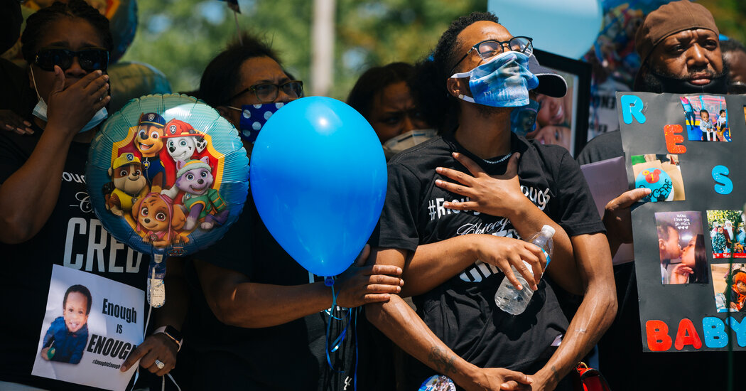
Gunshots are the top cause of death for children and teenagers in the U.S. Fatal or not, the wounds reverberate through communities and the health care system.With each mass shooting, Americans look to one grim indicator — the number of dead — as a measure of the destructive impact. But damage left behind by gunshot wounds reverberates among survivors and families, sending mental health disorders soaring and shifting huge burdens onto the health care system, a new analysis of private health insurance claims shows.In 2020, gunshot wounds became the leading cause of death for children and adolescents in the United States. Though the government does not systematically track nonfatal gunshot wounds, existing evidence suggests that they are two to three times as common as fatal ones. These wounds can be especially catastrophic in children, whose bodies are so small that the amount of tissue destroyed is greater.“What comes after the gunshot is so often not talked about,” said Dr. Chana Sacks, co-director of the Gun Violence Prevention Center at Massachusetts General Hospital and an author of the new study, published on Monday in the journal Health Affairs. The study, which analyzed thousands of insurance claims, maps out lasting damage to families and communities.For families in which a child died of a gunshot wound, surviving family members experienced a sharp increase in psychiatric disorders, taking more psychiatric medications and making more visits to mental health professionals: Fathers had a 5.3-fold increase in treatment for psychiatric disorders in the year after the death; mothers had a 3.6-fold increase; and surviving siblings had a 2.3-fold increase.Children and teenagers who survive gunshot wounds become, as Dr. Sacks put it, “more like lifelong patients.” During the year after the injury, their medical costs rose by an average of $34,884, a 17-fold increase from baseline, driven by hospitalizations, emergency room visits and home health care, the study found.Children and adolescents who survived the most severe gunshot wounds, requiring treatment in an intensive care unit, struggled considerably. In that group, diagnoses of pain disorders increased 293 percent, and psychiatric disorders increased by 321 percent.The study examined medical records from 2,052 children who survived gunshots, 6,209 family members of children who survived, and 265 family members of children who died from gunshot wounds, comparing each with five controls. Because the study was based on private insurance claims, it did not reflect the experience of families who were uninsured or on public insurance.Rising costs linked to firearms injuries make it “increasingly an economic issue,” said Dr. Zirui Song, an associate professor at Harvard Medical School and co-author of the study. The prevalence of gunshot wounds has quadrupled over the last 12 years in the population covered by private insurance, he said.In a paper published last year in the Journal of the American Medical Association, Dr. Song calculated the annual cost of firearms injuries in lost wages and medical spending as $557 billion, or 2.6 percent of gross domestic product. The new study is the first to focus on the cost of nonfatal gunshot wounds, he said.“The cruel reality is that if one dies from a firearm injury, one is free to society — there’s no more health care spending, no more taxpayer dollars, no more resources used,” he said. “But actually surviving a firearm injury is quite expensive to society. The magnitude of that was previously not known.”National data on nonfatal gunshot wounds is “disturbingly unreliable,” but many survivors face long-term disability, said Dr. Megan Ranney, an emergency room physician and the dean of the Yale School of Public Health, who was not involved in the study.“It may be that they have been shot in the intestine, or through a major blood vessel, it could be a bullet has gone through their lung,” Dr. Ranney said. “It can also be that they’ve been shot through the head or the spine.”Trauma physicians have long observed the ripple effect of shootings on the health of family members and communities, she said, often because of repeated visits to the emergency room for nightmares, anxiety or depression, but “we’ve never been able to measure it.”Clementina Chery, a Boston woman whose 15-year-old son was fatally shot in crossfire in 1993, and who founded the Louis D. Brown Peace Institute, an organization to support families who have lost members to gun violence, said she had often seen survivors struggle with addictive behavior, job loss, suicidal or homicidal thoughts in the years after a young person dies.“In that immediate aftermath, I just felt that I was having an out-of-body experience,” Ms. Chery said. She turned to alcohol, she said — “a little wine here, a little wine there” — and found it difficult to leave her house. Her marriage ended. What finally woke her up, she said, was realizing that her younger children were starved of attention.“I literally was going through the motions,” she said. “I was not living. It was like, what do you call it, a mechanical robot.”The ripple effect of gunshot wounds is important because these injuries tend to be concentrated in specific communities, usually communities of color, where many young people know someone who has been shot, Dr. Sacks said.She traced her interest in the subject to the 2012 mass shooting at Sandy Hook Elementary School in Newtown, Conn., where the 7-year-old son of her cousin was one of 20 children killed. The child’s death “changed my life” and has continued to shape extended families and communities in the years that followed, she said.“We can’t think about this as a problem that starts and ends with the bullet going in and then the acute surgical care,” Dr. Sacks said. “Leaving the hospital is just the beginning of that family’s journey, and I think we need to treat it that way.”

Read more →

Male-Killing Virus Is Discovered in Insects

The chance finding in a Japanese university’s greenhouse could help researchers find ways to control agricultural pests or even insects that spread disease.Scientists in Japan have identified a virus that selectively kills males — and it happens to be inheritable, creating generation upon generation of all females.The discovery, made in caterpillars and described Monday in The Proceedings of the National Academies of Sciences, is “robust” evidence that “more than one virus has evolved to selectively kill male insects,” said Greg Hurst, a symbiont specialist at the University of Liverpool in England who wasn’t involved in the study. That could one day help control populations of pest insects and disease vectors like mosquitoes.“I expect there are a lot more cases like this that will be discovered in the near future,” said Daisuke Kageyama, a researcher at the National Agriculture and Food Research Organization in Japan and one of the study’s authors.The virus was found by chance. Misato Terao, a research technician at Minami Kyushu University, was straightening up the campus greenhouse when she found unwelcome intruders — fat green caterpillars — nibbling on the impatiens. She scooped them up and, on a whim, dropped them off in the lab of Yoshinori Shintani, an insect physiologist who is Minami Kyushu’s resident bug guy.Dr. Shintani decided the caterpillars — tobacco cutworms, a ravenous pest species and scourge of Asian agriculture — might be useful to feed to other insects. “It was almost a miracle” they didn’t end up in the trash, he said. By the time he remembered them several days later, he had about 50 adult moths, and unexpectedly, all of them were female.On a hunch, he bred the females from the greenhouse with male tobacco moths he found fluttering around the lights in his own home. The greenhouse moths only had daughters — and so did their daughters, and their daughters’ daughters. Over 13 generations of the moths’ descendants, only three had males.Dr. Shintani and his colleague Dr. Kageyama quickly realized they had a “male-killer” on their hands.For decades, scientists have known that microbial hitchhikers, usually bacteria, can take up residence in the jellylike cytoplasm of insects’ cells. And through a process that’s not very well understood, those microbes can be passed from mother to offspring.Sometimes these microbial symbionts tamper with the host’s reproduction. From the symbiont’s perspective, “males are useless” because they can’t help propagate the microbe, Dr. Kageyama said. So the symbiont simply eliminates them. The bacteria Wolbachia can prevent male butterflies from being born. Other bacteria kill developing males before they hatch, reducing competition for the females and giving them a fortifying snack: the eggs that held their brothers.Dr. Shintani’s team found that antibiotics didn’t knock out the male-killing effect in the greenhouse moth’s progeny, so bacteria couldn’t be responsible. Genetic analysis turned up telltale signs of a virus, but unlike any male-killer ever seen before. Only two male-killing viruses have ever been documented; the virus found by the Japanese researchers, which they named SlMKV, seems to have evolved separately.To confirm the male-killer was actually infectious and inheritable, Dr. Shintani needed to juice some tobacco moths. He and his team blended the bodies of pupae and adult moths with SlMKV and injected the resulting slurry into the bodies of uninfected pupae and moths. That did the trick — the next generation heavily favored females, and in subsequent generations males vanished altogether.Further experiments revealed just how lucky the researchers were to find this male-killer. While cool weather can be lethal to tobacco cutworms, SlMKV is vulnerable to heat, and the researchers found that the virus’s effect was diminished and eventually neutralized at higher temperatures. The tobacco cutworm’s native range is in subtropical parts of China and Taiwan.The scientists suspect the balmy climate in the caterpillar’s home acts like a perpetual fever, suppressing the male-killing effect. It was pure chance that Japan’s mild temperatures fell in the “Goldilocks zone” in which SlMKV is active, and that scientists could therefore notice the sex imbalance in the greenhouse.Outside experts say the team’s discovery is a sign that viral male-killers are more common than anticipated. And the find could have implications for controlling other important agricultural pests to which the tobacco cutworm is closely related, Dr. Hurst said.Anything researchers can learn about male-killers helps advance the quest for the pest controller’s holy grail: a “female-killer,” which could help fight invasive pests or disease-carrying species such as mosquitoes.According to Anne Duplouy, an evolutionary biologist at the University of Helsinki who studies microbial symbionts in insects, time is running out for humans to learn from these temperature-sensitive microbes. As the climate changes, she said, “we are likely to be losing many of these interactions” before they can be documented

Read more →