Doctors question science behind blood sugar diet trend

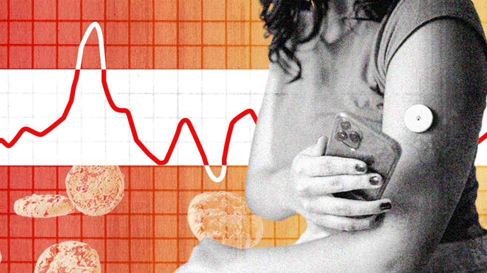
Published6 minutes agoShareclose panelShare pageCopy linkAbout sharingBy Rachel SchraerHealth and disinformation reporterBlood sugar-monitors are unnecessary for people without diabetes and could, in extreme cases, fuel eating disorders, leading doctors have warned. They are part of a personalised diet trend, promoted on social media and spearheaded by companies including ZOE. But NHS national diabetes advisor Prof Partha Kar said there is no strong evidence the gadgets help people without the condition. ZOE said research is at an early stage but is “cutting edge”. In people with diabetes, blood sugar – also known as blood glucose – can remain high for several hours after eating. At very high levels this can cause organ damage if it’s not monitored and kept in check. ZOE – previously involved with the Covid symptom-tracking app – is one of the leading companies bringing the use of blood sugar monitors to people without the condition. Now it offers a programme, starting at around £300, which it advertises widely, including across social media platforms.Participants log their food intake and wear a continuous glucose monitor (CGM) for two weeks to measure blood sugar levels after eating.In separate tests, they also have their responses to fat, and their gut bacteria tested.ZOE says all of these tests have helped it identify that even two healthy people can have wildly different responses to the same food – for example one person’s blood sugar might spike and dip more after eating carbohydrates than another person’s. It suggests this could guide individual food decisions.But other researchers argue that what, if anything, those numbers mean – including bigger spikes and dips in blood sugar within the non-diabetic range – is still not properly understood.Dr Nicola Guess, a dietitian and diabetes researcher at the University of Oxford, said the majority of evidence linking high, and highly varied, blood sugar to health problems is based on glucose levels only seen in people with diabetes or prediabetes.High blood sugar is a symptom, not directly a cause, of diabetes, she explains. Prof Kar said “the evidence base is nothing” for understanding what the swings in blood sugar mean in people without diabetes. ZOE points to some evidence – including some very small studies – suggesting that even before it reaches prediabetic or diabetic levels, having higher blood sugar and big variations through the day may be linked to some worse outcomes. Most data is still in diabetic patients, though.It says it is investigating gut bacteria and starting to see links between gut microbes, diet and health. Gut microbiome expert and colorectal surgeon at Imperial College London, James Kinross, said while the microbiome was very important, direct-to-consumer testing was “problematic” because “this is such a young science and there are many unanswered questions about how the microbiome influences our health.”ZOE’s chief scientist Dr Sarah Berry told the BBC its programme used “decades” of existing nutrition research, and their own studies on links between blood sugar and health. But she acknowledges that “we don’t have all of the evidence”. But given the risks of poor diet we already understand, she said, “it would actually be irresponsible to wait” decades to understand long-term outcomes like heart disease and death.Dr Ran Crooke, a GP who founded a company providing health services to start-ups, praised the company for trying to gather the evidence, and said not having all the evidence on blood sugar shouldn’t be “stifling innovation”. He and others, including some of ZOE’s critics, agree CGMs could potentially be a helpful tool for some people to motivate them and change their diet. However, people have been sounding the alarm over diet-related illness for decades. Yet hundreds of diet programmes have failed to resolve the challenge of getting people to stick to habits when their environment – a modern prevalence of high-sugar foods, for example – and biology seems to be stacked against them. And they aren’t without risks themselves.The company said: “ZOE is scientifically rigorous in its approach, unrivalled by others in the industry in terms of clinical trials, robust research and a dedicated team of scientists and nutrition professionals looking to improve health through useful, evidence-based advice.” But Dr Guess is concerned she is seeing patients who use ZOE’s products cutting out foods she believes are good for their health, because they seem to spike their blood sugar. That in itself can lead to health problems, and is not recommended by the company.She adds people who avoid carbohydrates will get a temporarily “exaggerated glucose response” the next time they eat them – which she says is “perfectly normal,” but that could potentially lead them to think they are unable to tolerate carbs at all. Prof Kar thinks using CGMs when there’s no health reason to do so can drive an obsessive focus on numbers which, in the most extreme cases, “can translate into eating disorders”. According to eating disorder charity Beat, “people with eating disorders often fixate on numbers… as part of their illness, so we’d never recommend that anybody affected uses glucose monitors”. ZOE does attempt to screen out people with a history of eating disorders, and Dr Berry told the BBC the company takes “the wellbeing of our members very seriously,” and that customers have access to trained nutrition coaches, who can support them with food anxiety and refer them on if they feel there’s a problem.The company has published research based on the data it has collected from participants to try to find patterns in areas like food choices, hunger and blood test results. But it can’t show which aspects are actually causing changes to health and which are coincidences. ZOE has carried out a study to understand changes caused by the programme, but it has not yet been published. Critics are concerned this study won’t be able to unpick the effects of the programme’s different elements, such as personalised diets based on test results versus support and coaching.Dr Berry argues ZOE’s programme is, “a very holistic product that doesn’t involve just microbiome testing or just continuous glucose monitoring”. However, Dr Guess believes since these elements remain unproven, without them, it’s just a “sciencey-sounding way of having people eat more fruit and veg”.A lot of ZOE’s advice, including around eating more whole foods and fewer processed foods, is sensible, she thinks but she believes this message is “not compelling enough” to sell a £300 product.Are you affected by the issues raised in this story? Share your experiences by emailing haveyoursay@bbc.co.uk.Please include a contact number if you are willing to speak to a BBC journalist. You can also get in touch in the following ways:WhatsApp: +44 7756 165803Tweet: @BBC_HaveYourSayUpload pictures or videoPlease read our terms & conditions and privacy policy

If you are reading this page and can’t see the form you will need to visit the mobile version of the BBC website to submit your question or comment or you can email us at HaveYourSay@bbc.co.uk. Please include your name, age and location with any submission. More on this storyPlant-based diets good for the heartPublished25 May 2023Can green ‘super powders’ really make you healthy?Published3 MarchThe quest to find healthy and cheap sweetenersPublished15 December 2023

Read more →

Speaking without vocal cords, thanks to a new AI-assisted wearable device

People with voice disorders, including those with pathological vocal cord conditions or who are recovering from laryngeal cancer surgeries, can often find it difficult or impossible to speak. That may soon change.
A team of UCLA engineers has invented a soft, thin, stretchy device measuring just over 1 square inch that can be attached to the skin outside the throat to help people with dysfunctional vocal cords regain their voice function. Their advance is detailed this week in the journal Nature Communications.
The new bioelectric system, developed by Jun Chen, an assistant professor of bioengineering at the UCLA Samueli School of Engineering, and his colleagues, is able to detect movement in a person’s larynx muscles and translate those signals into audible speech with the assistance of machine-learning technology — with nearly 95% accuracy.
The breakthrough is the latest in Chen’s efforts to help those with disabilities. His team previously developed a wearable glove capable of translating American Sign Language into English speech in real time to help users of ASL communicate with those who don’t know how to sign.
The tiny new patch-like device is made up of two components. One, a self-powered sensing component, detects and converts signals generated by muscle movements into high-fidelity, analyzable electrical signals; these electrical signals are then translated into speech signals using a machine-learning algorithm. The other, an actuation component, turns those speech signals into the desired voice expression.
The two components each contain two layers: a layer of biocompatible silicone compound polydimethylsiloxane, or PDMS, with elastic properties, and a magnetic induction layer made of copper induction coils. Sandwiched between the two components is a fifth layer containing PDMS mixed with micromagnets, which generates a magnetic field.
Utilizing a soft magnetoelastic sensing mechanism developed by Chen’s team in 2021, the device is capable of detecting changes in the magnetic field when it is altered as a result of mechanical forces — in this case, the movement of laryngeal muscles. The embedded serpentine induction coils in the magnetoelastic layers help generate high-fidelity electrical signals for sensing purposes.

Measuring 1.2 inches on each side, the device weighs about 7 grams and is just 0.06 inch thick. With double-sided biocompatible tape, it can easily adhere to an individual’s throat near the location of the vocal cords and can be reused by reapplying tape as needed.
Voice disorders are prevalent across all ages and demographic groups; research has shown that nearly 30% of people will experience at least one such disorder in their lifetime. Yet with therapeutic approaches, such as surgical interventions and voice therapy, voice recovery can stretch from three months to a year, with some invasive techniques requiring a significant period of mandatory postoperative voice rest.
“Existing solutions such as handheld electro-larynx devices and tracheoesophageal- puncture procedures can be inconvenient, invasive or uncomfortable,” said Chen who leads the Wearable Bioelectronics Research Group at UCLA, and has been named one the world’s most highly cited researchers five years in a row. “This new device presents a wearable, non-invasive option capable of assisting patients in communicating during the period before treatment and during the post-treatment recovery period for voice disorders.”
How machine learning enables the wearable tech
In their experiments, the researchers tested the wearable technology on eight healthy adults. They collected data on laryngeal muscle movement and used a machine-learning algorithm to correlate the resulting signals to certain words. They then selected a corresponding output voice signal through the device’s actuation component.
The research team demonstrated the system’s accuracy by having the participants pronounce five sentences — both aloud and voicelessly — including “Hi, Rachel, how are you doing today?” and “I love you!”
The overall prediction accuracy of the model was 94.68%, with the participants’ voice signal amplified by the actuation component, demonstrating that the sensing mechanism recognized their laryngeal movement signal and matched the corresponding sentence the participants wished to say.

Going forward, the research team plans to continue enlarging the vocabulary of the device through machine learning and to test it in people with speech disorders.
Other authors of the paper are UCLA Samueli graduate students Ziyuan Che, Chrystal Duan, Xiao Wan, Jing Xu and Tianqi Zheng — all members of Chen’s lab.
The research was funded by the National Institutes of Health, the U.S. Office of Naval Research, the American Heart Association, Brain & Behavior Research Foundation, the UCLA Clinical and Translational Science Institute, and the UCLA Samueli School of Engineering.

Read more →

Fatty food before surgery may impair memory in old, young adults

Eating fatty food in the days leading up to surgery may prompt a heightened inflammatory response in the brain that interferes for weeks with memory-related cognitive function in older adults — and, new research in animals suggests, even in young adults.
The study, building upon previous research from the same lab at The Ohio State University, also showed that taking a DHA omega-3 fatty acid supplement for a month before the unhealthy eating and surgical procedure prevented the effects on memory linked to both the high-fat diet and the surgery in aged and young adult rats.
Three days on a high-fat diet alone was detrimental to a specific type of fear-related memory in aged rats for as long as two weeks later — the same type of impairment seen in younger rats that ate fatty food and had a surgical procedure. The team has traced the brain inflammation behind these effects to a protein that activates the immune response.
“These data suggest that these multiple insults have a compounding effect,” said senior author Ruth Barrientos, an investigator in Ohio State’s Institute for Behavioral Medicine Research and associate professor of psychiatry and behavioral health and neuroscience in the College of Medicine.
“We’ve shown that an unhealthy diet, even in the short term, especially when it’s consumed so close to a surgery, which in and of itself will cause an inflammatory response, can have damaging results,” Barrientos said. “The high-fat diet alone might increase inflammation in the brain just a little bit, but then you have surgery that does the same thing, and when put together in a short amount of time you get a synergistic response that can set things in motion toward a longer-term memory issue.”
The study was published recently in the journal Brain, Behavior, and Immunity.
Barrientos’ lab studies how everyday life events might trigger inflammation in the aging brain as the nervous system responds to signals from the immune system reacting to a threat. Decades of research has suggested that with aging comes long-term “priming” of the brain’s inflammatory profile and a loss of brain-cell reserve to bounce back.

Researchers fed young adult and aged rats a diet high in saturated fat for three days before a procedure resembling exploratory abdominal surgery — an event already known to cause about a week of cognitive issues in an older brain. Control rats ate regular food and were anesthetized, but had no surgery. (Barrientos’ lab has determined anesthesia alone does not cause memory problems in rats.)
In this study, as in previous research on aged rats treated with morphine after surgery, the team showed that an immune system receptor called TLR4 was the culprit behind the brain inflammation and related memory problems generated by both surgery and the high-fat diet, said first author Stephanie Muscat, assistant clinical professor of neuroscience at Ohio State.
“Blocking the TLR4 signaling pathway prior to the diet and surgery completely prevented that neuroimmune response and memory impairments, which confirmed this specific mechanism,” Muscat said. “And as we had found before in another model of an unhealthy diet, we showed that DHA supplementation did mitigate those inflammatory effects and prevent memory deficits after surgery.”
There were some surprising memory findings in the new work. Different behavioral tasks are used to test two types of memory: contextual memory based in the hippocampus and cued-fear memory based in the amygdala. In contextual memory tests, rats with normal memory freeze when they re-enter a room in which they had an unpleasant experience. Cued-fear memory is evident when rats freeze in a new environment when they hear a sound connected to that previous bad experience.
For aged rats in this study, as expected, the combination of a high-fat diet and surgery led to problems with both contextual and cued-fear memory that persisted for at least two weeks — a longer-lasting effect than the researchers had seen before.
The high-fat diet alone also impaired the aging rats’ cued-fear memory. And in young adult rats, the combination of the high-fat diet and surgery led to only cued-fear memory deficits, but no problems with memory governed by the hippocampus.

“What this is telling us in aged animals, along with the fact we’re seeing this same impairment in young animals after the high-fat diet and surgery, is that cued-fear memory is uniquely vulnerable to the effects of diet. And we don’t know why,” Barrientos said. “One of the things we’re hoping to understand in the future is the vulnerability of the amygdala to these unhealthy diet challenges.”
With increasing evidence suggesting that fatty and highly processed foods can trigger inflammation-related memory problems in brains of all ages, the consistent findings that DHA — one of two omega-3 fatty acids in fish and other seafood and available in supplement form — has a protective effect are compelling, Barrientos said.
“DHA was really effective at preventing these changes,” she said. “And that’s amazing — it really suggests that this could be a potential pretreatment, especially if people know they’re going to have surgery and their diet is unhealthy.”
This work was supported by grants from the National Institute on Aging and the National Institute of Neurological Disorders and Stroke.
Co-authors included Michael Butler, Menaz Bettes, James DeMarsh, Emmanuel Scaria and Nicholas Deems, all of Ohio State.

Read more →

Researchers identify a signaling molecule in neuroblastoma immunosuppression and aggressiveness

The MYCN oncoprotein (proteins related to the growth of cancer cells)plays a key role in starting, advancing and making it difficult to treat various human cancers. When MYCN is overactive, especially in high-risk neuroblastoma (childhood cancer often found in the adrenal glands), the tumors become less responsive to immunotherapy — a treatment that uses the body’s immune system to fight cancer. Still, recognition of this problem has not led to any effective strategies to tackle this problem.
In a new study from Boston University Chobanian & Avedisian School of Medicine, researchers found that MYCN selectively increases the levels of a signaling molecule, CKLF, in neuroblastoma cells to suppress anti-tumor immune responses and promote tumor aggressiveness.
“As scientists, we are looking for ways to make these less responsive tumors more receptive to immunotherapy to increase its effectiveness. Understanding how tumor cells utilize this molecule to communicate with immune cells will facilitate the development of effective immunotherapeutic strategies to provide more effective treatments with fewer toxicities for children with high-risk neuroblastoma,” explained corresponding author Hui Feng, MD, PhD, associate professor of pharmacology, physiology & biophysics at the school.
The researchers studied an experimental model, clinical patient samples, and in-vitro cell culture. The experimental models of neuroblastoma with and without overexpression of the signaling molecule, CKLF, were compared for their ability to increase tumor aggression through the suppression of anti-tumor immune responses.
According to Xiaodan Qin, PhD, the first author of the study and a research scientist in the Feng Lab, the research is critical to understanding the mechanisms by which tumor cells induce a tumor microenvironment that compromises the function of immune cells, and it uncovered additional therapeutic approaches to evoke anti-tumor immune responses. “The long-term goal of this research is to uncover effective drugs that are much less toxic than chemotherapy and radiotherapy for treating high-risk neuroblastoma and perhaps other types of MYCN-driven childhood cancers,” Feng says.
The findings appear online in Science Advances.

Read more →

Key metabolic process responsible for rapid immune responses discovered

Researchers from Children’s Hospital of Philadelphia (CHOP) identified a key metabolite in cells that helps direct immune responses and explains at a single cell level why immune cells that most efficiently recognize pathogens, vaccines, or diseased cells grow and divide faster than other cells. The findings also indicate that a better understanding of this metabolite and its role in immune response could improve the design of immunotherapies and create longer-lived responses against different types of cancer as well as enhance vaccine strategies.
The findings were published online today by the journal Science Immunology.
Antigens are foreign substances that our immune system recognizes and responds to by producing more T and B cells. These cells each have unique receptors that recognize specific antigens and can respond appropriately, and they can “remember” and respond similarly when exposed to the same antigen again. How well a T or B cell to sees its antigen is known as its affinity. This fundamental concept of immunology is how vaccines work. When those T and B cells encounter a pathogen, the body needs the ones that recognize their antigen the best, with high affinity, to divide more quickly to produce more daughter cells and “attack” the invader.
However, the underlying mechanisms as to why high affinity immune cells respond more efficiently have remained a mystery for researchers. After seeing an antigen, the chemistry inside T and B cells needs to change to allow them to properly respond. The researchers in this study wanted to look at metabolism to understand what causes high affinity cells to know that they need to divide more quickly to respond appropriately.
“We wanted to see if specific metabolites were sensitive to T cell receptor affinity and controlled T cell expansion during immune responses,” said senior study author Will Bailis, PhD, Assistant Professor of Pathology and Laboratory Medicine at CHOP and the Perelman School of Medicine of the University of Pennsylvania.
The researchers identified nicotinamide adenine dinucleotide (NAD) as a key, affinity-dependent component of T cell receptor metabolic reprogramming during the early stages of a T cell activation. Using flow cytometry, the researchers could look at NAD in single cells immediately after activation and show how it dictates the number of times T cells can divide in the future. Therefore, researchers could essentially predict how T cells behave and how many times they divide based on how much NAD they started with.
Additionally, the researchers found that manipulating how much NAD a cell was allowed to make could control when that cell went from a resting state to wanting to divide, suggesting that the metabolite could be used to improve response in certain T cell-driven therapies or vaccines.
“We believe this work shows how single cell differences in metabolism are a key reason why similar cells sometimes display strikingly different behaviors and that this may provide insight into underlying processes that drive disease and dysfunction that cannot simply be explained by gene regulation or signaling,” Bailis said. “With more work, we also believe that this information could potentially be used to improve vaccine strategies and the response and durability of cell-based therapies used to treat cancer and other diseases.”
This study was supported by National Institutes of Health grants K22AI141758, R35GM138085, R01DK098656, R01HL165792, P30ES013508, R01AI165706, and F31CA261156, a Children’s Hospital of Philadelphia Cell and Gene Therapy Collaborative SEED Award, a Children’s Hospital of Philadelphia Junior Faculty Pilot Grant, Transfusion Medicine Research Training Program grant 2T32HL00777528, Microbial Pathogenesis and Genomics Training Grant 5T32AI141393, and Immunobiology of Normal and Neoplastic Lymphocytes Training Grant T32CA009140.

Read more →

Gut bacteria make neurotransmitters to shape the newborn immune system

Weill Cornell Medicine investigators discovered that unique bacteria colonize the gut shortly after birth and make the neurotransmitter serotonin to educate gut immune cells. This prevents allergic reactions to food and the bacteria themselves during early development.
The preclinical study, published in Science Immunology on Mar. 15, showed that bacteria abundant in the guts of newborns produce serotonin, which promotes the development of immune cells called T-regulatory cells or Tregs. These cells suppress inappropriate immune responses to help prevent autoimmune diseases and dangerous allergic reactions to harmless food items or beneficial gut microbes.
“The gut is now known as the second human brain as it makes over 90 percent of the neurotransmitters in the human body. While neurotransmitters such as serotonin are best known for their roles in brain health, receptors for neurotransmitters are located throughout the human body,” explained the study’s senior author, Dr. Melody Zeng, an assistant professor of immunology in the Gale and Ira Drukier Institute for Children’s Research and the Department of Pediatrics at Weill Cornell Medicine.
Gut Bacteria in Babies Provide a Helping Hand
The researchers observed that the neonatal mouse gut had much higher levels of neurotransmitters, including serotonin, than the adult gut. “So far, almost all studies of gut neurotransmitters were conducted in adult animals or human subjects, where a specific gut cell type called enterochromaffin cells produce neurotransmitters,” said Dr. Zeng. “However, we discovered that this isn’t the case in the newborn gut where most of the serotonin is made by bacteria that are more abundant in the neonatal gut.”
This was also confirmed in babies through a human infant stool biobank that the Zeng lab has established in collaboration with the Neonatal Intensive Care Unit in the NewYork-Presbyterian Alexandra Cohen Hospital for Women and Newborns. These samples were obtained with parental consent and deidentified.
The study results suggest that before the neonatal gut is mature enough to make its own neurotransmitters, unique gut bacteria may supply neurotransmitters that are needed for critical biological functions during early development.

“We found that gut bacteria in young mice not only directly produce serotonin but also decrease an enzyme called monoamine oxidase that normally breaks down serotonin, thus keeping gut serotonin levels high,” said the study’s lead author Dr. Katherine Sanidad, postdoctoral associate in pediatrics at Weill Cornell Medicine.
The high serotonin levels shift the balance of immune cells by increasing the number of Tregs, which helps prevent the immune system from overreacting and attacking gut bacteria or food antigens. “The neonatal gut needs these serotonin-producing bacteria to keep the immune system in check,” Dr. Sanidad added.
Healthy Immune System Helps Later in Life
Dr. Zeng noted that this work underscores the importance of having the right types of beneficial bacteria soon after birth. Babies in developed countries have better access to antibiotics, less exposure to diverse microbes in their clean environments and potentially unhealthy diets that may significantly impact the abundance of serotonin-producing bacteria in their intestines.
As a result, these babies may have fewer Tregs and develop immune reactions to their own gut bacteria, or allergies to food. This may be one reason food allergies have become increasingly common in children, particularly in developed countries. “If educated properly, the immune system in babies would recognize that things like peanuts and eggs are okay, and it doesn’t have to attack them,” she said. This may also have an impact on developing autoimmune diseases — when the immune system attacks the body’s own healthy cells — later in life.
The team next plans to look at bacteria in human infant stool samples to measure their production of serotonin, other neurotransmitters and molecules that may help train the immune system to prevent future immune-related diseases, such as allergies, infections and cancer.
“It’s essential to understand how the immune system is trained during early life, but this is understudied in newborns and children. Further studies of these developmental periods may hopefully lead us to mitigation approaches to reduce the risk of inflammatory diseases like food allergies and inflammatory bowel disease later in life,” Dr. Sanidad said.
Dr. Melody Zeng’s lab is supported in part by the National Institutes of Health grants R01HD110118, R01HL169989, R21CA270998, and K01DK114376; The Starr Cancer Consortium; the Hartwell Foundation; and the Jill Roberts Center for Inflammatory Bowel Disease, the Children’s Health Council, and the Drukier Institute for Children’s Health at Weill Cornell Medicine.

Read more →

Why killer T cells lose energy inside of solid tumors

T cells are often called “assassins” or “killers” because they can orchestrate and carry out missions to hunt down bacteria, viruses, and cancer cells throughout the body. Mighty as they may be, recent research has shown that once T cells infiltrate the environment of a solid tumor, they lose the energy needed to combat the cancer.
A research team led by Jessica Thaxton, PhD, MsCR, associate professor of cell biology and physiology and co-leader of the Cancer Cell Biology Program at the UNC Lineberger Comprehensive Cancer Center, aimed to understand why T cells do not sustain energy in tumors. Using their expertise in tumor immunity and metabolism, the Thaxton Lab, led by the Katie Hurst, MPH, and 4th year graduate student Ellie Hunt, found that a metabolic enzyme called Acetyl-CoA Carboxylase (ACC) causes T cells to store fat rather than burning fat for energy.
“Our discovery fills a long-standing gap in knowledge regarding why T cells in solid tumors don’t appropriately generate energy,” said Thaxton. “We inhibited the expression of ACC in mouse cancer models, and we observed that T cells were able to persist much better in solid tumors.”
The new findings and immunotherapeutic strategies, which were published in Cell Metabolism, could be used to make multiple types of T-cell therapies more effective for patients, possibly encompassing both checkpoint and chimeric antigen receptor (CAR) T-cell therapies.
In the field of cancer immunotherapy, it has long been known that T cells are not able to create their cellular energy, called adenosine triphosphate or ATP, when they are inside of a solid tumor.
In 2019, Thaxton’s lab studied a T cell with optimal antitumor function. In a publication in Cancer Immunology Research, Hurst and Thaxton used a proteomics screen to identify enzymes associated with the optimal antitumor metabolism of these T cells. Through this screen, the two discovered that ACC expression may limit the ability of T cells to make ATP in tumors. ACC, a key molecule that is involved in many metabolic pathways, blocks cells from breaking down fat and using it as fuel for energy in mitochondria.
“Acetyl-CoA carboxylase can drive the balance between storing lipids versus breaking down those lipids and feeding them into the citric acid cycle for energy,” said Thaxton. “If ACC is flipped ‘on’, cells generally store lipid. If ACC is ‘off’, cells tend to use the lipid in their mitochondria to make ATP.”
Using Hunt’s expertise in confocal imaging, the research team was able to observe lipid stores in T cells isolated from multiple types of cancers. The observation, as well as other experiments, confirmed the team’s hypothesis that T cells were storing lipids instead of breaking them down.

Thaxton’s team then used CRISPR Cas9-mediated gene deletion to see what would happen if they “deleted” ACC from the picture. There was a rapid reduction in the amount of lipid storage in T cells, and the team was able to visualize fat relocating to the mitochondria to be used to generate energy.
Thaxton now hypothesizes that T cells may need a “delicate balance” of lipids to persist in solid tumors with a certain amount of lipid dedicated to cancer cell assassination and low levels of fats being maintained in stores.
The latest findings could prove to be useful in enhancing chimeric antigen receptor (CAR) T-cell therapies. This cutting-edge technology takes T cells out of cancer patients, modifies them in the lab to hunt down tumor cells, and then re-infuses the cells to fight the patient’s cancer. Preliminary data from Thaxton’s lab demonstrates that even the manufactured T cells contain excess lipid stores.
The lab is starting to look in patient samples to understand how researchers can possibly flip the ACC metabolic switch directly in patient tumors, negating the need to take out and reinfuse cells back into the body. But researchers must first determine how this could affect other immune cell populations in the body, such as macrophages.

Read more →

Sleep apnea solution could be right under your nose

Australian researchers have discovered that a bedtime nasal spray has the potential to reduce the severity of sleep apnea in people and lower their blood pressure.
The new research published in the American Journal of Physiology-Heart and Circulatory Physiology offers hope to millions of people around the world affected by sleep apnoea, a common and debilitating chronic respiratory condition.
“Obstructive sleep apnea (OSA) is a sleep disorder where the muscles in the back of the throat relax and the upper airway narrows or collapses, restricting oxygen intake and causing people to wake repeatedly throughout the night,” says Professor Danny Eckert, College of Medicine and Public Health.
“It has been linked to a variety of medical conditions including cardiovascular disease, stroke, obesity, diabetes, anxiety and depression.
“Treatment options are limited and while continuous positive airway pressure (CPAP) machines are a proven treatment for OSA, around 50 per cent of people struggle to tolerate them,” he says.
The aim of the study was to determine the effects of a new potassium channel blocker nasal spray on OSA severity and to investigate the potential influence of different breathing approaches such as restricted ‘nasal only’ breathing and the physiological characteristics of those who had a favourable response.
“Potassium channel blockers are a class of drugs that block the potassium channel in the central nervous system.

When used in a nasal spray, the blockers have the potential to increase the activity of the muscles that keep the upper airway open and reduce the likelihood of the throat collapsing during sleep,” says lead author Dr Amal Osman.
Using a randomised, blind trial, 10 people with OSA were given either the potassium blocker nasal spray, a placebo nasal spray or the potassium nasal spray in combination with restricted ‘nasal only’ breathing.
Seven out of the 10 people responded to the potassium channel blocker nasal spray showing a reduction in the frequency of upper airway collapsing episodes during sleep and lower blood pressure the next morning. The use of the spray with restricted ‘nasal only’ breathing did not improve quality of sleep in this trial.
“What we have discovered is that the nasal spray application of the potassium channel blocker that we tested is safe, well tolerated. Those who had a physiological improvement in their airway function during sleep also had between 25-45% reductions in markers of their OSA severity including improved oxygen levels as well as a reduction in their blood pressure the next day,” says Dr Osman.
“These insights provide a potential pathway for development of new therapeutic solutions for those people with OSA who are unable to tolerate CPAP machines and/or upper airway surgery, and those with a desire for alternatives to existing therapies,” says Professor Eckert.
“Right now, there are no approved drugs for treating OSA, but through these findings and future research we are getting closer to developing new and effective drugs that are safe and easy to use,” says Professor Eckert.

Read more →

Abnormal brain structure identified in children with developmental language problems

A rigorous analysis of numerous studies concludes that a part of the brain traditionally associated with movement is abnormal in children with developmental language impairments, according to Georgetown University Medical Center neuroscientists. The discovery has the potential to improve both the diagnosis and treatment of the language difficulties.
The researchers investigated brain abnormalities in developmental language disorder. This condition, which impacts the development of various aspects of language, is about as common as attention-deficit/hyperactivity disorder (ADHD) and dyslexia, and more prevalent than autism. The scientists found that abnormalities occurred specifically in the anterior neostriatum within the basal ganglia, a structure found deep in the brain. They describe their findings in Nature Human Behaviour on March 15.
To better understand why the language impairments occur, the researchers analyzed the results of 22 articles examining brain structures in people with the disorder, and then employed a new computational method to identify common patterns of abnormalities across the studies. They determined that the anterior neostriatum was abnormal in 100% of the studies that examined the structure, with fewer abnormalities in all other parts of the brain.
“We hope that by identifying the neural bases of developmental language difficulties we may help increase awareness of a major, but also rather unrecognized, disorder,” says the study’s lead author Michael T. Ullman, PhD, professor of neuroscience and director of the Brain and Language Laboratory at Georgetown University Medical Center. “We caution, however, that further research is necessary to understand exactly how the anterior neostriatum might lead to the language difficulties.”
Ullman says the findings underscore the potential utility of drugs that are known to improve movement impairments due to basal ganglia dysfunction, such as those that act on dopamine receptors. Interventions that encourage compensation by intact brain structures may also be useful. Additionally, basal ganglia abnormalities could potentially serve as early biomarkers of an increased likelihood of developmental language problems. Such early warning signs could trigger further diagnostic procedures, potentially leading to early therapy.
“Continuing research efforts to further understand the neurobiology of developmental language disorder, especially the role of the basal ganglia, could help the many children who are affected by these problems,” concludes Ullman.
In addition to Ullman, other authors at Georgetown include Mariel Pullman, Jarrett Lovelett, Xiong Jiang, and Peter Turkeltaub. Gillian Clark was at Deakin University, Melbourne, Australia. Elizabeth Pierpont is at the University of Minnesota Medical Center, Minneapolis.
This work was supported by NIH grants R01 HD049347 and R21 HD 087088; NSF grants BCS 1439290 and BCS 1940980; and funding from the Mabel H. Flory Trust.
The authors declare no personal financial interests related to the study.

Read more →

DNA origami-based vaccines toward safe and highly-effective precision cancer immunotherapy

Therapeutic cancer vaccines are a form of immunotherapy in the making that could not only destroy cancer cells in patients, but keep a cancer from coming back and spreading. Multiple therapeutic cancer vaccines are being studied in clinical trials, but despite their promise, they are not routinely used yet by clinical oncologists to treat their patients.
The central ingredient of therapeutic cancer vaccines is antigens, which are preferentially produced or newly produced (neoantigens) by tumor cells and enable a patient’s immune system to search and destroy the cancerous cells. In most cases, those antigens cannot act alone and need the help of adjuvant molecules that trigger a general alarm signal in immune cells known as antigen-presenting cells (APCs). APCs internalize both antigen and adjuvant molecules and present the antigens to different types of T cells. Those T cells then launch an immediate attack against the tumor, or preserve a longer-lasting memory of the tumor for future defense.
A cancer vaccine’s effectiveness depends on the level and duration of the “alarm” its adjuvants can ring in APCs. Previously, researchers found that delivering adjuvant and antigen molecules to APCs simultaneously using nanostructures like DNA origami can increase APC activation. However, none of these approaches systematically investigated how the number and nanoscale arrangement of adjuvant molecules affect downstream tumor-directed immunity.
Now, a research team at the Wyss Institute at Harvard University, Dana-Farber Cancer Institute (DFCI), Harvard Medical School (HMS), and Korea Institute of Science and Technology (KIST) has created a DNA origami platform called DoriVac, whose core component is a self-assembling square block-shaped nanostructure. To one face of the square block, defined numbers of adjuvant molecules can be attached in highly tunable, nanoprecise patterns, while the opposite face can bind tumor antigens. The study found that molecules of an adjuvant known as CpG spaced exactly 3.5 nanometers apart from each other resulted in the most beneficial stimulation of APCs that induced a highly-desirable profile of T cells, including those that kill cancer cells (cytotoxic T cells), those that cause beneficial inflammation (Th-1 polarized T cells), and those that provide a long-term immune memory of the tumor (memory T cells). DoriVac vaccines enabled tumor-bearing mice to better control the growth of tumors and to survive significantly longer than control mice. Importantly, the effects of DoriVac also synergized with those of immune checkpoint inhibitors, which are a highly successful immunotherapy that is already widely used in the clinic. The findings are published in Nature Nanotechnology.
“DoriVac’s DNA origami vaccine technology merges different nanotechnological capabilities that we have developed over the years with an ever-deepening knowledge about cancer-suppressing immune processes,” said Wyss Core Faculty member William Shih, Ph.D., who led the Wyss Institute team together with first-author Yang (Claire) Zeng, M.D., Ph.D.. “We envision that in the future, antigens identified in patients with different types of tumors could be quickly loaded onto prefabricated, adjuvant-containing DNA origami to enable highly effective personalized cancer vaccines that can be paired with FDA-approved checkpoint inhibitors in combination therapies.” Shih is also a Professor at HMS and DFCI’s Department of Cancer Biology and, as some of the other authors, a member of the NIH-funded cross-institutional “Immuno-engineering to Improve Immunotherapy” (i3) Center based at the Wyss.
DNA origami rationale
The CpG adjuvant is a synthetic strand of DNA made up of repeated CpG nucleotide motifs that mimic the genetic material from immune cell-invading bacterial and viral pathogens. Like its natural counterparts, CpG adjuvants bind to a “danger receptor” called TLR9 in immune cells, which in turn induces an inflammatory (innate) immune response that works in concert with the antigen-induced (adaptive) immune response.

“We knew from previous work that to trigger strong inflammatory responses, TLR9 receptors need to dimerize and aggregate into multimeric complexes binding to multiple CpG molecules. The nanoscale distances between the CpG-binding domains in effective TLR9 assemblies revealed by structural analysis fell right into the range of what we hypothesized we could mirror with DNA origami structures presenting precisely spaced CpG molecules,” explained Zeng, who was an Instructor in Medicine at the time of the study and now is a senior scientist at DFCI and Harvard Medical School (HMS). In addition to Shih, Zeng was also mentored on the project by senior authors Ju Hee Ryu, Ph.D., a Principal Researcher at KIST, and Wyss Founding Core Faculty member David Mooney, Ph.D., who also is Professor at Harvard John A. Paulson School of Engineering and Applied Sciences (SEAS), and one of the i3 Center’s Principal Investigators.
Zeng and the team fabricated DoriVac vaccines in which different numbers of CpG strands were spaced at 2.5, 3.5, 5, or 7 nanometers apart from each other on one face of the square block, and a model antigen was attached to the opposite face. They protected their structures from being degraded in the body using a chemical modification method that Shih’s group had developed earlier. When internalized by different types of APCs, including dendritic cells (DCs), which orchestrate tumor-directed T cell responses, the DoriVac vaccines improved the uptake of antigens compared to controls consisting of free antigen molecules. A CpG spacing of 3.5 nanometers produced the strongest and most beneficial responses in APCs, and significantly outperformed a control vaccine containing only free CpG molecules. “We were excited to find that the DoriVac vaccine preferentially induced an immune activation state that supports anti-tumor immunity, which is what researchers generally want to see in a good vaccine,” said Zeng.
Besides spacing, the numbers of CpG molecules in DoriVac vaccines also mattered. The team tested vaccines containing between 12 to 63 optimally spaced CpG molecules and found that 18 CpG molecules provided the best APC activation. This meant that their approach can also help limit the dosage of CpG molecules and thus minimize commonly observed toxic side effects observed with adjuvants.
Gained in (tumor) translation
Importantly, these in vitro trends translated to in vivo mouse tumor models. When prophylactically injected under the skin of mice, DoriVac vaccines accumulated in the closest lymph nodes where they stimulated DCs. A vaccine loaded with a melanoma antigen prevented the growth of subsequently injected aggressive melanoma cells. While all control animals had succumbed to the cancer by day 42 of the experiment, DoriVac-protected animals all were alive. DoriVac vaccines also inhibited tumor growth in mice in which the formation of melanoma tumors was already underway, with a 3.5 nanometer spacing of 18 CpG molecules again providing maximum effects on DC and T cells, and the strongest reduction in tumor growth.
Next, the team asked whether DoriVac vaccines could also boost immune responses produced by small “neoantigens” emerging in melanoma tumors. Neoantigens are ideal targets because they are exclusively made by tumor cells. However, they often are not very immunogenic themselves, which make highly effective adjuvants an important component in neoantigen vaccines. A DoriVac vaccine customized with four neoantigens enabled the researchers to significantly suppress growth of the tumor in mice that produced the neoantigens.

Finally, the researchers asked whether DoriVac could synergize with immune checkpoint therapy, which reactivates T cells that have been silenced in tumors. In mice, the two therapies combined resulted in the total regression of melanoma tumors, and prevented them from growing back when the animals were exposed to the same tumor cells again four months later. The animals had built up an immune memory of the tumor. The team obtained a similar vaccination efficiency in a mouse lymphoma model.
“We think that DoriVac’s value for determining a sweet spot in adjuvant delivery and enhancing the delivery and effects of coupled antigens can pave the way to more effective clinical cancer vaccines for use in patients with a variety of cancers,” said Zeng. The team is currently translating the DoriVac platform toward its clinical application, which is supported by the study’s assessment of vaccine distribution and vaccine-directed antibodies in mice, as well as cytokines produced by immune cells in response to the vaccines in vivo.
“The DoriVac platform is our first example of how our pursuit of what we call Molecular Robotics — synthetic bioinspired molecules that have programmable shape and function — can lead to entirely new and powerful therapeutics. This technology opens an entirely new path for development of designer vaccines with properties tailored to meet specific clinical challenges. We hope to see its rapid translation into the clinic,” said Wyss Institute Founding Director Donald Ingber, M.D., Ph.D., who is also the Judah Folkman Professor of Vascular Biology at HMS and Boston Children’s Hospital, and the Hansjörg Wyss Professor of Bioinspired Engineering at SEAS.
Other authors on the study are Olivia Young, Christopher Wintersinger, Frances Anastassacos, James MacDonald, Giorgia Isinelli, Maxence Dellacherie, Miguel Sobral, Haiqing Bai, Amanda Graveline, Andyna Vernet, Melinda Sanchez, Kathleen Mulligan, Youngjin Choi, Thomas Ferrante, Derin Keskin, Geoffrey Fell, Donna Neuberg, Cathrine Wu, and Ick Chan Kwon. The study was funded by the Wyss Institute’s Validation Project and Institute Project programs, Claudia Adams Barr Program at DFCI, Korean Fund for Regenerative Medicine (award #21A0504L1), Intramural Research Program of KIST (award #2E30840), and National Institutes of Health (under the i3 Center supporting U54 grant (award #CA244726-01).

Read more →