To Live Past 100, Mangia a Lot Less: Italian Expert’s Ideas on Aging

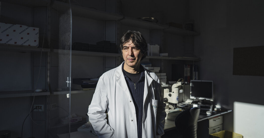
Valter Longo, who wants to live to a healthy 120 or 130, sees the key to longevity in diet — legumes and fish — and faux fasting.Most members of the band subscribed to a live-fast-die-young lifestyle. But as they partook in the drinking and drugging endemic to the 1990s grunge scene after shows at the Whiskey a Go Go, Roxy and other West Coast clubs, the band’s guitarist, Valter Longo, a nutrition-obsessed Italian Ph.D. student, wrestled with a lifelong addiction to longevity.Now, decades after Dr. Longo dropped his grunge-era band, DOT, for a career in biochemistry, the Italian professor stands with his floppy rocker hair and lab coat at the nexus of Italy’s eating and aging obsessions.“For studying aging, Italy is just incredible,” said Dr. Longo, a youthful 56, at the lab he runs at a cancer institute in Milan, where he will speak at an aging conference later this month. Italy has one of the world’s oldest populations, including multiple pockets of centenarians who tantalize researchers searching for the fountain of youth. “It’s nirvana.”Dr. Longo, who is also a professor of gerontology and director of the U.S.C. Longevity Institute in California, has long advocated longer and better living through eating Lite Italian, one of a global explosion of Road to Perpetual Wellville theories about how to stay young in a field that is itself still in its adolescence.In addition to identifying genes that regulate aging, he has created a plant and nut-based diet with supplements and kale crackers that mimics fasting to, he argues, allow cells to shed harmful baggage and rejuvenate, without the down side of actually starving. He has patented and sold his ProLon diet kits; published best-selling books (“The Longevity Diet”); and been called an influential “Fasting Evangelist” by Time magazine.Last month, he published a new study based on clinical trials of hundreds of older people — including in the Calabria town from which his family hails — that he said suggests that periodic cycles of his own faux-fasting approach could reduce biological age and stave off illnesses associated with aging.We are having trouble retrieving the article content.Please enable JavaScript in your browser settings.Thank you for your patience while we verify access. If you are in Reader mode please exit and log into your Times account, or subscribe for all of The Times.Thank you for your patience while we verify access.Already a subscriber? Log in.Want all of The Times? Subscribe.

Read more →

Patients Hate ‘Forever’ Drugs. Are Ozempic and Wegovy Different?

Most people, study after study shows, don’t take the medicines prescribed for them. It doesn’t matter what they are — statins, high blood pressure drugs, drugs to lower blood sugar, asthma drugs. Either patients never start taking them, or they stop.It’s a problem that doctors call nonadherence — the common human tendency to resist medical treatment — and it leads to countless deaths and billions of dollars of preventable medical costs each year.But that resistance may be overcome by the blockbuster obesity drugs Wegovy and Zepbound, which have astounded the world with the way they help people lose weight and keep it off. Though it’s still early days, and there is a paucity of data on compliance with the new drugs, doctors say they are noticing another astounding effect: Patients seem to take them faithfully, week in and week out.Some patients may have to get over an initial reluctance to start. A national survey showed that when people were told they would gain weight back if they stopped taking the drugs, most lost interest in starting them.In one small study, patients stopped refilling prescriptions for months at a time, perhaps because of side effects, lack of availability, or insurance and cost issues.But anecdotally, doctors and patients say, those who begin taking the drugs are continuing.“I don’t intend to ever stop taking this medicine,” said Kimberly DelRosso of Pembroke, Mass., who takes Wegovy.We are having trouble retrieving the article content.Please enable JavaScript in your browser settings.Thank you for your patience while we verify access. If you are in Reader mode please exit and log into your Times account, or subscribe for all of The Times.Thank you for your patience while we verify access.Already a subscriber? Log in.Want all of The Times? Subscribe.

Read more →

The man in the iron lung: How Paul Alexander lived life to the full

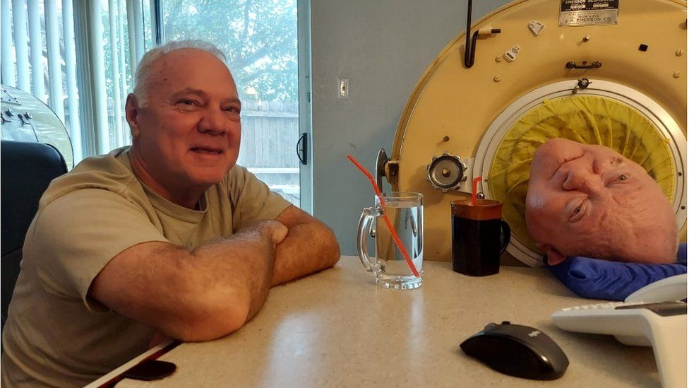
Published45 minutes agoShareclose panelShare pageCopy linkAbout sharingImage source, Philip AlexanderBy Catherine SnowdonBBC NewsPaul Alexander was six years old when he woke, terrified, to find himself inside a large metal tube, with only his head sticking out.He couldn’t move to feel what was trapping him, and when he tried to call for help, he discovered he couldn’t make a sound.Paul had survived a serious bout of polio, but had been left quadriplegic. After an emergency tracheostomy operation, he was unable to breathe without the iron lung machine that now encased his small body.When he died recently at the age of 78, Paul had spent more than seven decades using his iron lung, longer than anyone else in history. Image source, GoFundMeBut what was it that marked him out from so many of his peers, and kept him going?Polio ran riot in the late 19th and early 20th centuries, killing and maiming scores of children.In hospital in Texas in 1952, Paul was surrounded by other children in similar predicaments. According to the World Health Organization, one in 200 polio infections leads to irreversible paralysis. Among those paralysed, 5-10% die when their breathing muscles become immobilised.Uncertain futureAfter two years in hospital, doctors began to cast doubt on Paul’s future.That led his parents to make the brave decision to take him and his iron lung home, to live out his days in peace. But instead of dying, Paul went from strength to strength, once he was home in his parents’ care.The iron lung uses a negative pressure system. Powered by a motor, its bellows suck air out of the cylinder, creating a vacuum around the patient’s body and forcing the lungs to expand and take in air. When the air is let back in, the same process in reverse makes the lungs deflate. The device needs a source of energy to function.During power cuts, the bellows had to be pumped by hand; neighbours would come to help with the job. Paul’s father designed an alarm bell that Paul could ring using his mouth if he needed urgent attention. Over time Paul learnt to consciously breathe by gulping down air, using his throat muscles to force air into his lungs. He called it frog-breathing.His younger brother, Philip, explained to the BBC that the promise of a puppy gave Paul the incentive to be brave, and attempt to spend time out of his iron lung to learn the complicated glossopharyngeal breathing technique, the medical term for frog-breathing.”He was scared of course of choking to death,” says Philip. “They told him if you last three minutes, you’ll get the dog that you want.” And he did.Image source, Rotary Club of Park Cities DallasAs his confidence and strength grew, he was able to spend increasingly longer periods out of the iron lung. This allowed him to begin to experience a bit more of life. He was allowed to venture into the neighbourhood in his wheelchair with childhood friends, returning to the iron lung when he was tired.”He was just a normal brother to me. We fought, we played, we loved, we partied, we went to concerts together – he was just a normal brother,” says Philip.Paul finished school at home and went on to earn a college degree before setting his sights on law school.Image source, University of TexasPhilip recalls Paul’s time at the University of Texas in Austin as being “incredible”. His parents helped Paul move in with his iron lung, and then he was on his own – with limited help at first, after the carer he had hired failed to show up.”He didn’t really have any caregivers. He was in the dorm and different people just took care of him accidentally. They pushed him around campus [in his wheelchair],” says Philip.Paul went on to practise law in downtown Dallas. He had to deal with the surprise of clients on entering his office and seeing him in his iron lung.”It’s not an easy thing to see, just a head sticking out,” says Philip. “People immediately go into shock. I saw that happen a lot.”Paul lived alone for much of his adult life, no mean feat for someone unable to take care of his basic human needs like using the toilet or getting a drink on his own.This video can not be playedTo play this video you need to enable JavaScript in your browser.Philip says he became the master of his own domain, helping people to help him.”He needed a unique type of care. Not even professionals are trained to take care of a quadriplegic in an iron lung,” says Philip of the huge responsibility that came with supporting his brother.”Most of the care was basic – shaving and feeding for example. But to move him you needed to be careful not to jam his finger and so on.” There was no instruction manual for those who answered Paul’s adverts for caregivers. “They learned as they went,” says Philip. “And many left after a day or two. I remember going around once and asking a couple of assisted living centres if they could care for him, and the looks on their faces were priceless.”Paul had one carer who was in his life for decades. When Kathy Gaines died, her passing left Paul bereft.Philip says he always saw himself as the back-up carer for his brother, but he admired the support system Paul built: “He had a lot of wonderful friends, some really beautiful people in his life.”Help neededOne of those people came into Paul’s life in a moment of dire need.In 2015, his iron lung began to leak. As the machines were by now extremely rare, it was a race against time to find someone able to carry out the vital repair job to make it airtight once more.After a plea was posted on social media, responses came in from around the world. But the solution turned out to be much closer to home.Ten miles down the road from Paul’s apartment in Dallas, was the mechanical durability business Environmental Testing Laboratory, owned by Brady Richards.Mr Richards had come into possession of two iron lung machines at a building clearance and he recalls how one day a paramedic walked in asking: “Is this the place I can find iron lungs?”The medic had been transporting Paul to and from hospital, as carers fought to keep him alive in the failing iron lung. As the situation became more desperate, word of mouth had brought her to Mr Richards.Image source, Brady Richards”I did not have any idea about Paul Alexander at that time,” says Mr Richards. On learning about the emergency, he immediately set about rebuilding one of the broken machines he had in his warehouse. He built some parts from scratch and scavenged others from other devices, learning as he went along: “Iron lungs are very robust machines. They are built to last. “It’s a simple machine so I managed to work it out. You can increase and decrease the breathing rate and the pressure. Paul always liked the settings up high.”Once the refurbished machine was ready, Mr Richards swapped it for the damaged one at Paul’s apartment. He wouldn’t accept any money for his work.But that wasn’t the end of the story. Later that night Mr Richards received a call from Paul’s carer to say the machine wasn’t working.Mr Richards returned to Paul’s apartment and quickly realised the neck collar wasn’t on properly and had come loose.”Paul was saying ‘I’m fine, I’m OK’ as we worked to correct the problem,” recalls Mr Richards. “The truth was, he was turning blue.”In moments of desperation like these, Paul had access to more portable breathing devices, which used a different sort of technology to the iron lung.”Positive pressure respirators can make the patient feel like they have their head stuck out of the car window. Not everyone likes that sensation” explains Dr Patrick Murphy, the clinical lead consultant at the Lane Fox respiratory unit at St Thomas’ Hospital, in London.Paul never moved full-time to these mask-based devices. Another polio survivor, living in York, in northern England, did make the switch.Image source, James PorteousJames Porteous, 78, was infected with polio in the same year as Paul. He was initially placed in an iron lung. He recovered to a point where he could live his life without significant breathing support, but as is often the case for polio survivors, as he aged he began to need more help. He now uses a respirator mask for about 17 hours a day: “I don’t remember much about being in the iron lung. I was also placed in a head-to-toe plaster cast initially as the thought was that this would prevent my limbs from becoming deformed,” says Mr Porteous, who is the regional president of the British Polio Fellowship. Mr Porteous had a long career, first in stockbroking and then in various senior roles for Rowntree’s, which became part of Nestlé. He married and has four daughters. He travelled the world and in 2001 he was awarded an MBE for services to the community.Image source, James Porteous”I could have sat all my life with a rug over my knees, but I decided to just get on with it. I’m tired these days and I don’t get out much any more, but life is good. I have a nice family and good friends. One thing I’d still like to see achieved in my lifetime is the complete eradication of polio around the world,” he says.Ending polio globally was also Paul’s ambition. He wrote about it in his 2020 memoir, which he typed himself, using a pencil attached to a stick gripped in his mouth to reach the computer keyboard.Philip says it was after the book was published that he fully realised what an inspiration his brother was to people around the world: “His personality had a lot to do with how much he was admired. He had that great big smile and he was such a welcoming, warm person. He made people comfortable.”Brady Richards remained involved in Paul’s life over the years. He helped Paul move apartments when needed, and serviced the iron lung regularly: “It was always a pleasure to be around Paul – he had a very upbeat and positive attitude.” Image source, NHSDr Murphy is full of admiration for the people who cared for Paul.”His parents took their son home in a machine that was at the time relatively high-end technology. He also had a tracheostomy, which calls for special care. They will have needed to be engineers, nurses and doctors. “If you speak to lots of trained doctors or nurses they won’t be confident in managing a patient with complex respiratory failure, and yet patients like Paul and their families do it at home on their own. The bravery cannot be underestimated,” says Dr Murphy.World recordLast year Paul was recognised by Guinness World Records as the person who had lived the longest in an iron lung.Philip’s admiration for his brother runs deep: “I saw him go through a lot of struggles in his life. I’m going to miss him. I called him when I needed to talk to someone about any kind of problem I had.”He says that while the rest of the world might be amazed by how long his brother survived in his iron lung, his parents would not have been: “They believed in him. They gave him so much strength and love. They wouldn’t have been shocked.” More on this story’Man in the iron lung’ Paul Alexander dies at 78Published13 MarchSuper-engineered vaccines created to help end polioPublished14 June 2023What is polio and how can you protect yourself?Published10 August 2022

Read more →

Stem cell model offers first glimpse of early human embryonic development

It’s one of life’s most defining moments — that crucial step in embryonic development, when an indistinct ball of cells rearranges itself into the orderly three-layered structure that sets the stage for all to come. Known as gastrulation, this crucial process unfolds in the third week of human development. “Gastrulation is the origin of our own individualization, the emergence of our axis,” says Rockefeller’s Ali Brivanlou. “It is the first moment that separates our heads from our behinds.”
Observing the molecular underpinnings of this pivotal event would go a long way toward helping scientists prevent miscarriages and developmental disorders. But studying human gastrulation has proven both technologically difficult and ethically complicated, and thus current approaches have had limited success in expanding our understanding of early human development. Now Brivanlou and colleagues have demonstrated how a stem cell model system known as a blastoid can allow the study of the nuances of human gastrulation in the presence of pre-implantation extra-embryonic cell types. Their study, published in Stem Cell Reports, describes the scientific and clinical potential of this new platform.
“Gastrulation was a tremendous black box. We had never seen ourselves at that stage,” Brivanlou says. “This moves us closer to understanding how we begin.”
A better blastocyst
Prior to implantation, an embryo is a ball of about 250 cells organized as a blastocyst. This elusive ball of cells was difficult to study directly, so scientists developed blastoids — stem-cell-based blastocyst models. Blastoids can be cloned, experimentally manipulated, and programmed, allowing scientists to study identical blastoids over and over again.
The question was whether blastoids could gastrulate in vitro. Unlike a blastocyst in vivo, which rolls around in the uterus until it attaches to maternal tissue, blastoids were good at modeling the ball of cells from which life emerges, but it remained unclear whether this in vitro model could model later stages of human development. That is, until Brivanlou developed a platform to allow blastoids to attach in vitro, and thereby progress toward gastrulation.
“We were then able to see epiblast symmetry breaking, marked by BRA expression, for the first time with the high molecular resolution,” says Riccardo De Santis, a research associate in the Brivanlou lab and lead author on the study. “This allowed us to start asking more detailed questions about the earliest moments of life.”
With this unprecedented clarity, the team directly observed two key moments in gastrulation: the first epiblast symmetry-breaking event and the emergence of the molecular markers of the primitive streak and mesoderm upon in vitro attachment.

The primitive streak is a structure that marks the beginning of gastrulation and lays the foundation for the three primary layers of the embryo. One of those layers, the mesoderm, forms during gastrulation and gives rise to muscles, bones, and the circulatory system. The team discovered that, as early as seven days after attachment, they were already able to use molecular markers to detect the earliest signature of a nascent primitive streak and mesodermal cells.
To confirm their findings, the team also compared the blastoid results with data from in vitro attached human embryos and demonstrated that blastoids express the same genes in vitro that a regular embryo would at that stage in vivo, a strong demonstration of the power of blastoids as models for human embryonic development. Further highlighting the power of the lab’s in vitro attached blastoid system, the team then used it to demonstrate that pathways that regulate the rise of the primitive streak and mesoderm in vivo also regulate blastoids symmetry breaking in vitro — all with nothing but stem-cell-derived blastoid models.
Along the way, the team also demonstrated that gastrulation in vitro can begin at day 12, earlier than once thought. “This will change textbooks,” Brivanlou says. “We’ve contributed to redefining the molecular signature and timing of the onset of gastrulation upon in vitro attachment.”
Therapeutic possibilities
The results demonstrate that blastoids, when combined with the Brivanlou lab’s unique attachment platform, are now capable of conveying insights into early human development that have long been inaccessible. De Santis envisions a future in which blastoid-based research leads to advancements in diagnosing and treating developmental disorders, or offers insights into potential causes of early miscarriages during gastrulation.
“Many couples can’t have babies because the embryo doesn’t attach properly, and many miscarriages occur in the first few weeks of pregnancy,” De Santis explains. “We now have a model system that can help us understand the molecular mechanism that defines whether a pregnancy will be successful or not.” In the near future, De Santis hopes to combine this method with machine learning to help predict pregnancy outcomes and the trajectories of developmental disorders by observing how model blastoids built with particular genetic makeups fare in vitro.
“A better understanding of gastrulation — and the ability to study it with a reliable model system — impacts everything from survival of the fetus to autism to neurodegeneration.”

Read more →

Living With a Messy Partner? Tips for Handling Different Organization Habits

When people with different tolerances for clutter live together, things can get tense. Experts offer ways to find peace amid the piles.Last year, Tracy McCubbin — who has been a professional declutterer for two decades and lives by the motto “don’t put it down, put it away” — married a man she described as “very messy.”Both acknowledged the “cosmic joke” of their unlikely pairing. Ms. McCubbin put blue painter’s tape on every drawer and cupboard in the kitchen when the pair first moved in together, offering a map to what goes where. But she has also learned to practice what she preaches to her clients, staying cool and calm about messes that don’t affect her day-to-day functioning. Like his night stand, which is buried under books, charging cables and remotes to TVs she is fairly certain they no longer own.Or the jumble of tools her husband, an avid gardener, tends to leave in the yard. “It’s all over the place,” Ms. McCubbin sighed. “But you know what? We have a beautiful garden. Our fruit trees are fruiting. It’s really been about understanding: This part doesn’t matter.”Ms. McCubbin, and other experts in organizing as well as psychology, said there were a few practical strategies that could help pack rats and neatniks cohabitate in relative harmony.Improve your organization systems.“Oftentimes when one person is more cluttered, the underlying thesis is that they’re wrong, that they’re doing it the wrong way, that they’re bad,” Ms. McCubbin said. But in many cases, household clutter is simply an indication you don’t have solid systems in place.Some of the solutions she offers to clients are almost too obvious, she said. For instance, she has worked with frustrated parents whose children toss backpacks and coats in what she calls the “landing strip” just inside the front door. Hanging a few hooks that they can easily reach helps.We are having trouble retrieving the article content.Please enable JavaScript in your browser settings.Thank you for your patience while we verify access. If you are in Reader mode please exit and log into your Times account, or subscribe for all of The Times.Thank you for your patience while we verify access.Already a subscriber? Log in.Want all of The Times? Subscribe.

Read more →

Diagnosis of Princess Kate’s Cancer Followed Familiar Pattern, Doctors Say

Although it is not known what type of cancer Princess Catherine has, oncologists say that what she described in her public statement that was released on Friday — discovering a cancer during another procedure, in this case a “major abdominal surgery” — is all too common.“Unfortunately, so much of the cancer we diagnose is unexpected,” said Dr. Elena Ratner, a gynecologic oncologist at Yale Cancer Center who has diagnosed many patients with ovarian cancer, uterine cancer and cancers of the lining of the uterus.Without speculating on Catherine’s procedure, Dr. Ratner described situations where women will go in for surgery for endometriosis, a condition in which tissue similar to the lining of the uterus is found elsewhere in the abdomen. Often, Dr. Ratner says, the assumption is that the endometriosis has appeared on an ovary and caused a benign ovarian cyst. But one to two weeks later, when the supposedly benign tissue has been studied, pathologists report that they found cancer.In the statement, Princess Catherine said she was is getting “a course of preventive chemotherapy.”That, too, is common. In medical settings it is usually called adjuvant chemotherapy.Dr. Eric Winer, director of the Yale Cancer Center, said that with adjuvant chemotherapy, “the hope is that this will prevent further problems,” and avoid a recurrence of the cancer.It also means that “you removed everything” that was visible with surgery, said Dr. Michael Birrer, director of the Winthrop P. Rockefeller Cancer Institute at the University of Arkansas for Medical Sciences. “You can’t see the cancer,” he added because microscopic cancer cells may be left behind. The chemotherapy is a way to attack microscopic disease, he explained.Other parts of Catherine’s statement also hit home for Dr. Ratner, particularly her concern for her family.“William and I have been doing everything we can to process and manage this privately for the sake of our young family,” the statement said.And, “it has taken us time to explain everything to George, Charlotte, and Louis in a way that is appropriate for them, and to reassure them that I will be okay.”Those are sentiments that Dr. Ratner hears on a regular basis and reveal, she says, “how hard it is for women to be diagnosed with cancer.”“I see this day in and day out,” she said. “Women always say, ‘Will I be there for my kids? What will happen with my kids?’”“They don’t say, ‘What will happen to me?’”

Read more →

What we know about Kate’s cancer diagnosis

Published24 minutes agoShareclose panelShare pageCopy linkAbout sharingImage source, Getty ImagesBy Michelle Roberts, Philippa Roxby and Smitha MundasadBBC NewsCatherine, Princess of Wales, says she is having treatment for cancer which was discovered following major abdominal surgery. She started preventative chemotherapy in late February. When was the cancer found?The princess underwent surgery in January. The palace did not disclose details at the time but said the procedure was planned and her condition was “not cancer-related”.Her video message says that tests carried out after the operation found cancer had been present.What type of cancer does the princess have?Kensington Palace has not said what type of cancer she has.A spokesperson said: “We will not be sharing any further private medical information. The princess has a right to medical privacy as we all do.”What is preventative chemotherapy?Catherine said her medical team advised she should undergo “preventative chemotherapy” and she is in the early stages of this treatment.Chemotherapy is a catch-all term for medication to kill cancer cells – it can be given via a drip or in pill form.How much you need and how often depends on the type of cancer and how it is growing. The aim is to reduce the risk of cancer remaining or coming back.Bob Phillips, professor of paediatric oncology at the University of York, said chemotherapy can be used to “mop up” cancer cells if there are any still present.How did doctors find the cancer?After the surgery in January, which Kensington Palace says was successful, doctors performed tests which revealed cancer had been present.Commonly, tissue removed during surgery would be sent to a lab for analysis and to see what cells are there.Is she getting treatment privately or on the NHS?Catherine’s original operation took place at the London Clinic, a private facility.The Palace is not commenting on where she is now receiving care. In her video message Catherine thanked the “fantastic medical team” who have been treating her.What is cancer?Cancer occurs when cells in a specific part of the body divide in an uncontrolled way.Untreated, these cells can potentially spread to other tissues in the body, including organs, which is known as secondary or metastatic cancer.How many people get cancer?In the UK, one in two people develop some kind of cancer during their lifetime.There are more than 200 different types of cancer – the most common ones in the UK are breast, lung, prostate and bowel, according to the NHS UK website.Each cancer is diagnosed and treated in a particular way.Anyone can develop cancer, but the risk goes up the older we become because there’s more time for cell damage to build up.Most cases of cancer are in people aged 50 and over. In the UK, a third of all cases are in people aged 75 and over.Survival from cancer has doubled in the last 50 years.What should you do if you think you have cancer?If you notice something that isn’t normal for you, see a doctor. That might include:unexplained bleeding or painan unusual lump or swellingunexplained tiredness and weight lossa persistent coughThe symptoms you are experiencing may not be cancer, but it is important to get checked. Finding cancer early can often make it easier to treat.More on this storyPrincess of Wales in hospital after abdominal surgeryPublished17 JanuaryKing Charles diagnosed with cancerPublished6 FebruaryEnlarged prostate: What is it and how to treat itPublished29 JanuaryRelated Internet LinksMacmillan Cancer SupportCancer Research UKNHS: Cancer signs and symptomsThe BBC is not responsible for the content of external sites.

Read more →

Bioelectronic mesh capable of growing with cardiac tissues for comprehensive heart monitoring

A team of engineers led by the University of Massachusetts Amherst and including colleagues from the Massachusetts Institute of Technology (MIT) recently announced in the journal Nature Communications that they had successfully built a tissue-like bioelectronic mesh system integrated with an array of atom-thin graphene sensors that can simultaneously measure both the electrical signal and the physical movement of cells in lab-grown human cardiac tissue. In a research first, this tissue-like mesh can grow along with the cardiac cells, allowing researchers to observe how the heart’s mechanical and electrical functions change during the developmental process. The new device is a boon for those studying cardiac disease as well as those studying the potentially toxic side-effects of many common drug therapies.
Cardiac disease is the leading cause of human morbidity and mortality across the world. The heart is also very sensitive to therapeutic drugs, and the pharmaceutical industry spends millions of dollars in testing to make sure that its products are safe. However, ways to effectively monitor living cardiac tissue are extremely limited.
In part, this is because it is very risky to implant sensors in a living heart, but also because the heart is a complex kind of muscle with more than one thing that needs monitoring. “Cardiac tissue is very special,” says Jun Yao, associate professor of electrical and computer engineering in UMass Amherst’s College of Engineering and the paper’s senior author. “It has a mechanical activity — the contractions and relaxations that pump blood through our body — coupled to an electrical signal that controls that activity.”
But today’s sensors can typically only measure one characteristic at a time, and a two-sensor device that could measure both charge and movement would be so bulky as to impede the cardiac tissue’s function. Until now, there was no single sensor capable of measuring the heart’s dual properties without interfering with its functioning.
The new device is built of two critical components, explains lead author Hongyan Gao, who is pursuing his Ph.D. in electrical engineering at UMass Amherst. The first is a three-dimensional cardiac microtissue (CMT), grown in a lab from human stem cells under the guidance of co-author Yubing Sun, associate professor of mechanical and industrial engineering at UMass Amherst. CMT has become the preferred model for in vitro testing because it is the closest analog yet to a full-size, living human heart. However, because CMT is grown in a test tube, it has to mature, a process that takes time and can be easily disrupted by a clumsy sensor.
The second critical component involves graphene — a pure-carbon substance only one atom thick. Graphene has a few surprising quirks to its nature that make it perfect for a cardiac sensor. Graphene is electrically conductive, and so it can sense the electrical charges shooting through cardiac tissue. It is also piezoresistive, which means that as it is stretched — say, by the beating of a heart — its electrical resistance increases. And because graphene is impossibly thin, it can register even the tiniest flutter of muscle contraction or relaxation and can do so without impeding the heart’s function, all through the maturation process. Co-author Jing Kong, professor of electrical engineering at MIT, and her group supplied this critical graphene material.
“Although there have already been many applications for graphene, it is wonderful to see that it can be used in this critical need, which takes advantage of graphene’s different characteristics,” says Kong.

Gao, Yao and their colleagues then embedded a series of graphene sensors in a soft, stretchable porous mesh scaffold they developed that has close structural and mechanical properties to human tissue and which can be applied non-invasively to cardiac tissue.
“No one has ever done this before,” says Gao. “Graphene can survive in a biological environment without degrading for a very long time and not lose its conductivity, so we can monitor the CMT across its entire maturation process.”
“This is crucial for a number of reasons,” adds Yao. “Our sensor can give real-time feedback to scientists and drug researchers, and it can do so in a cost-effective way. We take pride in using the insights of electrical engineering to help build tools that can be useful to a wide range of researchers.”
In the future, Gao says, he hopes to be able to adapt his sensor to grander scales, even to in vivo monitoring, which would provide the best-possible data to help solve cardiac disease.
This research was supported by the Army Research Office, the National Institutes of Health, the U.S. National Science Foundation, the Semiconductor Research Corporation, and the Link Foundation, as well as the Institute for Applied Life Sciences at UMass Amherst.

Read more →

If faces look like demons, you could have this extraordinary condition

Imagine if every time you saw a face, it appeared distorted. Well, for those who have a very rare condition known as prosopometamorphopsia (PMO), which causes facial features to appear distorted, that is reality.
As the Dartmouth-based website about prosopometamorphopsia explains, “‘Prosopo’ comes from the Greek word for face ‘prosopon’ while ‘metamorphopsia’ refers to perceptual distortions.”’ Specific symptoms vary from case to case and can affect the shape, size, color, and position of facial features. The duration of PMO also varies; it “can last for days, weeks, or even years.”
A new Dartmouth study published in the “Clinical Pictures” section of The Lancet reports on a unique case of a patient with PMO. The research is the first to provide accurate and photorealistic visualizations of the facial distortions experienced by an individual with PMO.
The patient, a 58-year-old male with PMO, sees faces without any distortions when they are viewed on a screen and on paper, but he sees distorted faces that appear “demonic” when viewed in-person. Most PMO cases however, see distortions in all contexts, so his case is especially rare and presented a unique opportunity to accurately depict his distortions.
For the study, the researchers took a photograph of a person’s face. Then, they showed the patient the photograph on a computer screen while he looked at the real face of the same person. The researchers obtained real-time feedback from the patient on how the face on the screen and the real face in front of him differed, as they modified the photograph using computer software to match the distortions perceived by the patient.
“In other studies of the condition, patients with PMO are unable to assess how accurately a visualization of their distortions represents what they see because the visualization itself also depicts a face, so the patients will perceive distortions on it too,” says lead author Antônio Mello, a PhD student in the Department of Psychological and Brain Sciences at Dartmouth. In contrast, this patient doesn’t see distortions on a screen. This means that the researchers were able to modify the face in the photograph, and the patient could accurately compare how similar his perception of the real face was to the manipulated photograph. “Through the process, we were able to visualize the patient’s real-time perception of the face distortions,” says Mello.
In their research with other PMO cases, the co-authors state that some of their PMO participants have seen health professionals who wanted to help but diagnosed them with another health condition, not PMO.
“We’ve heard from multiple people with PMO that they have been diagnosed by psychiatrists as having schizophrenia and put on anti-psychotics, when their condition is a problem with the visual system,” says senior author Brad Duchaine, a professor of psychological and brain sciences and principal investigator of the Social Perception Lab at Dartmouth.
“And it’s not uncommon for people who have PMO to not tell others about their problem with face perception because they fear others will think the distortions are a sign of a psychiatric disorder,” says Duchaine. “It’s a problem that people often don’t understand.”
Through their paper, the researchers hope to increase public awareness of what PMO is.

Read more →

The aging brain: Protein mapping furnishes new insights

For the neurons in the brain to work smoothly and be able to process information, the central nervous system needs a strictly regulated environment. This is maintained by the blood-brain barrier, whereby specialized brain endothelial cells lining the inner walls of blood vessels regulate the exchange of molecules between the circulatory and nervous systems. Earlier studies have shown that various functions that are dependent on these cells, such as the integrity of the blood-brain barrier or the regulation of blood supply to the brain, decline over the course of a person’s life. This dysregulation leads to a dysfunction of the brain vasculature and is therefore a major contributor to medical conditions such as strokes and dementia.
However, the molecular changes that underlie this loss of function have remained largely obscure. To improve our mechanistic understanding, researchers carry out molecular profiling studies to investigate the different components of brain endothelial cells and collect their findings in large databases. “The transcriptome — that is to say, the RNA contained in endothelial cells — has since been quite comprehensively mapped,” says LMU professor Martin Dichgans, Director of the Institute for Stroke and Dementia Research at University of Munich Hospital and Principal Investigator at the SyNergy Cluster of Excellence. “What has been lacking is corresponding data on the complete set of proteins in the cells, the proteome.” A study recently published in the journal Nature Aging, which had major contributions by researchers from LMU and SyNergy, has now closed this knowledge gap.
Dysregulated metabolism
For the study, the team developed a protocol for enriching brain endothelial cells in mice, which makes it possible to resolve age-related changes in protein composition. Using an unsupervised (computer-aided) cluster analysis, the scientists then related these protein dynamics to biological functions. “Our results show a dysregulation of key molecules involved in the uptake of substances into cells, in receptor recycling, and in the degradation of molecules within specific cellular compartments called lysosomes,” says Dichgans.
One of the most striking changes concerned a decrease in proteins involved in vesicle-mediated transport. In addition, the study provides evidence that deficiency of apolipoprotein E, a protein involved in lipid metabolism results in a signature of accelerated endothelial aging. “The results complement and expand findings from studies on the RNA sequencing of brain endothelial cells during aging,” summarizes Dichgans. “Our proteomic approach captures processes that are not detected at the RNA level.” Overall, the study offers a framework for understanding important endothelial signaling pathways during aging and serves as a data basis for future analyses of brain endothelial function. The researchers are making their data on age-related protein abundance of the mouse endothelium available in a publicly accessible database for further use.

Read more →