Unregulated sales of a toxic and hallucinogenic mushroom endanger public health

Americans’ interest in a potentially harmful “magic mushroom” is soaring, with Google searches skyrocketing 114 percent from 2022 to 2023, according to a new study by researchers at the University of California San Diego Herbert Wertheim School of Public Health and Human Longevity Science. In a paper published in the American Journal of Preventive Medicine, the scientists suggest that the growing market for Amanita muscaria may be sparked in part by emerging clinical research supporting the safety and efficacy of psilocybin as a treatment for depression.
Like psilocybin mushrooms, Amanita muscaria mushrooms have psychotropic effects. These include a feeling of weightlessness, visual and auditory hypersensitivity, space distortion, unawareness of time, and colored hallucinations. The psychotropic effects are produced by compounds that naturally occur in the mushroom called muscimol and ibotenic acid, its biosynthetic precursor.
However, in addition to being psychotropic, these compounds can also be more toxic than fentanyl, cocaine, and PCP, according to the scientists’ review of estimates from published mouse studies. Nevertheless, gummies and chocolates containing these compounds are being marketed with health-related claims such as mitigation of anxiety, depression, and other conditions, often by vague references to clinical studies related to psilocybin, which is not as toxic and produces different psychotropic effects.
“There is a lot of interest in the therapeutic potential for psilocybin and for good reason. But at the same time, a growing industry may be trying to capitalize on this interest by marketing other mushrooms. For example, some manufacturers are calling Amanita muscaria products ‘magic mushroom gummies’ and not disclosing what mushroom they contain, or not making it clear Amanita muscaria is a different mushroom than psilocybin and has essentially no clinical evidence supporting its use as a therapy,” said Eric Leas, Ph.D., M.P.H., assistant professor in the UC San Diego Herbert Wertheim School of Public Health and Human Longevity Science and senior author on the paper.
Psilocybin and muscimol work in different ways. Psilocybin is an antidepressant that primarily binds to serotonin receptors, activating a neural pathway that mediates happiness and optimism. Amanita muscaria however is a depressant, similar to alcohol and benzodiazepines, which suppress the central nervous system. Leas believes that marketing Amanita muscaria as a psilocybin-type product violates consumers’ right to informed consent.
“There may be some pharmaceutical potential to Amanita muscaria, but muscimol does not have the same effects on the body as psilocybin, so it probably would not have the same treatment applications if it ever went through drug development. For this reason, it is misleading not to clearly distinguish between muscimol and psilocybin. If someone is consenting to a psychedelic experience, they have a right to know what substance they are taking and receive accurate information about its potential health benefits and health risks.”
False marketing may be enabled by lack of federal regulation of Amanita muscaria. Under the 1970 Controlled Substances Act, psilocybin is a Schedule 1 drug, making its manufacture, distribution, import/export, possession and use illegal. In 2017, the FDA designated psilocybin as a “breakthrough therapy” and in 2023 loosened restrictions to allow drug developers and scientists to conduct clinical trials with psilocybin, including some that are taking place at UC San Diego. Nevertheless, it continues to be a Schedule 1 controlled substance, and, therefore its use is disallowed out of the context of clinical trials.

Not so for Amanita muscaria. Although there are several published case studies of hospitalizations and deaths resulting from Amanita muscaria consumption, to date it is not included on a Controlled Substances list (except for the State of Louisiana, where sales are restricted). However, it is often marketed as a dietary supplement, products covered by regulations enforced by the U.S. Food and Drug Administration and the Federal Trade Commission.
“We have found that many manufacturers use supplement labeling, including ‘Supplement Facts’ panels,” said Leas. “However, there is a process for bringing a supplement to market that involves presenting safety data and filing an application, and we cannot find any evidence that any of these manufacturers have gone through this process, and this makes the current products sold in this manner illegal.
“In my view, if a manufacturer wanted to develop a dietary supplement from Amanita muscaria, the application probably would not be approved because of muscimol and ibotenic acids’ inherent risks,” he added. “But right now it is the ‘Wild West,’ and companies are profiting from delayed enforcement while putting consumers at risk.”
The authors are making several general recommendations. The most restrictive would be to put Amanita muscaria on the Controlled Substances list, where it could first be evaluated for its medical potential and abuse liability before it is widely sold. However, if Amanita muscaria is not placed on a drug schedule, they recommend commonsense precautions, such as setting age restrictions, accurate dosing standards, childproof packaging, and marketing aimed at adults rather than children, all now required for legal sales of recreational cannabis. The authors would also like to see mental health professionals help their patients distinguish between psilocybin and Amanita muscaria.
The authors’ key takeaway is that “companies who are making these products are pushing the limits of our regulations. They are getting away with making a buck until someone tells them they can’t. Given the substantial risks associated with using Amanita muscaria products, it is a buyer beware marketplace where consumers are at risk and are not accurately informed. The time for a public health first response is now.”
Co-authors include: Nora Satybaldiyeva, Wayne Kepner, Kevin H. Yang, Raquel M. Harati, Jamie Corroon, and Matthieu Rouffet, of UC San Diego.
The work is supported in part by grant T32IP4684 from the California Tobacco Related Disease Research Program and grant K01DA054303 from the U.S. National Institute on Drug Abuse.
Read more →

Protein study could help researchers develop new antibiotics

A bacterial enzyme called histidine kinase is a promising target for new classes of antibiotics. However, it has been difficult to develop drugs that target this enzyme, because it is a “hydrophobic” protein that loses its structure once removed from its normal location in the cell membrane.
Now, an MIT-led team has found a way to make the enzyme water-soluble, which could make it possible to rapidly screen potential drugs that might interfere with its functions.
The researchers created their new version of histidine kinase by replacing four specific hydrophobic amino acids with three hydrophilic ones. Even after this significant shift, they found that the water-soluble version of the enzyme retained its natural functions.
No existing antibiotics target histidine kinase, so drugs that disrupt these functions could represent a new class of antibiotics. Such drug candidates are badly needed to combat the growing problem of antibiotic resistance.
“Each year, more than 1 million people die from antibiotic-resistant infections,” says Shuguang Zhang, a principal research scientist in the MIT Media Lab and one of the senior authors of the new study. “This protein is a good target because it’s unique to bacteria and humans don’t have it.”
Ping Xu and Fei Tao, both professors at Shanghai Jiao Tong University, are also senior authors of the paper, which appears today in Nature Communications. Mengke Li, a graduate student at Shanghai Jiao Tong University and a former visiting student at MIT, is the lead author of the paper.
A new drug target
Many of the proteins that perform critical cell functions are embedded in the cell membrane. The segments of these proteins that span the membrane are hydrophobic, which allows them to associate with the lipids that make up the membrane. However, once removed from the membrane, these proteins tend to lose their structure, which makes it difficult to study them or to screen for drugs that might interfere with them.

In 2018, Zhang and his colleagues devised a simple way to convert these proteins into water-soluble versions, which maintain their structure in water. Their technique is known as the QTY code, for the letters that represent the hydrophilic amino acids that become incorporated into the proteins. Leucine (L) becomes glutamine (Q), isoleucine (I) and valine (V) become threonine (T), and phenylalanine (F) becomes tyrosine (Y).
Since then, the researchers have demonstrated this technique on a variety of hydrophobic proteins, including antibodies, cytokine receptors, and transporters. Those transporters include a protein that cancer cells use to pump chemotherapy drugs out of the cells, as well as transporters that brain cells use to move dopamine and serotonin into or out of cells.
In the new study, the team set out to demonstrate, for the first time, that the QTY code could be used to create water-soluble enzymes that retain their enzymatic function.
The research team chose to focus on histidine kinase in part because of its potential as an antibiotic target. Currently most antibiotics work by damaging bacterial cell walls or interfering with the synthesis of ribosomes, the cell organelles that manufacture proteins. None of them target histidine kinase, an important bacterial protein that regulates processes such as antibiotic resistance and cell-to-cell communication.
Histidine kinase can perform four different functions, including phosphorylation (activating other proteins by adding a phosphate group to them) and dephosphorylation (removing phosphates). Human cells also have kinases, but they act on amino acids other than histidine, so drugs that block histidine kinase would likely not have any effect on human cells.
After using the QTY code to convert histidine kinase to a water-soluble form, the researchers tested all four of its functions and found that the protein was still able to perform them. This means that this protein could be used in high-throughput screens to rapidly test whether potential drug compounds interfere with any of those functions.

A stable structure
Using AlphaFold, an artificial intelligence program that can predict protein structures, the researchers generated a structure for their new protein and used molecular dynamics simulations to investigate how it interacts with water. They found that the protein forms stabilizing hydrogen bonds with water, which help it keep its structure.
They also found that if they only replaced the buried hydrophobic amino acids in the transmembrane segment, the protein would not retain its function. The hydrophobic amino acids have to be replaced throughout the transmembrane segment, which helps the molecule maintain the structural relationships it needs to function normally.
Zhang now plans to try this approach on methane monooxygenase, an enzyme found in bacteria that can convert methane into methanol. A water-soluble version of this enzyme could be sprayed at sites of methane release, such as barns where cows live, or thawing permafrost, helping to remove a large chunk of methane, a greenhouse gas, from the atmosphere.
“If we can use the same tool, the QTY code, on methane monooxygenase, and use that enzyme to convert methane into methanol, that could deaccelerate climate change,” Zhang says.
The research was funded, in part, by the National Natural Science Foundation of China.

Read more →

Study reveals target for reversing scar tissue after heart attack

A new study by investigators at Massachusetts General Hospital (MGH), a founding member of the Mass General Brigham (MGB) healthcare system, reveals an important step to help the human heart regenerate after myocardial infarction (MI).
For the first time, the researchers compared the formation of scar tissue in the injured hearts of zebrafish and mice, discovering how to potentially reverse the permanent scarring that is so damaging to mammal hearts. The results of the study are published in Nature Communications.
“We are the first to directly compare and show very fundamental differences in the formation of scar tissue between zebrafish and mammals,” says lead author Eman A. Akam-Baxter, PhD, an investigator at the MGH Cardiovascular Research Center and instructor in Medicine at Harvard Medical School. “The results of our study point to a possible new target to reverse scarring after myocardial infarction, which has never been shown before.”
A myocardial infarction causes the death of massive numbers of heart cells. To repair the injury, the body replaces the damaged and dead cells with scar tissue. Initially, the scar tissue is beneficial in keeping the heart intact. But eventually the scar tissue becomes a permanent part of the cardiac muscle, causing the heart to pump blood less effectively. An overworked heart causes the scar tissue to expand, leading to permanent heart damage.
The formation of permanent scar tissue after a heart injury is characteristic of all mammals. But zebrafish have a remarkable ability to completely remove scar tissue after an injury, allowing space for cardiac cells to regenerate and fully regrow a healthy heart.
“For many years investigators have focused on the properties of the cardiomyocytes (heart muscle cells) and immune cells in the zebrafish heart to explain this phenomenon” explained David Sosnovik, MD, the senior author of the paper.
“However, no studies characterizing the nature of the collagen scar in zebrafish have been performed. The expertise of Dr. Akam-Baxter in synthetic and analytical chemistry allowed us to approach this problem from a new angle.”
Until now, it was not possible to examine scar tissue formation in the tiny zebrafish heart by imaging. For this study, the researchers first had to develop a molecular imaging probe they named TMR-O, which allowed them to see details of the scarring inside the hearts of zebrafish and mouse models of cardiac injury.

Scar tissue is comprised of collagen, long strands of protein that bind to each other to form a fiber that gives scar tissue its structure and stability. The process of binding of collagen molecules is called cross-linking.
“Think of cross-linked collagen as a network of long strands of protein linked together,” explains Akam-Baxter. “Hands on multiple points of each protein strand clasp the opposite strand like a handshake.”
The probe the researchers developed bound to each hand, providing a fluorescent readout of how the collagen was cross-linked.
Researchers have long believed that the extent of collagen cross-linking is the key to whether a scar is resorbable or permanent. But when the MGH investigators tested that hypothesis, they found that the amount of cross-linking was similar in the zebrafish and the mice after cardiac injury. However, the type of cross-link was different.
“In the mouse heart, the chemical nature of the collagen cross-links was highly mature, and formed a structure that cannot be broken down by the antifibrotic enzymes in the body.
In contrast, the cross-links in the zebrafish resembled a looser handshake,” says Akam-Baxter. “The cross-links in the zebrafish heart persisted in a chemically immature form that can be broken down, and this allowed the fibrotic scars to be resorbed and replaced with regenerated heart cells.”
The authors further showed that the cross-links that form in the mouse hearts are a result of chemical modification (lysine hydroxylation) of the collagen strands in mice, and that this doesn’t occur to the same extent in the zebrafish heart.

This modification is made by an enzyme called lysyl hydroxylase 2; this enzyme is linked to permanent scarring in other organs in diseases of fibrosis.
“No one has studied the effect of blocking this enzyme in the context of heart attack,” says Akam-Baxter. Her research team is studying whether inhibition of this enzyme can effectively prevent permanent scarring in the heart after an MI. The researchers will also investigate whether scar tissue can be reversed in other organs.
“The numbers of deaths and amount of heart failure that results from scarring after MI are staggering,” says Akam-Baxter. “And fibrotic diseases also account for a huge number of deaths. If we can find a common feature of reversing scar tissue in multiple organs, we can potentially save many lives.”
“This study was highly multidisciplinary and was made possible through the unique research landscape that exists at the MGH,” explained Dr. Sosnovik, who directs the Program in Cardiovascular Imaging in the Martinos Center for Biomedical Imaging. “Researchers from the Cardiovascular Research Center, the Institute for Innovation in Imaging and the Martinos Center for Biomedical Imaging all brought different skills to the table that allowed us to collaboratively address this challenging scientific question.”

Read more →

Novel radiotracer produces high quality images of ‘Alzheimer’s disease of the heart’

A newly developed radiotracer can generate high quality and readily interpretable images of cardiac amyloidosis, a condition referred to as the “Alzheimer’s disease of the heart.” As the first amyloid-specific and pan-amyloid binding radiotracer designed for planar and SPECT/CT imaging, 99mTc-p5+14 could play an important role in early detection and treatment of cardiac amyloidosis. This research was presented at the 2024 Society of Nuclear Medicine and Molecular Imaging Annual Meeting.
Systemic amyloidosis is an incurable disease in which abnormal amounts of proteins build up within the body’s tissues and organs. About 20 percent of patients who have amyloid build-up in the heart experience early deaths. While recent progress in the treatment of cardiac amyloidosis has greatly improved patient prognosis, median survival remains low at approximately three to five years.
“Therapies that slow the progression of amyloid deposition have been developed; however, they are not effective in patients with late-stage disease. Therefore, the ability to detect cardiac amyloidosis early is critical,” noted Jonathan Wall, PhD, director of the Amyloidosis and Cancer Theranostics Program and professor at the University of Tennessee Graduate School of Medicine in Knoxville, Tennessee. “Unfortunately, there are currently no Food and Drug Administration (FDA)-approved imaging agents that detect cardiac amyloidosis.”
To address this issue, researchers developed a novel technetium-99m labeled variant of the pan-amyloid reactive peptide p5+14 (99mTc-p5+14). In the first-in-human study, five healthy volunteers and 30 patients newly diagnosed with light chain or transthyretin amyloidosis underwent 99mTc-p5+14 imaging with standard planar gamma scintigraphy and SPECT/CT. Blood was collected to assess serum biomarkers, and a transthoracic echocardiogram was performed. Standard 99mTc-pyrophosphate imaging was also performed on most patients at 72 hours after 99mTc-p5+14 image acquisition.
The planar and SPECT/CT images generated using 99mTc-p5+14 were of high quality and readily interpretable at both one and three hours post-injection. Patients with amyloid cardiomyopathy had significant 99mTc-p5+14 uptake in the heart, whereas no cardiac uptake was observed in healthy subjects.
“Early and accurate diagnosis of cardiac amyloidosis is crucial to ensure the most positive outcomes for patients,” said Wall. “Imaging with 99mTc-p5+14 could provide an easy to use and interpret technology that could be employed in the community cardiology setting, where SPECT imaging is common, as a rapid screen for amyloid cardiomyopathy in the future.”
The 99mTc-p5+14 radiotracer is currently in ongoing early-stage clinical evaluation at the University of Tennessee Graduate School of Medicine in conjunction with Attralus Inc., to assess safety and efficacy in patients with cardiac amyloidosis and healthy subjects. The data and insights obtained from this research will support initiation of a pivotal Phase 3 study and approval submissions to the FDA in the coming years.
Abstract 241277. “Preliminary Evaluation of 99mTc-Labeled Peptide p5+14 for the Detection of Cardiopulmonary Amyloidosis Using SPECT/CT and Planar Gamma Scintigraphic Imaging,” Jonathan Wall, Emily Martin, Alan Stuckey, Bryan Whittle, Joseph Jackson, Angela Williams, Trevor Hancock, R. Eric Heidel, Muddassir Mehmood, Anne Kassira, Ronald Lands, Hannah Watson, Rebecca Hung, Stephen Kennel, University of Tennessee Graduate School of Medicine, Knoxville, Tennessee.

Read more →

Early life exposure to weed pollen could increase childhood asthma risk

A study published in the European Respiratory Journal has found children who are exposed to tree and weed pollen in urban environments are at increased risk of respiratory health problems, including asthma. While green areas in urban settings decrease exposure to air pollution, allow kids to be active, and offer positive contact to a diverse microbiota — which in turn may help the positive development of a child’s immune system — they can also lead to the development of childhood asthma. Thankfully, trees can help mitigate this effect to some degree, thanks to their canopy.
“We found that tree canopy offers some protective effects towards the development of asthma in children. However, this protective effect goes away when there is increased exposure to weed pollen,” says lead author Éric Lavigne, an Adjunct Professor in the School of Epidemiology and Public Health in the Faculty of Medicine at the University of Ottawa.
Researchers tapped into the Ontario ASTHMA cohort database, analyzing data over an eight-year period to identify cases of childhood asthma between birth and the age of six. Just over 13 percent of the 214,000 mother-child pairs included in the study were diagnosed with childhood asthma. Researchers measured environmental exposure with the normalized difference vegetation index (NDVI) plus tree canopy estimates within a 250-meter radius from the child’s postal code at birth.
The surprising findings show the development of green spaces in urban environments increase pollen exposure and lessen the protection garnered from tree canopies.
With the increased focus on increasing urban vegetation, Lavigne says the public should be aware of the effect trees in cities can have, including the consequences of pollen on children’s respiratory health, particularly if it is the cause of increased risk of asthma.
“This could have an impact for urban planners and public health officials that work on redesigning our urban centres,” adds Lavigne, who is also a Senior Epidemiologist at Health Canada. “Along with weed pollen control programs and deciding when to plan specific trees, it will be important to consider tree species that minimize allergic pollen in the air.”

Read more →

Study shows first evidence of male-female differences in how pain can be produced

Research suggests that males and females differ in their experience of pain, but up until now, no one knew why. In a recent study published in BRAIN, University of Arizona Health Sciences researchers became the first to identify functional sex differences in nociceptors, the specialized nerve cells that produce pain.
The findings support the implementation of a precision medicine-based approach that considers patient sex as fundamental to the choice of treatment for managing pain.
“Conceptually, this paper is a big advance in our understanding of how pain may be produced in males and females,” said Frank Porreca, PhD, research director of the Comprehensive Center for Pain & Addiction at UArizona Health Sciences and professor and associate department head of pharmacology at the UArizona College of Medicine — Tucson. “The outcomes of our study were strikingly consistent and support the remarkable conclusion that nociceptors, the fundamental building blocks of pain, are different in males and females. This provides an opportunity to treat pain specifically and potentially better in men or women, and that’s what we’re trying to do.”
Porreca and the research team focused their study on the excitability of nociceptor cells located near the spinal cord in the dorsal root ganglion. Nociceptors, when activated by damage or injury, send a signal through the spinal cord to the brain that results in the perception of pain. Nociceptors are also adaptable in their response to injury.
For example, touching a hot stove is a high-intensity stimulus, while a shirt collar rubbing a sunburn is low-intensity, yet both produce the perception of pain. In injury settings such as sunburn, pain medications, including nonsteroidal anti-inflammatory drugs such as ibuprofen, work by normalizing the threshold for nociceptor activation, thereby blocking pain produced by low-intensity stimuli such as the rubbing of a shirt.
Following up on prior research on the relationship between chronic pain and sleep, unexpected sex differences led Porreca to choose two substances — prolactin and orexin B — for this study. Prolactin is a hormone responsible for lactation and breast tissue development; orexin is a neurotransmitter that helps to promote staying awake. However, both prolactin and orexin have many other functions that are only now being revealed.
The research team used tissue samples from male and female mice, nonhuman primates and humans to test the effect of prolactin and orexin B on nociceptor activation thresholds that can allow low-intensity stimuli to produce pain.

“What we found is that in males and females — animals or humans — what changes the thresholds of the nociceptors can be completely different,” Porreca said. “When we added the sensitizing substances that lower these thresholds for activation, we found that prolactin only sensitizes female cells and not male cells, and orexin B only sensitizes male cells and not female cells. The startling conclusion from these studies is that there are male nociceptors and female nociceptors, something that has never previously been recognized.”
Taking the research one step further, they then blocked prolactin signaling and orexin B signaling and examined the effect on the threshold for activation of the nociceptors. As anticipated, blocking prolactin signaling reduced nociceptor activation in females and had no effect in males, while blocking orexin B signaling was effective in males and not in females.
“Until now, the assumption has been that the driving mechanisms that produce pain are the same in men and women,” Porreca said. “What we found is that the basic, underlying mechanisms that result in the perception of pain are different in male and female mice, in male and female nonhuman primates, and in male and female humans.”
The findings suggest a new way to approach treating pain conditions, many of which are female prevalent. Migraine and fibromyalgia, for example, have female-to-male ratios of 3:1 and 8 or 9:1, respectively.
Porreca believes preventing prolactin-induced nociceptor sensitization in females may represent a viable approach for the treatment of female-prevalent pain disorders, while targeting orexin B-induced sensitization might improve the treatment of pain conditions associated with nociceptor activation in males.
Moving forward, Porreca and his team will continue looking for other sexually dimorphic mechanisms of pain while building on this study to seek viable ways to prevent nociceptor sensitization in females and males. He is encouraged by his recent discovery of a prolactin antibody, which could prove useful in females, and the availability of orexin antagonists that are already Food and Drug Administration-approved for the treatment of sleep disorders.
“We are bringing the concept of precision medicine — taking a patient’s genetics into account to design a therapy — to the treatment of pain,” Porreca said. “The most basic genetic difference is, is the patient male or female? Maybe that should be the first consideration when it comes to treating pain.”
Porreca’s University of Arizona Health Sciences co-authors include associate professor Edita Navratilova, PhD; assistant professor Laurent Martin, PhD; postdoctoral research associate Grace Lee, PhD; doctoral student Mahdi Dolatyari; research program manager Stefanie Mitchell; researcher Xu Yue and former doctoral student Harrison Stratton, PhD; all of the College of Medicine — Tucson Department of Pharmacology; and Mohab Ibrahim, MD, PhD, professor in the College of Medicine — Tucson Department of Anesthesiology and medical director of the Comprehensive Center for Pain & Addiction. Other co-authors include assistant professor Aubin Moutal, PhD, research assistant professor Liberty François-Moutal, PhD, doctoral student Nicolas Dumaire and graduate research assistant Lyuba Salih, all from Saint Louis University; and Andre Ghetti and Tamara Cotta of Anabios in San Diego.

Read more →

Advisory Panel of Experts Endorses F.D.A. Approval of New Alzheimer’s Drug

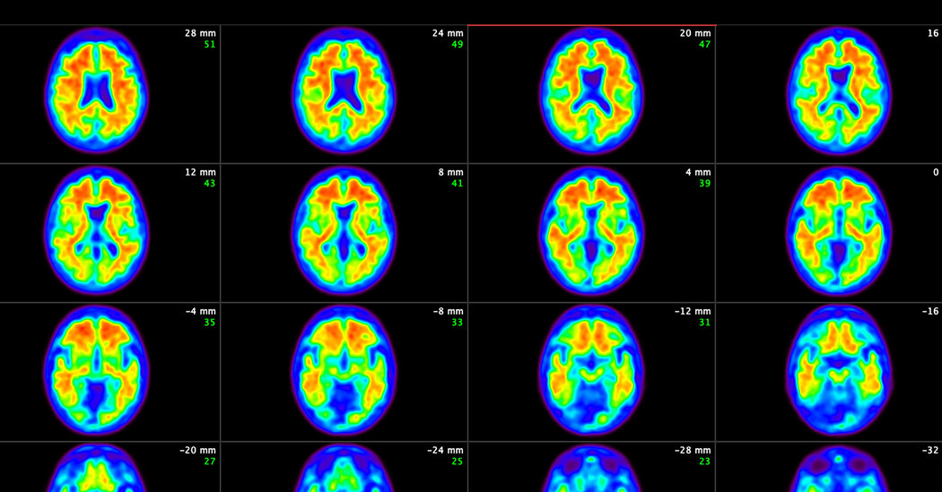
The modest benefits of the treatment, donanemab, made by Eli Lilly, outweigh the risks, the panel concluded unanimously.A committee of independent advisers to the Food and Drug Administration voted unanimously on Monday that the benefits outweigh the risks of the newest experimental drug for Alzheimer’s disease.Alzheimer’s afflicts more than six million Americans. It has no cure, and there is no treatment or lifestyle modification that can restore memory loss or reverse cognitive decline.The drug, made by Eli Lilly, is donanemab. It modestly slowed cognitive decline in patients in the early stages of the disease but also had significant safety risks, including swelling and bleeding in the brain.The committee concluded, though, that the consequences of Alzheimer’s are so dire that even a modest benefit can be worthwhile.The F.D.A. usually follows the advice of the agency’s advisory committees but not always.The drug is based on a long-held hypothesis that Alzheimer’s disease begins when rough hard balls of amyloid, a protein, pile up in patients’ brains, followed by a cascade of reactions leading to the death of neurons.The idea is to treat Alzheimer’s by attacking amyloid, clearing it from the brain. Two similar amyloid-fighting drugs were approved recently: Leqembi, made by Eisai and Biogen, was approved last year. That drug’s risks and modest benefits are similar to those of donanemab. Aduhelm, made by Biogen, is the other drug and was approved in 2021 but was discontinued because there was insufficient evidence that it could benefit patients.We are having trouble retrieving the article content.Please enable JavaScript in your browser settings.Thank you for your patience while we verify access. If you are in Reader mode please exit and log into your Times account, or subscribe for all of The Times.Thank you for your patience while we verify access.Already a subscriber? Log in.Want all of The Times? Subscribe.

Read more →

Disturbed blood flow can damage the vessel wall in cases of aortic dilation

Abnormal blood flow in the aorta is linked to inflammation and breakdown of the vessel wall in conditions where the aorta is dilated. This is shown in a study led by researchers at Linköping University. The findings can contribute to better diagnosis and open up new ways to assess the risk of serious and usually fatal complications, such as rupture of the aorta.
All blood in the body passes through the heart about once a minute. With every heartbeat, blood from the heart is pumped up into the aorta.
Dilation can occur in all parts of the aorta. It is unclear exactly how this happens, but one explanation may be weakening of the connective tissue in the vessel wall. Factors such as elevated blood pressure, age and various hereditary conditions increase the risk. A person affected by aortic dilation usually does not notice anything. But more and more people with aortic dilation are being discovered by healthcare — often by chance.
“In healthcare, we’re doing more and more imaging where we look at, for example, the heart or lungs, and then we can find that the patient’s aorta is a little larger than normal in some parts. There’s a need for more tools to guide doctors on how to handle these patients. In a minority of them, the aorta will unfortunately dilate more, and, in the worst case, the aorta will rupture, which is usually fatal,” says Filip Hammaréus, PhD student at the Department of Health, Medicine and Caring Sciences at Linköping University and intern physician at Ryhov County Hospital in Jönköping.
Most people with aortic dilation are offered regular surveillance of the aortic diameter — the larger the diameter, the higher the risk. Sometimes the aorta is operated on preventively to avoid acute complications, but it can rupture before dilation has become so pronounced that surgery is considered appropriate. At the same time, many examinations are made of people whose aortic diameter does not change. New methods for early identification of patients at elevated risk of growth could contribute to more individualised and cost-effective care. Therefore, the researchers behind the current study, published in the journal European Heart Journal — Cardiovascular Imaging, are investigating new methods for understanding both the risk and the condition itself.
“We can be onto something new, which can say something more about the severity of the disease than what the aortic diameter shows,” says Petter Dyverfeldt, professor at Linköping University and affiliated to the Center for Medical Image Science and Visualization, who has led the study.
When blood is pushed out of the heart into the aorta, a frictional force occurs between the flowing blood and the vessel wall. This is important for a type of cell that is located in the vessel wall and can sense blood flow. When the frictional force is normal, the cells tend to be healthy. However, if the friction significantly changes direction, or becomes very low or abnormal in other ways, there seems to be a signalling into the vessel wall that can eventually lead to a weakening.

Blood flow through the body’s large vessels and in the heart can be measured and visualised with magnetic resonance imaging, using an advanced technology called 4D flow MRI that is available in a small number of hospitals. This gives the researchers a picture of how blood flow affects the vessel wall.
In the current study, the researchers have carried out such measurements and also measured various proteins in the blood. It turned out that there existed interesting relationships between the effect of blood flow on the aortic wall and various proteins that relate to inflammation and the build-up and degradation of connective tissue.
“We see that in patients with dilated aorta, abnormal blood flow dynamics are associated with increased inflammation and turnover of connective tissue — something we believe can reflect processes in the vessel wall. This seems reasonable based on the mechanisms that have been shown in previous research, but it’s completely new to show the connections in the way we do now using a combination of magnetic resonance imaging and blood samples,” says Filip Hammaréus.
The findings strengthen previous research, but also bring new insights.
“What’s interesting about the findings in our study is that the measures of how blood flow affects the aortic wall were not linked to the diameter of the aorta. So, the traditional measure that’s often used in healthcare was not part of the relationship we see in the study between abnormal blood flow, inflammation and breakdown of the vessel wall,” says Petter Dyverfeldt.
The study was conducted on 47 men and women who participated in the Swedish CArdioPulmonary bioImage study (SCAPIS) and whose aortic diameter was over 40 mm. They were compared with 50 control subjects who were matched by sex and age.
The research has been funded with support from, among others, ALF Grants, Region Östergötland, Linköping University, Region Jönköping (Futurum) and the Swedish Research Council. The main funder of SCAPIS is the Swedish Heart-Lung Foundation.
Facts: The aorta is shaped like a walking stick with a curved handle. From the heart, the aorta goes up into a bend (the “handle”) and from there the long part of the vessel passes down through the chest to the abdomen. It is easier to find dilated aortas, or aneurysms, on the part of the aorta located in the abdomen. In Sweden, all men over the age of 65 are offered screening for abdominal aortic aneurysm. In the current study, the researchers focused on the dilation of another part of the aorta — the very first part after the heart — that is difficult to find with ultrasound and is not included in the screening programme.

Read more →

How human derived RNA fragments help the Hepatitis E virus

Why does Hepatitis E become chronic in some patients, and why do medications not work? To find out, an international research team led by scientists from Bochum observed a patient with chronic Hepatitis E infection over a year. Repeated sequencing of the virus RNA showed that the virus incorporated various parts of the host’s messenger RNA into its genome. This resulted in a replication advantage, which may have contributed to the infection becoming chronic. “The so-called insertion of host RNA can possibly predict the transition of an acute infection to a chronic condition,” says Dr Daniel Todt, head of the Computational Virology research group in the Department of Medical and Molecular Virology at Ruhr University Bochum, Germany. The researchers report in the journal Nature Communications on June 6, 2024.
Sequencing of the virus population
Around 20 million people worldwide contract Hepatitis E every year. Normally, the infection heals without consequences, but it can be life-threatening for pregnant women or people with suppressed immune systems. In some cases, it becomes chronic. There are no specific effective medications. The broad-spectrum antiviral drug Ribavirin is also used against Hepatitis E, but it does not always work.
How can the virus evade the immune system? Why does the infection become chronic and not heal? The researchers wanted to find out and analyzed for the first time all virus populations of a chronically infected patient over a period of more than a year. They examined more than 180 individual sequences from blood samples in detail.
Replication in Cell Culture Benefits from Host RNA
“The Hepatitis E virus has a so-called hypervariable region in its genetic information, into which it can incorporate various RNA sequences from host cells,” describes Daniel Todt. His team was able to show that the composition of this region changed massively during the observation period. Additionally, many different compositions occurred simultaneously. In cell culture experiments, it was shown that incorporating host RNA provided a replication advantage: The altered viruses could replicate better than others. “We assume that this is partly responsible for the infection becoming chronic and the therapy failing,” says Daniel Todt.
The researchers examined the composition of the host RNA incorporated into the virus to determine if there were any common features characterizing the gene segments. “However, we could not detect any meaningful similarities,” says Todt. The incorporated gene sequences are predominantly those that are very common in host cells, indicating a random selection.
“Possibly, during Hepatitis E infection, a race between the virus and the immune system occurs in the body,” speculates Daniel Todt. If the virus manages to incorporate host RNA before the immune system successfully combats the infection, it may lead to a chronic course. “Host RNA in the viral genome could, in any case, serve as a biomarker in the acute phase of an infection, indicating early on that it is likely to become chronic.”
The researchers plan to expand their studies to larger cohorts of patients.

Read more →

Study estimates that between 1980 and 2020, 135 million premature deaths could be linked to fine particulate matter pollution

A study led by researchers from Nanyang Technological University, Singapore (NTU Singapore) revealed that fine particulate matter from 1980 to 2020 was associated with approximately 135 million premature deaths globally.
In the study, premature deaths refer to fatalities that occur earlier than expected based on average life expectancy, resulting from preventable or treatable causes such as diseases or environmental factors.
The study found that the impact of pollution from fine particulate matter was worsened by climate variability phenomena such as the El Niño-Southern Oscillation, the Indian Ocean Dipole, and the North Atlantic Oscillation, and led to a 14 per cent rise in premature deaths.
The researchers explain that during such weather events, the increased temperature, changes in wind patterns, and reduced precipitation can lead to stagnant air conditions and the accumulation of pollutants in the atmosphere. These result in higher concentrations of PM2.5 particles that are particularly harmful to human health when inhaled.
Fine particulate matter, or PM2.5, refers to particulate matter 2.5 micrometres in diameter or smaller. These tiny particles come from vehicle emissions, industrial processes, and natural sources such as wildfires and dust storms.
As they are so small, PM2.5 particles can easily get into the air we breathe and penetrate deep into our lungs, leading to a range of health problems, especially for vulnerable groups like children, the elderly, and people with respiratory conditions.
The study estimated that a third of the premature deaths from 1980 to 2020 were associated with stroke (33.3 per cent); another third with ischemic heart disease (32.7 per cent), while chronic obstructive pulmonary disease, lower respiratory infections, and lung cancer made up the rest of premature deaths.

To understand how PM2.5 pollution affects mortality rates, the researchers studied satellite data from NASA on the levels of fine particulate matter in the Earth’s atmosphere. They also analysed statistics on the incidence and mortality of diseases linked to pollution from the Institute for Health Metrics and Evaluation, based in the US. Additionally, they considered information on climate patterns from the National Oceanic and Atmospheric Administration.
While previous studies have explored aspects of air quality and climate, this study had a global scope and analysed over 40 years of data. By examining how specific climate patterns affect air pollution in different regions, it offers fresh insights into the complex relationship between climate and air quality.
First author of the study, Associate Professor Steve Yim of NTU’s Asian School of the Environment and Lee Kong Chian School of Medicine (LKCMedicine), who led the study, said:”Our findings show that changes in climate patterns can make air pollution worse. When certain climate events happen, like El Niño, pollution levels can go up, which means more people might die prematurely because of PM2.5 pollution. This highlights the need to understand and account for these climate patterns when tackling air pollution to protect the health of the global population.” Assoc Prof Yim is also a Principal Investigator at NTU’s Earth Observatory of Singapore (EOS).
Study co-author Distinguished University Professor Joseph Sung, NTU’s Senior Vice President (Health and Life Sciences), and Dean of NTU’s LKCMedicine, said: “Our study highlights how climate patterns affect air pollution, and this is crucial for healthcare professionals because it directly impacts public health. The effects of climate change and the environment on human health are not lesser than those of genomics and lifestyle patterns, and they have been increasing over the past decades. By recognising these patterns, healthcare providers can better prepare for potential increases in patients seeking treatment for pollution-related ailments. Additionally, this knowledge underscores the importance of proactive measures to reduce pollution and mitigate its health impacts, ultimately helping healthcare systems manage and alleviate the burden of pollution-related illnesses on communities.”
The findings from this study, which represents an advance in understanding how environmental challenges affect the health of the global population, reflect NTU’s commitment to responding to the needs and challenges of healthy living and ageing, one of humanity’s grand challenges that the University seeks to address through its NTU 2025 strategic plan.
The study is also part of NTU’s S$50 million interdisciplinary climate research programme, the Climate Transformation Programme (CTP). Hosted by EOS and funded by Singapore’s Ministry of Education, the CTP aims to investigate climate change, develop, inspire, and accelerate knowledge-based solutions, and educate future leaders to establish the stable climate and environment necessary for a resilient and sustainable Southeast Asia.

Co-author of the study Professor Benjamin Horton, Director of Earth Observatory of Singapore, said: “Our study aligns seamlessly with NTU Singapore’s ambitious Climate Transformation Programme (CTP), which aims to tackle the pressing challenges of climate change through interdisciplinary research and collaboration. By investigating the intricate relationship between weather patterns and deadly air pollution, we contribute valuable insights that will inform evidence-based solutions and policies to safeguard public health and promote environmental resilience in Southeast Asia and beyond.” Prof Horton is also Professor in Earth Science at NTU’s Asian School of the Environment.
This study was also funded by several awards and a grant from the Ministry of Education, Singapore. It is also part of The Prudential EOS Climate Impacts Initiative, for which Prudential Services Singapore funded NTU’s EOS to conduct a two-year, two-phase study to understand better the impacts of climate change on air quality and its associated health impacts.
The study also saw participation from NTU President’s Chair in Genomics Professor Stephan Schuster, who is also Deputy Director at the Singapore Centre for Environmental Life Sciences Engineering (SCELSE). Healthcare professionals and researchers from The Chinese University of Hong Kong, Imperial College London, UK, Sun Yat-sen University, China, and Tan Tock Seng Hospital, Singapore were also involved. The findings were published in April in the peer-reviewed journal Environment International.
Assessing the interplay between climate phenomena, pollution, and deaths
This study used data from a dataset managed by NASA called MERRA-2 (Modern-Era Retrospective Analysis for Research and Applications, version 2). The dataset provides monthly information about the concentration of fine particulate matter on the Earth’s surface.
The PM2.5 data analysed for this 40-year study spans from January 1980 to December 2020 and gives detailed information about air pollution levels in specific areas.
The study looked at how changes in air quality are affected by climate patterns, such as the El Niño-Southern Oscillation, Indian Ocean Dipole, and North Atlantic Oscillation weather patterns, obtained from indices assessed by the US National Oceanic and Atmospheric Administration.
The researchers also used data from the Institute for Health Metrics and Evaluation based in the United States on global deaths and occurrences of pollution-linked diseases, which include lower respiratory infections, tracheal, bronchus, and lung cancer, chronic obstructive pulmonary disease, stroke, and ischemic heart disease.
The researchers found that 363 major air pollution episodes happened worldwide over the past four decades, with an average of nine episodes yearly. The duration of an air pollution episode ranged from two to nine months, with 2002 being the year with the highest number of air pollution episodes (15 episodes), followed by 2004 and 2006 (14 episodes each year).
The study estimated that Asia had the highest number of premature deaths attributable to PM2.5 pollution between 1980 and 2020, at 98.1 million, with China and India reaching 49.0 million and 26.1 million deaths, respectively. Pakistan, Bangladesh, Indonesia, and Japan also had significant numbers of PM2.5-attributable premature deaths, ranging from 2 to 5 million each.
The researchers estimate that the three weather phenomena simultaneously caused approximately 7,000 more global premature deaths annually, with the Indian Ocean Dipole phenomena having the largest impact on the number of deaths, followed by North Atlantic Oscillation, and then El Niño.
All three weather patterns coincided in 1994, 1997, 2002, and 2015, with the Southeast Asian region being the most vulnerable. Around 3,100 more deaths occurred in that region each year due to the higher impact of pollution worsened by the weather patterns.
Assoc Prof Yim added: “This study underscores the importance of prioritising public health when developing air quality strategies. Instead of solely focusing on pollutant levels, governments should also consider the health effects of air pollution. This means evaluating policies based on their impact on reducing pollution-related health issues, especially highlighting the need for targeted interventions to mitigate pollution during specific weather conditions.”
Prof Sung added: “As our study has shown that PM2.5 pollution could have significant health consequences, health agencies need to allocate resources accordingly. This includes ensuring that healthcare services are equipped to handle the demands related to PM2.5 pollution-related illnesses. By emphasising health outcomes in air quality management, governments can better protect public health and improve overall well-being.”
The team of researchers will be conducting more detailed studies for a deeper understanding of local air pollution patterns and further detailing the mechanisms behind how climate patterns influence the formation and reduction of PM2.5.

Read more →