Scientists launch $14.2 million project to map the body’s “hidden sixth sense”

How does your brain know when to take a breath, stabilize your blood pressure, or fight off an infection? The answer lies in interoception, a lesser-known process through which the nervous system constantly monitors the body’s internal signals to keep essential functions running.
Now, a collaborative team from Scripps Research and the Allen Institute has received the National Institutes of Health (NIH) Director’s Transformative Research Award to develop the first comprehensive atlas of this internal sensory system.
A Major Investment in Brain-Body Research
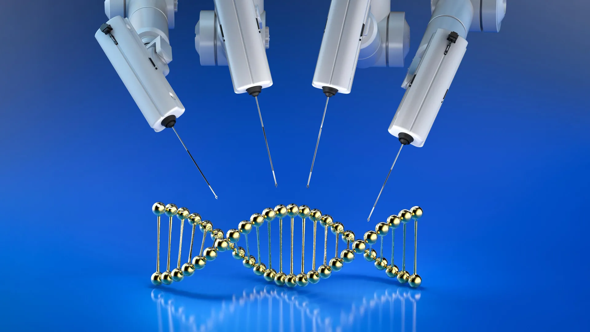
Leading the project is Nobel Prize-winning neuroscientist Ardem Patapoutian, joined by Li Ye, the N. Paul Whittier Chair in Chemistry and Chemical Biology at Scripps Research, and Bosiljka Tasic, Director of Molecular Genetics at the Allen Institute. Xin Jin, Associate Professor at Scripps Research, will serve as co-investigator, directing the genomic and cell-type identification work.
The NIH has awarded the team $14.2 million over five years to carry out this ambitious project.
“My team is honored that the NIH is supporting the kind of collaborative science needed to study such a complex system,” says Patapoutian, the Presidential Endowed Chair in Neurobiology at Scripps Research.
Patapoutian, who shared the 2021 Nobel Prize in Physiology or Medicine for his discovery of cellular sensors that detect touch, will now apply his expertise to understanding interoception.

“We hope our results will help other scientists ask new questions about how internal organs and the nervous system stay in sync,” adds Ye. Like Patapoutian, he’s also a Howard Hughes Medical Institute Investigator.
Established in 2009, the Transformative Research Award funds groundbreaking interdisciplinary projects that push beyond traditional scientific boundaries. It is part of the NIH Common Fund’s High-Risk, High-Reward Research Program, designed to support innovative ideas that could reshape our understanding of human health but might otherwise struggle to receive funding through conventional mechanisms.
What Makes Interoception Unique
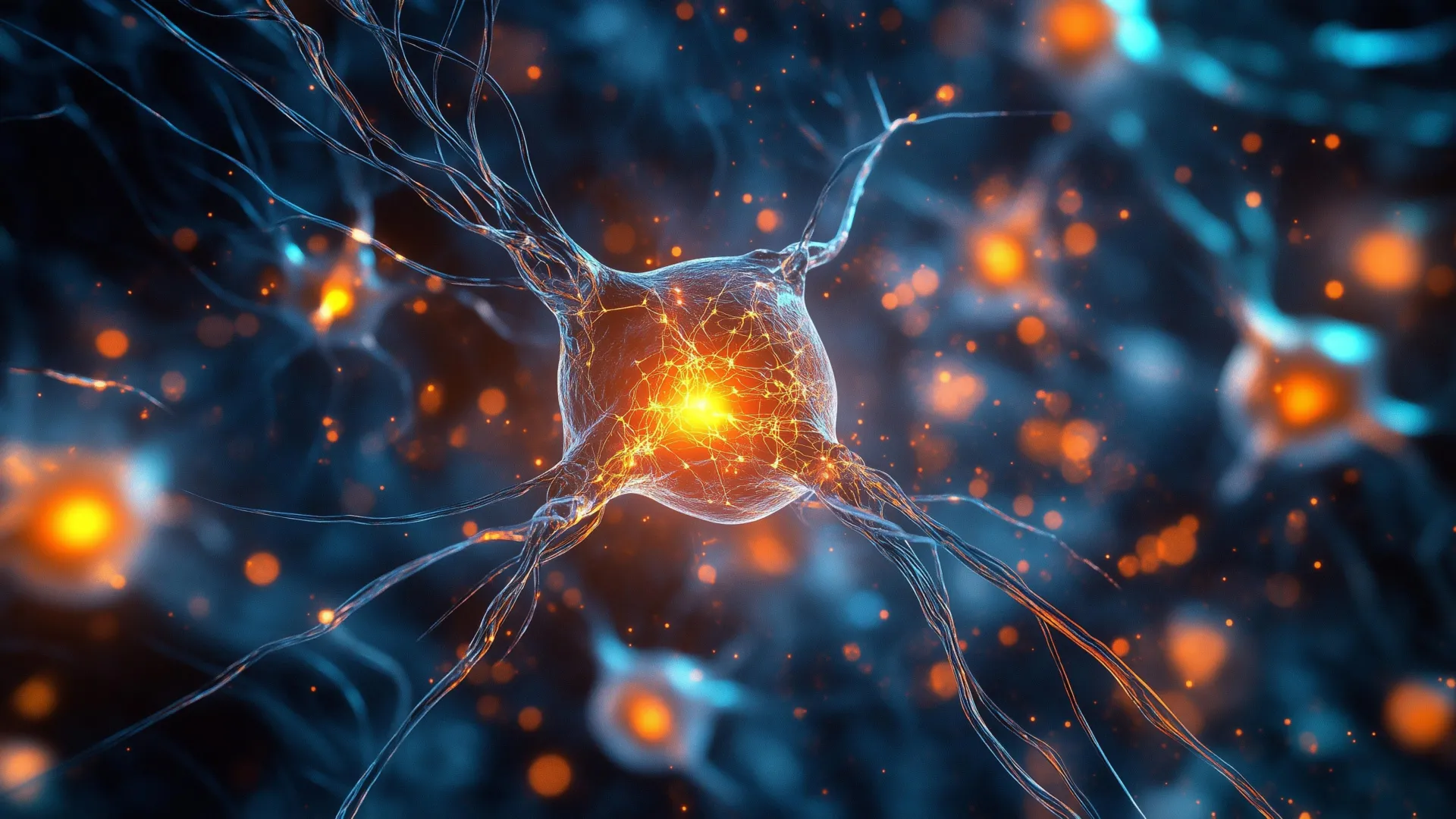
Unlike the classic senses — such as smell, sight, and hearing — which rely on specialized sensory organs that detect stimuli from the outside world, interoception involves a vast network of neurons that sense what’s happening inside the body. These neural circuits track critical processes including circulation, digestion, and immune activity.
Because interoceptive signals originate deep within the body and are often processed unconsciously, scientists often describe this system as our “hidden sixth sense.”
Despite its fundamental role, interoception has received little scientific attention. The signals it produces are complex, overlapping, and difficult to measure. The sensory neurons that carry them are distributed throughout organs such as the heart, lungs, stomach, and kidneys, making them hard to isolate and map precisely.

Mapping the Brain-Body Connection
With the NIH’s support, the Scripps and Allen Institute researchers plan to map how sensory neurons connect with a wide variety of internal organs, including the heart and gastrointestinal tract. Their goal is to create a detailed anatomical and molecular atlas that reveals how these neural pathways are organized.
To achieve this, one part of the project will label sensory neurons and use whole-body imaging to trace their routes from the spinal cord to different organs, producing a high-resolution 3D map. The second part will use genetic profiling to distinguish between different cell types, such as neurons that send signals from the gut, bladder, or fat tissue.
Together, these datasets will form the first standardized reference for understanding the body’s internal sensory wiring.
Why Interoception Matters for Health
By decoding how interoception works, scientists hope to uncover key principles of brain-body communication that could lead to new treatments for disease. Disruptions in these internal sensory pathways have been linked to a range of conditions, including autoimmune disorders, chronic pain, neurodegenerative diseases, and high blood pressure.
“Interoception is fundamental to nearly every aspect of health, but it remains a largely unexplored frontier of neuroscience,” says Jin, who’s a Howard Hughes Medical Institute Freeman Hrabowski Scholar. “By creating the first atlas of this system, we aim to lay the foundation for better understanding how the brain keeps the body in balance, how that balance can be disrupted in disease and how we might restore it.”

Read more →

Scientists just made gene editing far more powerful

Some inherited diseases, including cystic fibrosis, hemophilia, and Tay Sachs disease, involve multiple genetic mutations within a person’s DNA. Even two individuals with the same condition may have different sets of mutations. Because of this complexity, creating gene therapies that work broadly across all patients with a given disorder has been extremely difficult.
A New and More Efficient Gene Editing Breakthrough
Scientists at The University of Texas at Austin have developed a more precise and efficient gene-editing approach that can correct several disease-causing mutations at once in mammalian cells. The technique also successfully repaired mutations linked to scoliosis in zebrafish embryos.
This new approach is powered by retrons, genetic elements originally found in bacteria that help them defend against viral infections. Researchers have now used retrons for the first time to correct a disease-related mutation in vertebrates, offering fresh hope for the development of new gene therapies for human disorders.
“A lot of the existing gene-editing methods are restricted to one or two mutations, which leaves a lot of people behind,” said Jesse Buffington, a graduate student at UT and co-author of a new paper in Nature Biotechnology. “My hope, and what drives me, is to develop a gene-editing technology that’s much more inclusive of people who might have more unique disease-causing mutations, and that using retrons will be able to expand that impact onto a much broader patient population.”
Buffington led the research alongside Ilya Finkelstein, a professor of molecular biosciences at UT, with support from Retronix Bio and the Welch Foundation.
Replacing Faulty DNA With Healthy Sequences
The retron-based system can swap out long sections of defective DNA for healthy ones. This means a single retron “package” can potentially correct many mutations within the same stretch of DNA, rather than targeting one specific defect at a time.

“We want to democratize gene therapy by creating off-the-shelf tools that can cure a large group of patients in one shot,” Finkelstein said. “That should make it more financially viable to develop and much simpler from a regulatory standpoint because you only need one FDA approval.”
While retrons have been used before in mammalian cells, previous attempts were highly inefficient, correcting only about 1.5% of targeted cells. The UT Austin team’s method dramatically improved this efficiency, successfully inserting healthy DNA into around 30% of target cells. Researchers believe they can push this number even higher as the technique evolves.
Another key advantage is that the retron system can be delivered into cells as RNA enclosed in a lipid nanoparticle. These nanoparticles are specifically engineered to overcome the delivery problems faced by many traditional gene-editing systems.
Applying the Technique to Cystic Fibrosis
The research team is now adapting their approach to treat cystic fibrosis (CF), a life-threatening disorder caused by mutations in the CFTR gene. These mutations lead to thick mucus buildup in the lungs, resulting in chronic infections and long-term lung damage.
UT Austin recently received a grant from Emily’s Entourage, a non-profit organization dedicated to finding treatments for the roughly 10% of people with CF who do not benefit from current therapies. The researchers are beginning work on replacing faulty regions of the CFTR gene in laboratory models that mimic CF symptoms and, later, in airway cells derived from patients.

“Traditional gene-editing technologies work best with single mutations and are expensive to optimize, so gene therapies tend to focus on the mutations that are the most common,” Buffington said. “But there are over a thousand mutations that can cause CF. It’s not financially feasible for companies to develop a gene therapy for, say three people. With our retron-based approach, we can snip out a whole defective region and replace it with a healthy one, which can impact a much larger part of the CF population.”
A separate grant from the Cystic Fibrosis Foundation will support similar work targeting the region of the CFTR gene that includes the most common CF-causing mutations.
Along with Buffington and Finkelstein, the research team includes Hung-Che Kuo, Kuang Hu, You-Chiun Chang, Kamyab Javanmardi, Brittney Voigt, Yi-Ru Li, Mary E. Little, Sravan K. Devanathan, Blerta Xhemalçe, and Ryan S. Gray. Their work marks an important step toward gene therapies that are more adaptable, efficient, and inclusive for patients facing complex genetic diseases.

Read more →

Parents urged to vaccinate children over half-term as flu cases rise

Parents are being urged to get their children vaccinated against flu over half-term as the NHS warned of rising cases of the disease.There is an early flu season, health officials say, and the latest data for England showed cases and hospitalisations were increasing.NHS England said many school children will have already received the vaccine at school but there are still options for those who have not, including pop-up clinics.GP surgeries can give flu vaccines to school-aged children and those with certain health conditions, as well as two to three year olds, while preschoolers can also be taken to pharmacies for the vaccine.Most children are offered the vaccine in a nasal spray rather than an injection.NHS England’s weekly flu and Covid surveillance report said there was increased flu activity “particularly among children”.Duncan Burton, chief nursing officer for England, said it was concerning that the flu had hit early this year and the increase among children was “worrying”.”Flu can spread like wildfire across schools and can make children really unwell,” he said.”The virus changes each year, so vaccination remains the best way to shield your child from getting seriously ill.”He urged parents to make sure they had opted their child in for an in-school vaccine or find their nearest clinic.Pop-up clinics were being held in places like bowling alleys and fire stations, Mr Burton told BBC Radio 4’s Today programme, adding that the vaccine was “quick, easy and safe”.Dr Fari Ahmad told BBC Breakfast that while she was seeing cases, “I don’t think we’ve quite hit the peak yet”.”With flu, this is a war we have every year. Flu is unpredictable, it still kills people and every year we try to get enough people vaccinated so that when the big surge comes we are not decimated,” she said. Dr Ahmad said that getting enough school-age children vaccinated meant that it made that surge better, “because they are spreaders”.”It will be great for the kids individually but it will also be better for all of us,” she said.NHS England said more than 10 million vaccines have already been delivered in the latest campaign, including to almost 1.5 million school-aged children and more than 300,000 eligible two and three year olds.Free vaccines are available to those older than 65, with certain long-term health conditions, are pregnant, live in a care home, the main carer for an older or disabled person or live with someone who has a weakened immune system.

Read more →

Snake pee might hold the secret to ending gout pain and kidney stones

If you’ve never kept a reptile, you might be surprised to learn that many of them actually “pee” in crystal form. In a study published in the Journal of the American Chemical Society, researchers examined the solid urine of more than 20 different reptile species and found that all contained tiny spheres made of uric acid. This discovery highlights how reptiles have developed a unique method for safely storing and removing waste in a crystalline form. The findings could also lead to new ways of treating human conditions linked to uric acid buildup, such as kidney stones and gout.
How Reptiles Save Water With Solid Waste
Every living creature needs to get rid of waste, and reptiles are no exception. In humans, the body eliminates excess nitrogen by flushing it out through urine as urea, uric acid, and ammonia. Reptiles and birds, however, take a different approach. They transform some of those same nitrogen-based compounds into solids known as “urates,” which are expelled through a shared opening called the cloaca. Scientists think this solid form of waste evolved as an adaptation to conserve water, a valuable trait for animals that often live in dry environments.
What’s Dangerous for Humans Is Normal for Snakes
Although forming crystals in urine helps reptiles survive, the same process can cause serious health problems in people. When uric acid levels become too high in humans, the crystals can collect in the joints, leading to gout, or form in the urinary tract as kidney stones. To understand how reptiles manage to excrete these crystals safely, Jennifer Swift and her research team analyzed urates from more than 20 species.
“This research was really inspired by a desire to understand the ways reptiles are able to excrete this material safely, in the hopes it might inspire new approaches to disease prevention and treatment,” explains Swift, the corresponding author on the study.
Microscopic Spheres With Big Medical Potential
Using powerful microscopes, the researchers discovered that species such as ball pythons, Angolan pythons, and Madagascan tree boas produce urates made up of tiny textured spheres between 1 and 10 micrometers across. X-ray analysis revealed that these microspheres are built from even smaller nanocrystals made of uric acid and water. The team also found that uric acid helps transform ammonia, a toxic compound, into a safer solid form. They believe uric acid might play a similar protective role in humans. Although more research is needed, these findings suggest that the chemistry behind reptile waste could eventually help scientists develop better treatments for uric acid-related diseases.
Research Support and Collaboration
This study received support from the National Science Foundation, Georgetown University, the International Centre for Diffraction Data, and the Chiricahua Desert Museum.

Read more →

Scientists discover a hidden gene mutation that causes deafness—and a way to fix it

Mutations in a gene called CPD have been found to play a key role in a rare inherited form of hearing loss, according to an international research collaboration. Scientists from the University of Chicago, the University of Miami, and several institutions in Turkiye published the discovery in the Journal of Clinical Investigation. The study reveals that the CPD gene, which is typically known for modifying proteins, also affects the inner ear. Researchers not only identified the genetic mechanism behind this effect but also found two possible treatment strategies.
“This study is exciting because we found a new gene mutation that’s linked to deafness, and more importantly we have a therapeutic target that can actually mitigate this condition,” said lead author Rong Grace Zhai, PhD, Jack Miller Professor for the Study of Neurological Diseases of Neurology at UChicago. Although the study focused on individuals with a rare combination of mutations to the CPD gene, there could be broader implications if single mutations are linked to age-related hearing loss, she added.
The connection between CPD and hearing loss
Researchers began investigating CPD after identifying an unusual combination of mutations in three unrelated Turkish families affected by sensorineural hearing loss (SNHL), a congenital and hereditary condition that causes permanent deafness.
SNHL is typically diagnosed in early childhood and has long been considered irreversible. Hearing aids and cochlear implants can help improve perception of sound, but no direct medical treatment exists to repair the underlying damage.
When the scientists expanded their search through genetic databases, they discovered that individuals with other CPD mutations also showed signs of early-onset hearing loss, strengthening the link between this gene and auditory function.
How CPD protects sensory cells
To understand how CPD influences hearing, the team conducted experiments using mice. The CPD gene normally produces an enzyme responsible for generating the amino acid arginine, which then helps create nitric oxide, a key neurotransmitter involved in nerve signaling. In the inner ear, mutations in CPD disrupted this process, triggering oxidative stress and the death of delicate sensory hair cells that detect sound vibrations.

“It turns out that CPD maintains the level of arginine in the hair cells to allow a quick signaling cascade by generating nitric oxide,” Zhai explained. “And that’s why, although it’s expressed ubiquitously in other cells throughout the nervous system, these hair cells in particular are more sensitive or vulnerable to the loss of CPD.”
Fruit fly experiments reveal possible treatments
The researchers also used fruit flies as a model to explore how CPD mutations affect hearing. Flies with the defective gene exhibited behaviors consistent with inner ear dysfunction, such as impaired hearing and balance issues.
To test potential treatments, scientists tried two approaches. One was to provide arginine supplements to replace what was lost due to the gene defect. The other was to use sildenafil (Viagra), a drug known to stimulate one of the signaling pathways disrupted by reduced nitric oxide. Both treatments improved cell survival in patient-derived cells and reduced hearing-loss symptoms in the fruit flies.
“What makes this really impactful is that not only do we understand the underlying cellular and molecular mechanism for this kind of deafness, but we also found a promising therapeutic avenue for these patients. It is a good example of our efforts to repurpose FDA approved drugs for treating rare diseases,” Zhai said.
The study also demonstrates the value of fruit fly models for studying neurological diseases, including age-related conditions, Zhai noted. “They give us the capability to not only understand disease pathology, but also to identify therapeutic approaches,” she said.

Expanding the research to broader populations
The researchers plan to continue studying how nitric oxide signaling functions in the inner ear’s sensory system. They also aim to investigate how common CPD mutations are in larger populations and whether they might contribute to other forms of hearing loss.
“How many people carry variants in this gene and is there a susceptibility to deafness or age-dependent hearing loss?” she said. “In other words, is this a risk factor for other types of sensory neuropathy?”
The study included collaborators from multiple institutions, including the University of Miami, Ege University, Ankara University, Yüzüncü Yıl University, Memorial Şişli Hospital, the University of Iowa, and the University of Northampton (UK).

Read more →

Scientists just found the lung’s hidden self-healing switch

Researchers at Mayo Clinic have uncovered a molecular “switch” inside lung cells that determines when the cells focus on repairing tissue and when they shift to fighting infection. This important finding could pave the way for regenerative treatments for chronic lung conditions.
“We were surprised to find that these specialized cells cannot do both jobs at once,” says Douglas Brownfield, Ph.D., senior author of the study, which was published in Nature Communications. “Some commit to rebuilding, while others focus on defense. That division of labor is essential. And by uncovering the switch that controls it, we can start thinking about how to restore balance when it breaks down in disease.”
Understanding How Lung Cells Repair and Protect
The study focuses on alveolar type 2 (AT2) cells, which are unique because they both safeguard the lungs and act as reserve stem cells. AT2 cells produce proteins that keep the tiny air sacs open for breathing, while also regenerating alveolar type 1 (AT1) cells — the thin, flat cells that line the lung surface and enable oxygen exchange.
Scientists have long known that AT2 cells often struggle to regenerate properly in diseases such as pulmonary fibrosis, chronic obstructive pulmonary disease (COPD), and severe viral infections like COVID-19. What had remained unclear was how and why these cells lose their regenerative capacity.
Mapping the Life Cycle of Lung Cells
Using single-cell sequencing, advanced imaging, and preclinical models of lung injury, the Mayo Clinic team tracked the “life history” of AT2 cells. They discovered that new AT2 cells remain flexible for about one to two weeks after birth before they permanently adopt their specialized identity.

That critical transition is governed by a molecular circuit involving three key regulators — PRC2, C/EBPα, and DLK1. One of these, C/EBPα, acts as a clamp that keeps the cells from behaving like stem cells. To regenerate after injury, adult AT2 cells must release this clamp.
Why Infections Slow Lung Recovery
The same molecular switch also determines whether AT2 cells repair damaged tissue or fight infection. This dual role helps explain why infections can slow down or block recovery in chronic lung diseases.
“When we think about lung repair, it’s not just about turning things on — it’s about removing the clamps that normally keep these cells from acting like stem cells,” says Dr. Brownfield. “We discovered one of those clamps and how it times the ability of these cells to repair.”
Preventing Organ Failure
The findings open new possibilities for regenerative medicine. Drugs that fine-tune C/EBPα activity, for example, could help AT2 cells rebuild lung tissue more effectively or reduce scarring in conditions like pulmonary fibrosis.

“This research brings us closer to being able to boost the lung’s natural repair mechanisms, offering hope for preventing or reversing conditions where currently we can only slow progression,” says Dr. Brownfield.
The study may also help doctors identify early signs of disease by detecting when AT2 cells are trapped in one state and unable to regenerate. Such insights could lead to new biomarkers that detect lung disease in its earliest, most treatable stages.
Linking Discovery to Mayo Clinic’s Regenerative Initiatives
This work aligns with Mayo Clinic’s Precure initiative, which focuses on identifying diseases early — when treatments can have the greatest impact — and preventing progression before organ failure occurs.
It also advances the Genesis initiative, which aims to prevent organ failure and restore function through regenerative medicine. Building on these findings, the research team is now testing ways to release the repressive clamp in human AT2 cells, grow them in the lab, and explore their potential for future cell-based therapies.

Read more →

Magnesium: Can this ‘miracle mineral’ really help us sleep?

5 days agoShareSaveRuth CleggHealth and wellbeing reporterShareSaveGetty ImagesIt’s been described as the mineral of the moment.Millions of us are taking magnesium for a whole range of reasons. Can it help us sleep better? Sort our digestion problems? Give our busy brains a moment of peace?In the fast-moving world of supplements, it’s magnesium’s time to shine.And the industry is booming. The global magnesium market is worth almost £3bn and that’s set to nearly double over the next decade.In a small factory, nestled in the Yorkshire Dales, huge plastic barrels of white powder are stacked up next to giant whirring machines.Workers in hazmat suits carefully weigh out magnesium citrate – a compound made by mixing the mineral with citric acid – into shiny, steel containers.Ruth Clegg/BBC”We are sending our supplies all over the world,” Andrew Goring, manager director of Lonsdale Health, explains. “Around the UK, obviously, but also to Australia, parts of Asia, Kuwait, Iraq.”It’s one of our biggest sellers and the market just keeps growing.”He is shouting over the hum of the pill presser, a machine that resembles a Dalek, which pops out dozens of small, white magnesium tablets a second.”Do we actually need it?” I bellow back. “And why now? Why has it become so popular?””Influencers, social media – that’s what’s pushing it,” Mr Goring explains. “We’ve known about magnesium and its benefits for years and now, finally, it’s mainstream.”I can almost hear the eye roll when I contact Kirsten Jackson, an IBS Dietitian, who specialises in gut health.”Clever marketing schemes,” Ms Jackson says, “magnesium is involved in areas people are willing to invest in: their sleep, digestion, mental health.”But, she is keen to stress, this does not automatically mean we need supplements to improve those things.Magnesium is one of several minerals in our bodies. The recommended daily amount for women is 270mg and for men, it’s 300mg. We store about 25g.It might make up less than 1% of us but “it is involved in over 300 different processes”, Ms Jackson explains.It is “especially important for our brain and mood,” she says, because it helps nerves send messages properly and supports the building blocks of brain cell membranes.It also helps balance blood sugar levels, regulate blood pressure, and is an essential player in moving calcium and potassium in and out of our cells, which maintains the rhythm of our heartbeat.So, surely popping a pill full of the stuff should keep our bodies running smoothly?It’s more complex than that, says Ms Jackson. For a magnesium supplement to work, we need to be lacking the mineral in the first place – and it’s difficult to test for any deficiency because the vast majority of our magnesium is stored in our bones and tissues.But on an individual level, many say the supplement has made a difference.Katie CurranFor Katie Curran, a communications specialist who has worked with some of the biggest fashion brands, sleeping well was something she could only dream about.”A year ago, I was struggling,” she confides. “It would take so long to get to sleep, my brain was racing, and then I would get off only to wake a couple of hours later.”Katie decided to try magnesium glycinate – a combination of magnesium and glycine, an amino acid with limited evidence linking it to better sleep.After two weeks of taking 270mg a day, she says the noise in her head started to quieten. The racing thoughts slowed down, and she began to feel like she could function again.”My sleep definitely improved, I had more energy. I became more active. Other things changed in my life, so I can’t put it down to just one thing, but I think the magnesium supplements were an important part of the puzzle.”While being deficient in magnesium can definitely affect sleep patterns, there isn’t the evidence to say taking supplements will definitely improve your sleep.Social media is awash with eager supplement-takers, many with posts stamped with “commission paid” in the corner – meaning they could earn money from their story or reel.According to these influencers, it feels as though there is little magnesium can’t help with, as they recommend a variety of different products.Magnesium is often mixed with other compounds with the aim of helping support various parts of our bodies. For example, magnesium mixed with either L-threonate or glycinate is supposed to target brain health, which helps with sleep and stress relief.If magnesium is combined with chloride, it is recommended for muscle tension and pre-menstrual cramps, while citrate and oxide blends are aimed at digestion and help with constipation. The majority of us can probably relate to having at least one of those issues. But as nutritionist Kristen Stavridis stresses, the problem is there is not enough strong evidence to show the majority of these different magnesium supplements have a positive effect on the healthy population.And even if they did – we would need to be deficient in magnesium in the first place to see a benefit.”We have supplement companies shouting out at us: ‘We are all going to die’,” Ms Stavridis says, “‘Quick! Take my pill and – hey presto – there’s your solution’.”Many of us are not getting enough magnesium,” she continues, “around 10% of men and 20% of women are not getting the recommended daily intake.”But just taking a supplement is not the answer.”Getty ImagesTake sleep health, for example. Ms Stavridis says there are many conflicting studies on whether magnesium supplements really make a difference. Some trials say it can have some benefits, while some randomised controlled trials – the gold standard – are more sceptical.There is also the added complication of supplements potentially working against each other because of the way they interact in the body.Taking zinc, for example – a supplement often recommended for peri-menopausal women – can also affect the absorption rate of magnesium.Basically, Ms Stavridis says, it’s a minefield, and not just a simple case of “take this” and you’ll be fixed.She recommends looking at diet first. But if you are thinking about taking magnesium supplements, Ms Stavridis advises taking half the amount recommended on the packet on a daily basis and seeing how you feel.If healthy people take too much, their kidneys can get rid of it in “expensive urine”, but there are still risks, like diarrhoea, vomiting and nausea.For those with kidney disease, taking magnesium supplements can be dangerous and can cause hypermagnesemia – a potentially life-threatening condition that can leave someone with paralysis or in a coma.Dietitian Kirsten Jackson also says most people should “100% look at diet first”.Foods like seeds, nuts, whole-grain breads, greens and fruit are good sources of magnesium, she says.She warns that if you don’t regularly consume these types of foods, you’re probably also short of other essential nutrients like vitamin C, vitamin K, fibre and prebiotics too.”One magnesium supplement is not going to sort all that.”More weekend picks

Read more →

Your gut microbes might be turning fiber into extra calories

Deep within your gut lives a bustling world of microbes, each playing a role in digesting your food. Among them is one unusual microbe that produces methane — a gas more often associated with cows and landfills than humans. According to new research from Arizona State University (ASU), this methane-making microorganism may influence how many calories your body extracts from what you eat.
The collection of microbes living in your digestive tract is known as the gut microbiome. While everyone has one, some people’s microbiomes produce large amounts of methane, whereas others produce very little.
Microbes and the Energy Hidden in Fiber
The study found that people whose microbiomes generate more methane tend to extract more energy from high-fiber foods. This may help explain why the same meal can provide different calorie counts for different individuals once it reaches the colon.
Researchers emphasized that high-fiber foods remain beneficial. People generally absorb more calories from a typical Western diet high in processed foods, regardless of methane levels. Even so, calorie absorption on a fiber-rich diet varies depending on how much methane a person’s gut produces.
These findings suggest that gut methane could become a key factor in personalized nutrition — a future where diets are tailored to the unique microbial activity in each person’s digestive system
“That difference has important implications for diet interventions. It shows people on the same diet can respond differently. Part of that is due to the composition of their gut microbiome,” says Blake Dirks, lead author of the study and graduate researcher at the Biodesign Center for Health Through Microbiomes. Dirks is also a PhD student in ASU’s School of Life Sciences.

Meet the Methane Makers
Published in The ISME Journal, the study identifies the key players: methane-producing microbes known as methanogens. These microorganisms appear to be linked with more efficient digestion and higher energy absorption.
A major job of the microbiome is breaking down food that the body cannot digest on its own. Microbes ferment fiber into short-chain fatty acids (SCFAs), which provide a valuable energy source. During this process, hydrogen gas is released. Too much hydrogen can slow fermentation, but other microbes prevent this by consuming hydrogen — keeping the digestive chemistry in balance.
Methanogens are the hydrogen consumers. As they feed on hydrogen, they release methane as a byproduct. They are the only microbes in the human gut that produce this gas.
“The human body itself doesn’t make methane, only the microbes do. So we suggested it can be a biomarker that signals efficient microbial production of short-chain fatty acids,” says Rosy Krajmalnik-Brown, corresponding author of the study and director of the Biodesign Center for Health Through Microbiomes.
How Microbes May Shape Metabolism
The ASU researchers found that the interactions between these microbes may directly affect metabolism. Participants who produced more methane also had higher levels of short-chain fatty acids, indicating that more energy was being created and absorbed in the gut.

To test these effects, each participant followed two different diets. One included highly processed, low-fiber foods, while the other emphasized whole foods and fiber. Both diets contained equal proportions of carbohydrates, proteins, and fats.
The research was conducted in collaboration with the AdventHealth Translational Research Institute, which provided access to a specialized facility. Each participant spent six days in a sealed, hotel-like room called a whole-room calorimeter. This environment allowed researchers to precisely measure metabolism and methane output.
Unlike traditional methods that rely on a single breath test, this setup continuously captured methane released through both breath and other emissions (ahem), providing a more accurate view of microbial activity.
“This work highlights the importance of the collaboration between clinical-translational scientists and microbial ecologists. The combination of precise measures of energy balance through whole-room calorimetry with ASU’s microbial ecology expertise made key innovations possible,” says Karen D. Corbin, a co-author and associate investigator at the institute.
Tracking Energy and Microbial Activity
Data collected from blood and stool samples revealed how much energy participants absorbed from their food and how active their gut microbes were. Researchers then compared people with high methane production to those with lower levels.
Almost all participants absorbed fewer calories while eating the high-fiber diet compared to the processed-food diet. However, those with higher methane production absorbed more calories from the fiber-rich foods than those with less methane in their systems.
A Step Toward Personalized Health
The findings lay important groundwork for future studies and medical applications.
This research creates a foundation for future studies and medical treatments.
“The participants in our study were relatively healthy. One thing that I think would be worthy to look at is how other populations respond to these types of diets — people with obesity, diabetes or other kinds of health states,” Dirks says.
Although the study did not aim to induce weight loss, some participants did lose a small amount while following the high-fiber diet. Future research may explore how methanogens influence weight-loss efforts or specialized nutrition programs.
“You can see how important it is that the microbiome is personalized,” Krajmalnik-Brown says. “Specifically, the diet that we designed so carefully to enhance the microbiome for this experiment had different effects on each person, in part because some people’s microbiomes produced more methane than others.”
Other members of the ASU research team include Professor Bruce Rittmann and graduate researcher Taylor Davis.
This project was funded by the National Institute of Diabetes and Digestive and Kidney Diseases of the National Institutes of Health.

Read more →

Popular cholesterol drugs may help prevent dementia

Having lower cholesterol levels may help protect against dementia, according to a large-scale international study led by the University of Bristol. The research, involving data from more than one million participants, found that people with genetic traits that naturally reduce cholesterol are less likely to develop dementia.
The work was led by Dr. Liv Tybjærg Nordestgaard during her time at the University of Bristol and at the Department of Clinical Biochemistry at Copenhagen University Hospital — Herlev and Gentofte. The findings were published in Alzheimer’s & Dementia: The Journal of the Alzheimer’s Association.
Genetic Clues and Cholesterol-Lowering Effects
Some individuals are born with genetic variants that affect the same proteins targeted by cholesterol-lowering medications such as statins and ezetimibe. To explore whether these drugs might influence dementia risk, the team used a technique called Mendelian Randomization. This method allows scientists to study how specific genetic variants mimic the effects of a treatment while minimizing the influence of outside factors like weight, diet, or lifestyle.
By comparing people with and without these cholesterol-lowering genetic variants, the researchers observed a clear difference in dementia risk. A small decrease in cholesterol levels (about one millimole per liter) was associated with up to an 80% reduction in dementia risk for certain drug-related genetic targets.
Lower Cholesterol, Lower Dementia Risk
“What our study indicates is that if you have these variants that lower your cholesterol, it looks like you have a significantly lower risk of developing dementia,” said Dr. Nordestgaard, who now works in the Department of Clinical Biochemistry at Copenhagen University Hospital — Bispebjerg and Frederiksberg hospital.

The results suggest that keeping cholesterol levels low — whether through genetics or medical treatment — may protect against dementia. However, the research does not yet confirm that cholesterol-lowering drugs themselves directly prevent the disease.
Why Studying Dementia Is So Challenging
Because dementia often develops late in life, studying its causes requires tracking participants for decades. This makes it difficult to establish cause and effect in traditional clinical trials.
It also remains unclear why high cholesterol raises dementia risk. One explanation, according to Dr. Nordestgaard, is that high cholesterol contributes to atherosclerosis — the buildup of fatty deposits in blood vessels.
How Cholesterol May Harm the Brain
“Atherosclerosis is a result of the accumulation of cholesterol in your blood vessels,” Dr. Nordestgaard said. “It can be in both the body and the brain and increases the risk of forming small blood clots — one of the causes of dementia.

“It would be a really good next step to carry out randomised clinical trials over 10 or 30 years, for example, where you give the participants cholesterol-lowering medication and then look at the risk of developing dementia,” Dr. Nordestgaard added.
Global Collaboration and Funding
The study used data from the UK Biobank, the Copenhagen General Population Study, the Copenhagen City Heart Study, the FinnGen study, and the Global Lipids Genetics Consortium.
Funding was provided by the Medical Research Council, Independent Research Fund Denmark, and Research Council at the Capital Region of Denmark (LTN).

Read more →

Scientists finally see what sparks Parkinson’s

For the first time, researchers have directly seen and measured the protein clusters thought to spark Parkinson’s disease, marking a major milestone in understanding the world’s fastest-growing neurological condition.
These microscopic clusters, known as alpha-synuclein oligomers, have long been suspected as the starting point for Parkinson’s, but they have remained undetectable in human brain tissue — until now.
A team from the University of Cambridge, UCL, the Francis Crick Institute, and Polytechnique Montréal developed a powerful imaging approach that allows scientists to visualize, count, and compare these protein clumps in human brain tissue. One researcher described the breakthrough as “like being able to see stars in broad daylight.”
Published in Nature Biomedical Engineering, the findings could transform how scientists study Parkinson’s, offering new insights into how it spreads through the brain and paving the way for earlier diagnosis and more targeted treatments.
Parkinson’s: A Growing Global Health Challenge
More than 166,000 people in the UK currently live with Parkinson’s disease, and the global total is expected to reach 25 million by 2050. While existing drugs can ease symptoms such as tremors and stiffness, none can halt or slow the disease’s progression.
For over a century, doctors have identified Parkinson’s by the presence of large protein deposits known as Lewy bodies. Yet researchers have long believed that smaller, early-stage oligomers may actually cause the damage to brain cells. Until now, these microscopic structures, just a few nanometers long, were impossible to observe directly.

Seeing Parkinson’s at Its Earliest Stages
“Lewy bodies are the hallmark of Parkinson’s, but they essentially tell you where the disease has been, not where it is right now,” said Professor Steven Lee from Cambridge’s Yusuf Hamied Department of Chemistry, who co-led the research. “If we can observe Parkinson’s at its earliest stages, that would tell us a whole lot more about how the disease develops in the brain and how we might be able to treat it.”
To achieve this, the researchers created a method called ASA-PD (Advanced Sensing of Aggregates for Parkinson’s Disease). This ultra-sensitive fluorescence microscopy technique can detect and analyze millions of oligomers in post-mortem brain samples. Because the oligomers are so tiny, their signal is faint, but ASA-PD enhances that signal while reducing background noise, allowing scientists to clearly see individual alpha-synuclein clusters for the first time.
Illuminating the Invisible
“This is the first time we’ve been able to look at oligomers directly in human brain tissue at this scale: it’s like being able to see stars in broad daylight,” said co-first author Dr Rebecca Andrews, who conducted the work when she was a postdoctoral researcher in Lee’s lab. “It opens new doors in Parkinson’s research.”
The researchers examined brain tissue from people with Parkinson’s and compared it to samples from healthy individuals of similar age. They found that oligomers were present in both groups, but in those with Parkinson’s, the clusters were larger, brighter, and far more numerous. This difference suggests a strong connection between oligomer growth and disease progression.

Clues to the Earliest Signs of Disease
The team also identified a unique subset of oligomers found only in Parkinson’s patients, which may represent the earliest detectable signs of the disease — possibly appearing years before symptoms emerge.
“This method doesn’t just give us a snapshot,” said Professor Lucien Weiss from Polytechnique Montréal, wo co-led the research. “It offers a whole atlas of protein changes across the brain and similar technologies could be applied to other neurodegenerative diseases like Alzheimer’s and Huntington’s.
“Oligomers have been the needle in the haystack, but now that we know where those needles are, it could help us target specific cell types in certain regions of the brain.”
A New Window Into the Human Brain
“The only real way to understand what is happening in human disease is to study the human brain directly, but because of the brain’s sheer complexity, this is very challenging,” said Professor Sonia Gandhi from The Francis Crick Institute, who co-led the research. “We hope that breaking through this technological barrier will allow us to understand why, where and how protein clusters form and how this changes the brain environment and leads to disease.”
This research was made possible with support from Aligning Science Across Parkinson’s (ASAP), the Michael J. Fox Foundation, and the Medical Research Council (MRC), part of UK Research and Innovation (UKRI). The team expressed gratitude to the patients, families, and caregivers who donated brain tissue to research, enabling discoveries like this to advance understanding and potential treatment of Parkinson’s disease.

Read more →