Your bedroom glow might be quietly damaging your heart

Higher exposure to artificial light at night was linked to heightened stress activity in the brain, inflammation in the arteries, and a greater likelihood of heart disease in a small study of adults in Boston. Researchers reported that light pollution at night appeared to influence cardiovascular health, suggesting it is a modifiable environmental factor in communities with elevated nighttime brightness.Nighttime Light and Heart HealthA preliminary analysis to be presented at the American Heart Association’s Scientific Sessions 2025 found that greater artificial light at night correlated with increased brain stress signals, inflamed blood vessels, and higher heart disease risk. The conference will take place Nov. 7-10 in New Orleans and features new developments, research, and practice updates in cardiovascular science.
The investigators described artificial light at night, often called nighttime light pollution, as nearly universal in modern urban settings. This first-of-its-kind work combined brain imaging and satellite measurements to outline a biological pathway that may connect nighttime brightness with cardiovascular risk.
“We know that environmental factors, such as air and noise pollution, can lead to heart disease by affecting our nerves and blood vessels through stress. Light pollution is very common; however, we don’t know much about how it affects the heart,” said study senior author Shady Abohashem, M.D., M.P.H., head of cardiac PET/CT imaging trials at Massachusetts General Hospital and an instructor at Harvard Medical School in Boston.
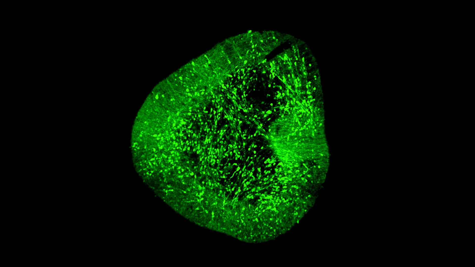
All participants had the same combined Positron Emission Tomography/Computed Tomography (PET/CT) scan. This study of 450 adults only included subjects without heart disease and no active cancer. “This is a routine imaging test at my hospital,” Abohashem said. “The CT portion provides detailed anatomy, while the PET portion reveals metabolic activity in tissues. Using both imaging techniques together allows for the measurement of brain stress activity and arterial inflammation in a single scan.”
Key Results: Higher Light Exposure, Higher Risk People exposed to higher levels of artificial light at night had higher brain stress activity, blood vessel inflammation and a higher risk of major heart events. This information was collected from medical records and evaluated by two cardiologists who were blinded, meaning they were unaware of any information that could influence their decisions. The greater the artificial night light exposure, the higher the risk of heart disease development. Every standard deviation increase in light exposure was associated with about 35% and 22% increased risk of heart disease over five- and 10-year follow-up periods, respectively. These associations remained after accounting for traditional risk factors and other socio-environmental exposures like noise pollution and socioeconomic status. In addition, these heart risks were higher among participants who lived in areas with additional social or environmental stress, such as high traffic noise or lower neighborhood income. Over a 10-year follow-up period, 17% of participants had major heart conditions.Stress Pathways and Vessel Inflammation

“We found a nearly linear relationship between nighttime light and heart disease: the more night-light exposure, the higher the risk. Even modest increases in night-time light were linked with higher brain and artery stress,” Abohashem said. “When the brain perceives stress, it activates signals that can trigger an immune response and inflame the blood vessels. Over time, this process can contribute to hardening of the arteries and increase the risk of heart attack and stroke.”
However, to counter the effects of artificial light at night, he suggested that cities could reduce unnecessary outdoor lighting, shield streetlamps or use motion-sensitive lights. On a personal level, “people can limit indoor nighttime light, keeping bedrooms dark and avoiding screens such as TVs and personal electronic devices before bed,” he said.
“These findings are novel and add to the evidence suggesting that reducing exposure to excessive artificial light at night is a public health concern,” said Julio Fernandez-Mendoza, Ph.D., DBSM, FAHA, who is on the writing committee of Multidimensional Sleep Health: Definitions and Implications for Cardiometabolic Health: A Scientific Statement From the American Heart Association.
Circadian Health: Recent Guidance and Broader Implications
Last week (Oct. 28), the Association issued a scientific statement on the Role of Circadian Health in Cardiometabolic Health and Disease Risk. The new statement also indicates that light pollution is a major factor disrupting body clocks and can suppress melatonin, delay sleep onset and even at low levels has been associated with increased risk of cardiovascular disease.
“We know too much exposure to artificial light at night can harm your health, particularly increasing the risk of heart disease. However, we did not know how this harm happened,” Fernandez-Mendoza said. “This study has investigated one of several possible causes, which is how our brains respond to stress. This response seems to play a big role in linking artificial light at night to heart disease.” Fernandez-Mendoza, who was not a part of the study, is also a professor of psychiatry, neuroscience and public health sciences and director of behavioral sleep medicine at Pennsylvania State University College of Medicine in Hershey, Pennsylvania.

The study had several strengths, including using state-of-the-art PET/CT imaging to measure brain stress and artery inflammation, combined with satellite light data and long-term follow-up for heart events.
However, the study also has several limitations. Importantly, it is an observational study, an analysis of previously collected information; therefore, it cannot prove a direct cause-and-effect relationship between any of the variables reviewed. Second, the study included participants who received health care at only one hospital system, which means the participant group may not represent a diverse group of people, and the findings cannot be generalized to represent a larger population group.
Study Details Researchers conducted a review of health records for 466 adults, comprising 43% men, with a median age of 55 years. Of the participants, 89.7% were white and 10.3% were non-white. All participants underwent a PET/CT scan at Massachusetts General Hospital in Boston between 2005 and 2008. Exposure data were obtained from the 2016 New World Atlas of Artificial Night Sky Brightness, which integrates upward radiance data from the Visible Infrared Imaging Radiometer Suite Day/Night Band aboard the Suomi National Polar-orbiting Partnership satellite, coupled with radiative transfer modeling to estimate ground-level zenith sky brightness. Artificial brightness is defined as the zenith luminance of the night sky attributable exclusively to human-generated light sources, excluding natural sources such as starlight, airglow and moonlight. The amount of nighttime light at each person’s home was measured, as well as stress signals in the brain and signs of artery inflammation on scans. The study participants were scanned between 2005 and 2008 and retrospectively followed up to the end of 2018. During follow-up, 79 participants, or 17%, had major heart problems.”This research indicates that light pollution is more than just an annoyance; it could also increase the risk of heart disease. We hope clinicians and policymakers will consider nighttime light exposure when developing prevention strategies,” Abohashem said. “We want to expand this work in larger, more diverse populations, test interventions that reduce nighttime light, and explore how reducing light exposure might improve heart health.”
Note: The study featured in this article is a research abstract. Abstracts presented at American Heart Association’s scientific meetings are not peer-reviewed, and the findings are considered preliminary until published as full manuscripts in a peer-reviewed scientific journal.

Read more →

A groundbreaking brain map could revolutionize Parkinson’s treatment

Scientists at Duke-NUS Medical School and partner institutions assembled one of the most complete single-cell maps of the developing human brain. The atlas identifies nearly every cell type, records their genetic signatures, and shows how these cells grow and interact. It also compares leading laboratory methods for producing high-quality neurons, advancing efforts to develop new therapies for Parkinson’s disease and other brain disorders.
Parkinson’s disease is Singapore’s second most common neurodegenerative condition, affecting about three in every 1,000 people aged 50 and above. The disorder harms midbrain dopaminergic neurons, which release dopamine to regulate movement and learning. Restoring these neurons could eventually ease symptoms such as tremors and difficulty with mobility.
To clarify how dopaminergic neurons form in laboratory settings, the team created a two-step mapping approach called BrainSTEM (Brain Single-cell Two tiEr Mapping). In collaboration with partners including the University of Sydney, they profiled nearly 680,000 cells from the fetal brain to chart the full cellular landscape.
A second, higher-resolution projection targets the midbrain with added precision and pinpoints dopaminergic neurons. This “comprehensive reference map” now serves as a global standard for evaluating how accurately midbrain models match real human biology.
Dr. Hilary Toh, an MD-PhD candidate from the Neuroscience & Behavioural Disorders program at Duke-NUS Medical School and one of the first authors of the paper, said:
“Our data-driven blueprint helps scientists produce high-yield midbrain dopaminergic neurons that faithfully reflect human biology. Grafts of this quality are pivotal to increasing cell therapy efficacy and minimizing side effects, paving the way to offer alternative therapies to people living with Parkinson’s disease.”
Published in Science Advances, the study reports that several methods used to grow midbrain cells also generated unwanted cells originating from other brain regions. These findings indicate that both experimental protocols and data analysis pipelines need refining to detect and remove such off-target populations.

Dr. John Ouyang, Principal Research Scientist from Duke-NUS’ Centre for Computational Biology and a senior author of the study, said:
“By mapping the brain at single-cell resolution, BrainSTEM gives us the precision to distinguish even subtle off-target cell populations. This rich cellular detail provides a critical foundation for AI-driven models that will transform how we group patients and design targeted therapies for neurodegenerative diseases.”
Assistant Professor Alfred Sun from Duke-NUS’ Neuroscience & Behavioural Disorders programme, who’s also a senior author of the paper, added:
“BrainSTEM marks a significant step forward in brain modelling. By delivering a rigorous, data-driven approach, it will speed the development of reliable cell therapies for Parkinson’s disease. We’re setting a new standard to ensure the next generation of Parkinson’s models truly reflects human biology.”
The researchers will release their brain atlases as open-source references and provide the multi-tier mapping approach as an out-of-the-box package. Because BrainSTEM can be applied to isolate any cell type in the brain, labs worldwide can use it to deepen insight, streamline workflows, and accelerate discovery across neuroscience.
Professor Patrick Tan, Senior Vice-Dean for Research at Duke-NUS, said:
“This study redefines the benchmark — establishing multi-tier mapping as essential for capturing cellular detail in complex biological systems. By revealing how the human midbrain develops in such detail, we will accelerate Parkinson’s research and cell therapy, delivering better care and offer hope to people living with the disease.”
This work received support from programs including the USyd-NUS Ignition Grant and the Duke-NUS Parkinson’s Research Fund through a generous donation by The Ida C. Morris Falk Foundation.
Duke-NUS remains a leader in medical research and education, committed to improving patient care through scientific innovation. The study advances ongoing efforts to understand fundamental brain mechanisms and to develop new therapeutic strategies for neurological conditions.

Read more →

Scientists discover the nutrient that supercharges cellular energy

Mitochondria are tiny structures inside cells that generate the energy required for the body to move, grow, and maintain health. Because energy needs change continuously, mitochondria must constantly fine-tune their activity to keep up. This adaptability depends in part on the nutrients available to the cell. Yet until recently, scientists did not fully understand how nutrients influence this process of energy adjustment.
A research group led by Professor Dr. Thorsten Hoppe at the University of Cologne’s Institute for Genetics and the CECAD Cluster of Excellence on Aging Research has now identified a new biological pathway showing how the amino acid leucine strengthens mitochondrial performance. Their work reveals that leucine stabilizes important mitochondrial proteins, enhancing the organelle’s ability to produce energy. The study, titled “Leucine inhibits degradation of outer mitochondrial membrane proteins to adapt mitochondrial respiration,” was published in Nature Cell Biology.
How Leucine Supports Energy Production
Leucine is one of the essential amino acids, meaning it must be obtained through food. It is a key component of protein synthesis and is abundant in protein-rich foods like meat, dairy products, and legumes such as beans and lentils. The researchers found that leucine helps protect certain proteins on the outer membrane of mitochondria from breaking down. These proteins are vital because they transport other metabolic molecules into the mitochondria to generate energy. By preserving them, leucine allows mitochondria to work more efficiently, resulting in higher energy output within the cell.
“We were thrilled to discover that a cell’s nutrient status, especially its leucine levels, directly impacts energy production,” said Dr. Qiaochu Li, the study’s first author. “This mechanism enables cells to swiftly adapt to increased energy demands during periods of nutrient abundance.”
The study also revealed that a protein called SEL1L plays an important role in this process. SEL1L is part of the cell’s quality control system that identifies and removes faulty or misfolded proteins. Leucine seems to reduce the activity of SEL1L, which decreases the breakdown of mitochondrial proteins and improves mitochondrial performance. “Modulating leucine and SEL1L levels could be a strategy to boost energy production,” said Li. “However, it is important to proceed with caution. SEL1L also plays a crucial role in preventing the accumulation of damaged proteins, which is essential for long-term cellular health.”
Broader Implications for Health and Disease
To explore the wider impact of their findings, the researchers studied the process in the model organism Caenorhabditis elegans. They discovered that problems in leucine metabolism can impair mitochondrial function and even cause fertility issues. When the team examined human lung cancer cells, they found that certain mutations affecting leucine metabolism actually help cancer cells survive, highlighting an important factor for future cancer treatment strategies.
These discoveries show that the nutrients we consume do more than simply fuel the body — they actively influence how energy is produced inside cells. By uncovering how leucine affects mitochondrial metabolism, the study points to potential new therapeutic targets for conditions linked to reduced energy production, including cancer and metabolic disorders.
This research received support through Germany’s Excellence Strategy as part of CECAD and from several Collaborative Research Centers funded by the German Research Foundation (DFG). Additional funding came from the European Research Council via the ERC Advanced Grant “Cellular Strategies of Protein Quality Control-Degradation” (CellularPQCD) and from the Alexander von Humboldt Foundation.

Read more →

Ancient viruses hidden inside bacteria could help defeat modern infections

For billions of years, bacteria have fought an unending battle with viruses, developing a wide range of survival strategies. Now, scientists say these age-old microbial defenses could inspire new antiviral tools for humans. Thomas Wood, a professor of chemical engineering at Penn State, and his team have uncovered a long-overlooked bacterial mechanism that helps protect against viral infection.
The researchers studied bacteria that carry extremely old, inactive viruses and found that these dormant invaders still play a protective role. Their findings, published in Nucleic Acids Research, suggest that this defense system could eventually help design stronger antiviral methods for use in medicine and food safety.
“There’s been a flurry of discoveries in the past few years related to antivirus systems in bacteria,” said Wood, who led the project. “Antibiotics are failing, and the most likely substitute is viruses themselves. Before using viruses as antibiotic replacements to treat human infections, however, we must understand how the bacterium defends itself from viral attack.”
How Dormant Viruses Help Bacteria Fight Back
According to Wood, scientists have long known that ancient, inactive viruses known as cryptic prophages can insert their genetic material into bacterial DNA. These genetic fragments allow bacteria to use specialized enzymes and proteins to prevent new viruses, called phages, from infecting the cell.
In this new study, the Penn State team found that a protein called recombinase (an enzyme that cuts and reconnects DNA strands) can modify bacterial DNA in response to viral threats, but only if a prophage is already embedded in the genome. This recombinase acts as a rapid-response defender when the cell detects danger.
The specific recombinase identified in this system is known as PinQ. When a virus approaches the bacterial cell, PinQ triggers a DNA inversion, flipping a section of genetic code inside the chromosome. This change creates two “chimeric proteins” composed of DNA from the prophage itself. Together, these proteins — collectively called Stf — block the virus from attaching to the bacterial surface and injecting its genetic material.

“It’s remarkable that this process actually produces new chimeric proteins, specifically from the inverted DNA — most of the time when you change DNA, you just get genetic mutations leading to inactive proteins,” Wood said. “These inversions and adaptations are clear evidence that this is a fine-tuned antivirus system that has evolved over millions of years.”
Implications for Antibiotic Resistance and Antiviral Research
The growing threat of antibiotic-resistant infections is partly due to the overuse of antibiotics, Wood explained. Viruses could offer a safer alternative because they target specific bacterial strains without harming others and evolve alongside their hosts. Understanding this natural bacterial defense could help researchers harness it to develop more precise treatments and reduce antibiotic dependence.
Although recombinase enzymes were previously detected near bacterial defense regions, this is the first study to show that they directly participate in virus defense.
“It’s not that researchers missed these enzymes, it’s that they saw them and overlooked them as mere markers of virus genes,” Wood said. “To defend against viruses, bacteria must have many different defense systems, and this is just yet another example of one of those systems.”
Testing the Ancient Defense System
To explore how this mechanism works, the team increased the production of Stf proteins in E. coli bacteria and then introduced viruses to the sample. After leaving the mixture overnight, they measured its turbidity, or cloudiness, to see whether the viruses had successfully infected the bacteria. The cloudier the solution, the fewer active viruses remained.

They also used computer models to simulate how viruses attach to bacterial surfaces, a process known as adsorption, confirming the accuracy of their simulations by comparing them to lab results.
“When we overproduce the protein, we initially stop the virus from landing on the cell surface,” Wood said. “After eight experimental iterations, however, the virus changes its landing proteins — how it identifies and attaches to the bacteria — and can get by this defense.”
Broader Benefits for Food and Health
This research has improved the team’s understanding of how antivirus systems operate, Wood said, which can help them more effectively cultivate the bacteria used to ferment foods like cheese and yogurt, as well as improve how bacterial infections are managed in health care settings. Looking forward, Wood said the team plans to continue researching the antivirus applications of eight additional prophages currently in their lab.
“This is a story about how a fossil protects its host from the outsider, and we have 10 other fossil-related stories that could offer their own defenses waiting to be tested,” Wood said. “Having a greater understanding of how these viruses interact with bacteria will give us incredible insight on how to effectively and safely harness bacteria in bioengineering.”
Other co-authors include Joy Kirigo, who recently received her doctorate in chemical engineering from Penn State; Daniel Huelgas-Méndez, a chemical engineering doctoral candidate from the National Autonomous University of Mexico (UNAM) who conducted a research stay at Penn State; Rodolfo García‐Contreras, a professor of microbiology at UNAM and adviser to Huelgas-Méndez; María Tomás, coordinator of the Genomic Diagnosis Unit at the University Hospital of A Coruña; and Michael J Benedik, Regents Professor of Biology at Texas A&M University.
This research was supported by the Biotechnology Endowment, the National Autonomous University of Mexico and the Secretariat of Science, Humanities, Technology and Innovation.

Read more →

Maldives bans smoking for younger generations

2 hours agoShareSaveMalu CursinoShareSaveGetty ImagesThe Maldives has banned young people born on or after 1 January 2007 from smoking tobacco, becoming the only country in the world to enforce a nationwide generational tobacco prohibition.The archipelago’s health ministry announced on Saturday that it would be illegal for younger generations to use, buy or sell tobacco within the country.The ban “reflects the government’s strong commitment to protecting young people from the harms of tobacco”, the ministry said. Ahmed Afaal, vice chair of the archipelago’s tobacco control board, told BBC World Service’s Newshour programme that the country’s general vaping ban last year had been a “good step towards a generation of tobacco-free citizens”. The new ban “applies to all forms of tobacco, and retailers are required to verify age prior to sale”, the health ministry said, adding that it aligned with the Maldives’ obligations under the World Health Organization Framework Convention on Tobacco Control.According to the UN’s health body, this convention “provides a global response to a global problem – namely, the tobacco epidemic”.Mr Afaal said the country’s crackdown on vaping had been an important first step because “these new stylish gadgets are tactics of the industry to approach the younger generations to uptake addictive processes, which definitely harms their health”.Last year, the Maldives made it illegal for anyone to import, sell, possess, use or distribute electronic cigarettes and vaping products, regardless of age.Tourists coming to visit the Maldives’ islands will also have to adhere to the law, but Mr Afaal argues the smoking ban will not have a detrimental impact on tourism.”People don’t come to the Maldives because they’re able to smoke. They come for the beaches, they come for the sea, they come for the sun, and they come for the fresh air,” he added.Quoting tourism data, Mr Afaal argued that despite the new regulations there had been no tourist cancellations and the number of arrivals had grown in the past year.”We’re projecting more than 2m [tourists] in the next year,” he said. Plans by New Zealand to pass a generational smoking ban were scrapped in 2023 after a new government took power.The move was seen as a blow to many health experts and Māori people in particular, who have one of the highest smoking rates.Last year, the UK’s then-Prime Minister, Rishi Sunak, had hoped to introduce a law that would ban young people born on or after 2009 from smoking.A new version of the legislation, introduced by the current government, has passed through the Commons and is now at the committee stage at the House of Lords – nearing its last hurdles before it gets royal assent.

Read more →

Firms ordered to reduce forever chemicals in drinking water sources for 6 million people

Water companies have been ordered to tackle potentially harmful levels of so-called forever chemicals in drinking water sources for more than six million people, the BBC can reveal.Forever chemicals, or PFAS, are a group of thousands of substances used in everyday products. They are persistent pollutants which build up in the environment, and a small number have been linked to increased risk of some serious illnesses.The BBC examined 23 enforcement notices issued by the Drinking Water Inspectorate over elevated levels of PFAS which could “constitute a potential danger to human health” to see how many people were affected.Industry body Water UK said it was confident drinking water was safe.But Water UK called for a ban on the chemicals to prevent accumulation. Amid growing worries about these chemicals, water companies have been required since 2021 to test for 47 of the most concerning ones in water supplied to customers’ homes and drinking water sources such as aquifers and reservoirs.In the last four years, 1.7 million tests for individual forever chemicals have been carried out across the network. At least 9,432 of those recorded PFAS levels above the level which the DWI says could constitute a potential danger to human health.When a test result is above or likely to breach this level – set at 0.01ug/L – (micrograms per litre) the Drinking Water Inspectorate issues enforcement notices to the water company requiring action to be taken to ensure water is safe.The BBC analysed the enforcement documents, highlighted by Watershed Investigations – a group of journalist campaigners – to identify all the water supply areas with sanctions in place. Using publicly available information, we matched each supply system to the number of customers it serves, identifying a minimum of six million people.Forever chemicals have been used prolifically since the 1940s in thousands of products from frying pans to medical equipment to school uniforms.Over time they have found their way into the environment – and the water that gets treated for drinking – through the washing of PFAS products, storm runoff and releases from industrial sites, according to Dr William Hartz, an environmental chemist specialising in PFAS at research institute NILU in Norway.He said this might include PFAS leaching out as rainwater filters through landfill sites or firefighting training sites, where the use of some firefighting foam directly releases forever chemicals into the environment.The study of PFAS is an emerging field but a small number of these chemicals have been identified as carrying significant risks to human health.Earlier this year the World Health Organization raised significant concerns about two specific compounds. It classified PFOA as carcinogenic, and PFOS as possibly carcinogenic – increasing the risk of thyroid, testicular and kidney cancers. Both substances are now banned.But Megan Kirton, senior projects officer at environmental charity Fidra, said the chemistry of PFAS meant they do not easily break down, so even if banned they remain in the environment unless treated by water companies.”It’s a very tough situation that we’re in, because PFAS is very hard to get out of water. It’s like trying to get milk out of your coffee once you’ve already poured it in there,” she said.The BBC assessed more than 2,000 individual test results from 2024, obtained via Freedom of Information Act requests, to identify which specific PFAS compounds were being found when levels breached the limits. This showed that both PFOS and PFOA were found in more than 350 of the drinking water tests.The Drinking Water Inspectorate has said that water quality remains safe because, once a sanction is issued, water companies are required to increase testing, change or strengthen treatment for PFAS, or remove a source of drinking water altogether.This process can take several years and requires months of monitoring before a sanction is removed.The inspectorate told the BBC that it “operates one of the world’s most comprehensive PFAS monitoring programmes”, ensuring the public can have “complete confidence in the safety of their drinking water”.However, environmental charities and the Royal Society of Chemistry have raised concerns that UK guidelines are not legally binding and that the limits, which are 2.5 times higher than those of the US, should be reduced.”I think we have a pretty good idea of both what PFAS are in the waters in the UK, and knowing that these health effects happen at very low levels, so we think it’s time that they put these guidance into law, to make sure water companies are fully held to account,” said Stephanie Metzger, policy adviser at the Royal Society of Chemistry.In July, an independent review into the England and Wales’ water system commissioned by the government found that “there is a need for stricter treatment requirements to protect public health and the environment”.The BBC understands that the government is currently preparing a white paper in response to the review which will include changes to the Drinking Water Inspectorate.But removing PFAS is very challenging for water companies with conventional water treatment infrastructure, the Environment Agency has said.Prof Peter Jarvis, professor of water science and technology at Cranfield University, said there were technologies such as nanofiltration occasionally deployed by the water industry which could be used more widely, but that these came with high costs and huge energy demands.”We have got to have a bit of a more mature conversation about how we go about implementing these types of technology, and how we pay for them,” he said.Water UK told the BBC that: “Regardless of where you are in the country, when you turn on your tap, you are enjoying the very best drinking water in the world”.But speaking to the Today programme, CEO David Henderson said that in light of the rising treatment costs the chemicals should be banned.”This £70m we are spending each and every year should be paid for by the chemical companies, it is really unfair for people through their water bills to have this added on top,” he said.In addition to the comment from Water UK, a spokesperson from South West Water said that the five notices against it were “precautionary” and that it was “investing £42 million over the next five years to upgrade the water supply system.”

Read more →

Breakthrough blood test finally confirms Chronic Fatigue Syndrome

Scientists from the University of East Anglia and Oxford BioDynamics have created a highly accurate blood test capable of diagnosing Chronic Fatigue Syndrome, also known as Myalgic Encephalomyelitis (ME/CFS).
The condition, which causes long-term and often debilitating exhaustion, affects millions of people around the world, including more than 400,000 individuals in the UK. Despite its prevalence, ME/CFS has remained poorly understood and notoriously difficult to diagnose.
This new test, which demonstrates 96 percent accuracy, offers renewed hope to patients who have struggled for recognition and reliable answers. Researchers believe the discovery could also lead to a similar test for identifying long Covid.
Validating an Illness Long Dismissed
Lead researcher Prof Dmitry Pshezhetskiy of UEA’s Norwich Medical School explained, “ME/CFS is a serious and often disabling illness characterized by extreme fatigue that is not relieved by rest.
“We know that some patients report being ignored or even told that their illness is ‘all in their head’. With no definitive tests, many patients have gone undiagnosed or misdiagnosed for years.
“We wanted to see if we could develop a blood test to diagnose the condition — and we did!

“Our discovery offers the potential for a simple, accurate blood test to help confirm a diagnosis, which could lead to earlier support and more effective management.”
“Post-Covid syndrome, commonly referred to as long Covid, is one example of ME/CFS, where a similar cluster of symptoms is triggered by the Covid-19 virus, rather than by other known causes such as glandular fever. We therefore hope that our research will also help pave the way for a similar test to accurately diagnose long Covid.”
Using DNA Folding to Detect Disease
To develop the test, researchers employed Oxford BioDynamics’ advanced EpiSwitch® 3D Genomics technology, which examines how DNA is folded within cells. The study analyzed blood samples from 47 people with severe ME/CFS and compared them to 61 healthy volunteers.
Each human cell contains around two metres of DNA, intricately folded in three dimensions. These folds are not random; they form deliberate patterns that help control how genes are activated or silenced, keeping the body functioning properly.
Alexandre Akoulitchev, Chief Scientific Officer at Oxford BioDynamics, said, “Chronic Fatigue Syndrome is not a genetic disease you’re born with. That’s why using EpiSwitch ‘epigenetic’ markers — which can change during a person’s life, unlike fixed genetic code — was key to reaching this high level of accuracy.

“The EpiSwitch platform behind this test, together with OBD’s vast 3D Genomic knowledgebase, has already been proven to deliver practical, rapid blood diagnostics accessible at scale.
“With this breakthrough, we are proud to enable a first-in-class test that can address an unmet need for a quick and reliable diagnostic for a complex, challenging-to-identify illness.”
Proven Technology Behind the Discovery
EpiSwitch technology has previously helped identify blood-based biomarkers for other complex conditions, including fast-progressing ALS (amyotrophic lateral sclerosis), rheumatoid arthritis, and several cancers. It also underpins the EpiSwitch PSE prostate cancer test, which delivers world-leading accuracy and is already in clinical use across the UK and US.
In the ME/CFS study, researchers found a distinctive genomic pattern present only in affected individuals and absent in healthy participants. This work looked beyond the linear DNA sequence explored in the large DecodeME study, which was the most extensive genetic investigation of ME/CFS to date.[1]
By examining the 3D architecture of DNA, the UEA and Oxford BioDynamics team uncovered hundreds of additional biological differences, including five of the eight genetic regions previously identified by DecodeME. This deeper insight could advance scientific understanding of the illness.
Uncovering Biological Clues for Future Treatments
The new analysis demonstrated exceptional precision, achieving 92 percent sensitivity (correctly identifying those with ME/CFS) and 98 percent specificity (correctly identifying those without it).
Researchers also observed signs of immune system involvement and inflammation pathways, suggesting potential biological targets for future therapies. These findings may help determine which patients are most likely to benefit from specific treatments.
Toward More Accurate Diagnosis and Personalized Care
“This is a significant step forward,” said UEA’s Prof Pshezhetskiy. “For the first time, we have a simple blood test that can reliably identify ME/CFS — potentially transforming how we diagnose and manage this complex disease.”
“Additionally, understanding the biological pathways involved in ME/CFS opens the door to developing targeted treatments and identifying which patients might benefit most from specific therapies.
“We hope that the Episwitch® CFS test could become a vital tool in clinical settings, paving the way for more personalized and effective care.”
Notes Genetics Discovery Team, et al (2025). Initial findings from the DecodeME genome-wide association study of myalgic encephalomyelitis/chronic fatigue syndrome. medRxiv, Preprint. https://doi.org/10.1101/2025.08.06.25333109This research was led by UEA and Oxford BioDynamics in collaboration with The London School of Hygiene & Tropical Medicine and Royal Cornwall Hospitals NHS Trust.
‘Development and validation of blood-based diagnostic biomarkers for Myalgic Encephalomyelitis/Chronic Fatigue Syndrome (ME/CFS) using EpiSwitch® 3-dimensional genomic regulatory immuno-genetic profiling’ is published in the Journal of Translational Medicine.

Read more →

Alzheimer’s might be powered by a broken sleep-wake cycle

Alzheimer’s disease often throws a person’s daily patterns into disarray. Difficulty sleeping through the night, restless tossing, and frequent daytime naps are common early warning signs. In later stages, patients often experience “sundowning,” a period of increased confusion and agitation that tends to occur later in the day. These patterns hint at a close relationship between Alzheimer’s progression and the circadian system — the internal body clock that regulates sleep, wakefulness, and other daily biological cycles. Until recently, however, scientists did not fully understand how deeply this connection ran.
A team of researchers at Washington University School of Medicine in St. Louis has now demonstrated, in mouse models, that Alzheimer’s disease disrupts circadian rhythms within specific brain cells. This disturbance alters how and when hundreds of genes switch on and off, changing key processes that help the brain function properly.
Their findings, published October 23 in Nature Neuroscience, suggest that restoring or stabilizing these internal rhythms could open up a new approach to treating Alzheimer’s.
“There are 82 genes that have been associated with Alzheimer’s disease risk, and we found that the circadian rhythm is controlling the activity of about half of those,” said Erik S. Musiek, MD, PhD, the Charlotte & Paul Hagemann Professor of Neurology at WashU Medicine, who led the study. In mice engineered to model the disease, these genes no longer followed their usual daily patterns. “Knowing that a lot of these Alzheimer’s genes are being regulated by the circadian rhythm gives us the opportunity to find ways to identify therapeutic treatments to manipulate them and prevent the progression of the disease.”
The Burden of Sleep Disruption
Musiek, who co-directs the Center on Biological Rhythms and Sleep (COBRAS) at WashU Medicine and specializes in aging and dementia, noted that disturbed sleep is one of the most frequent problems reported by caregivers of Alzheimer’s patients. His earlier work showed that sleep changes begin years before memory loss becomes evident. Beyond exhaustion for both patients and caregivers, these disruptions create stress that may accelerate the disease’s progression.
Breaking this cycle, he said, starts with identifying where it begins. The body’s circadian system regulates roughly 20% of all genes in the human genome, orchestrating vital processes such as digestion, immune response, and sleep-wake cycles.

In earlier research, Musiek identified a protein called YKL-40 that naturally fluctuates throughout the day and helps control normal levels of amyloid in the brain. Too much YKL-40, which is linked to Alzheimer’s risk in humans, can trigger the build-up of amyloid — a sticky protein that forms plaques, one of the hallmarks of the disease.
Amyloid’s Effect on the Brain’s Timing Mechanisms
Because Alzheimer’s symptoms follow a repeating daily pattern, the team suspected more circadian-regulated proteins and genes might be involved. In the new study, they examined gene activity in the brains of mice that developed amyloid build-up, as well as in healthy young mice and older mice without plaques. Samples were collected every two hours over a full 24-hour period to track how gene expression changed across the circadian cycle.
The researchers found that amyloid deposits disrupted the normal rhythm of hundreds of genes in two key types of brain cells — microglia and astrocytes. Microglia act as the brain’s immune cells, clearing away waste and harmful materials, while astrocytes help neurons communicate and maintain healthy function. Many of the affected genes are normally responsible for microglia’s ability to remove waste, including amyloid.
Although these genes were not entirely switched off, their usual order and timing became chaotic, weakening the brain’s coordinated system for removing toxins.
New Rhythms and Potential Therapies
The study also revealed that amyloid plaques appeared to create new rhythmic patterns in genes that do not usually follow a daily cycle. Many of these genes are involved in inflammation or the brain’s response to stress and imbalance.

According to Musiek, these discoveries suggest that therapies aimed at adjusting circadian rhythms in microglia and astrocytes could support healthier brain activity.
“We have a lot of things we still need to understand, but where the rubber meets the road is trying to manipulate the clock in some way, make it stronger, make it weaker or turn it off in certain cell types,” he said. “Ultimately, we hope to learn how to optimize the circadian system to prevent amyloid accumulation and other aspects of Alzheimer’s disease.”
This research was supported by the National Institute on Aging (R01AG054517, T32AG058518), the National Institute of Neurological Disorders and Stroke (R01NS102272), and the National Institutes of Health (R00AG061231). The authors note that the content reflects their findings and does not necessarily represent the official view of the NIH.

Read more →

A gene from 100-year-olds could help kids who age too fast

Scientists have uncovered a breakthrough in the fight against a rare genetic condition that causes children to age much faster than normal. The discovery involves “longevity genes” found in people who live exceptionally long lives, often beyond 100 years. Researchers from the University of Bristol and IRCCS MultiMedica found that these genes, which help maintain the health of the heart and blood vessels during aging, could reverse some of the damage caused by this devastating disease.
The study, published in Signal Transduction and Targeted Therapy, is the first to show that a gene from long-lived individuals can slow down heart aging in a model of Progeria. Known scientifically as Hutchinson-Gilford Progeria Syndrome (HGPS), this rare and fatal disorder causes children to exhibit signs of “accelerated aging.”
Progeria stems from a mutation in the LMNA gene, which leads to the creation of a harmful protein called progerin. This protein disrupts normal cell function, particularly in the heart and blood vessels. Most affected children die in their teenage years from heart complications, though some, like Sammy Basso — the oldest known person with Progeria — live longer. Sammy passed away on October 24, 2024, at the age of 28.
Progerin harms cells by destabilizing their nucleus, the “control center” that manages cell activity. This damage accelerates aging, especially in the cardiovascular system.
At present, the only drug approved by the United States Food and Drug Administration (FDA) is lonafarnib, which reduces the accumulation of progerin. Researchers are now testing a combination of lonafarnib with another experimental drug, Progerinin, to determine whether the two work better together.
Testing Longevity Genes from Supercentenarians
To explore new treatments, Dr. Yan Qiu and Professor Paolo Madeddu of the Bristol Heart Institute collaborated with Professor Annibale Puca’s team at IRCCS MultiMedica in Italy. Their goal was to determine if genes from people who live to extreme old age — supercentenarians — could protect against the cellular damage caused by Progeria.

The scientists focused on one particular gene, LAV-BPIFB4, which previous research has shown supports healthy heart and blood vessel function during aging.
Using genetically engineered mice that develop Progeria, the researchers observed early heart problems similar to those found in children with the disease. After a single injection of the LAV-BPIFB4 longevity gene, the mice showed improved heart function, particularly in the way the heart relaxes and fills with blood (a process known as diastolic function).
The gene treatment also reduced heart tissue damage, known as fibrosis, and lowered the number of “aged” cells in the heart. In addition, it promoted the growth of new small blood vessels, potentially improving heart health and resilience.
The team then tested the gene on human cells derived from Progeria patients. These experiments revealed that introducing the longevity gene reduced cellular aging and fibrosis without altering progerin levels directly. This suggests that the gene helps cells withstand the toxic effects of progerin rather than eliminating it. The approach strengthens the body’s natural defenses instead of attacking the defective protein itself.
A New Approach to Treating Progeria and Heart Aging
Dr. Yan Qiu, Honorary Research Fellow in the Bristol Heart Institute at the University of Bristol, said: “Our research has identified a protective effect of a “supercentenarian longevity gene” against progeria heart dysfunction in both animal and cell models.

“The results offer hope to a new type of therapy for Progeria; one based on the natural biology of healthy aging rather than blocking the faulty protein. This approach, in time, could also help fight normal age-related heart disease.
“Our research brings new hope in the fight against Progeria and suggests the genetics of supercentenarians could lead to new treatments for premature or accelerated cardiac aging, which might help us all live longer, healthier lives.”
Looking Ahead: Toward New Anti-Aging Therapies
Professor Annibale Puca, Research Group Leader at IRCCS MultiMedica and Dean of the Faculty of Medicine at the University of Salerno, added: “This is the first study to indicate that a longevity-associated gene can counteract the cardiovascular damage caused by progeria.
“The results pave the way for new treatment strategies for this rare disease, which urgently requires innovative cardiovascular drugs capable of improving both long-term survival and patient quality of life. Looking ahead, the administration of the LAV-BPIFB4 gene through gene therapy could be replaced and/or complemented by new protein- or RNA-based delivery methods.
“We are currently conducting numerous studies to investigate the potential of LAV-BPIFB4 in counteracting the deterioration of the cardiovascular and immune systems in various pathological conditions, with the goal of translating these experimental findings into a new biologic drug.”

Read more →

Private baby scan clinics ‘putting expectant mothers at risk’

9 hours agoShareSaveJim ReedHealth reporterShareSaveGetty ImagesSome high-street clinics are putting lives at risk by allowing unqualified non-specialists to carry out baby scans, the Society of Radiographers (SoR) has warned.The trade union says its members have seen examples of pregnant women being incorrectly diagnosed with serious health conditions and given dangerous advice.Other expectant mums have been sent to hospital after being told an abnormality meant they would need to end the pregnancy only to find their baby was completely healthy.It is concerned that anyone using an ultrasound machine can call themselves a sonographer and offer the service – often sold as a reassurance, souvenir or sexing scan – ahead of the routine 20-week NHS check.’Dangerous advice'”One time, we had a lady referred [to hospital] from a private clinic, who was eight or nine weeks pregnant,” says Elaine Brooks, Midlands regional officer at the SoR.”The sonographer at the private clinic said there was no heartbeat and that the baby was very, very malformed, and they sent her in for an induced miscarriage.”We started scanning the lady, who was in tears, and on the scan there was a clearly beautiful nine-week pregnancy with a heartbeat. It was absolutely fine.”A BBC investigation in 2020 uncovered similar failures in the diagnosis of serious medical issues during private baby scans.It heard reports of women who were bleeding and in pain being accepted for scans, rather than being told to contact their doctor.Bad practice and sexual misconductThe trade union, which is also the professional body for medical imaging, says it has seen other examples of bad practice by some private clinics including:Major foetal abnormalities such as spina bifida or polycystic kidneys being missedEctopic pregnancies, where the fertilised egg implants itself outside of the womb, either not being diagnosed or being falsely diagnosedA radiographer continuing to work as a private sonographer despite having been struck off and banned from working for the NHS due to sexual misconduct The president of the SoR, Katie Thompson, said there were some “really great” private services offering checks with properly trained staff, but she was particularly concerned about the growth of pop-up clinics in shopping centres and on high streets selling souvenir images or scans to reveal the sex of the baby.She said she was aware of another case where a private scan late in pregnancy did not record the baby was still breech or lying bottom first in the uterus. The mother was not immediately referred to the NHS and the baby later died. Getty ImagesThe SoR is now calling for sonographer to become a protected job title in the UK in the same way as dietician, podiatrist, art therapist or radiographer.That would mean only those who are properly qualified and registered with a regulatory body would be allowed to use that job description.Many private sonographers are already qualified midwives or radiographers, and accredited training courses are also available, although this is currently not a legal requirement.Individual sonographers can also decide to join the Register of Clinical Technologists, which the public can then search to see if certain standards have been met, but again participation is voluntary.The Professional Standards Authority for Health and Social Care recently carried out a public interest test of that register and has written to the government to say the “risks appear sufficiently high” that it should consider if stronger regulation of sonographers may be needed.The Care Quality Commission which inspects private clinics in England said many were providing good quality care, but it “remains concerned that some are not” with worries over staff training, consent policies and procedures for escalating unusual findings. Where its inspections identify concerns it said it would “hold providers to account and make clear that action is required to ensure staff are adequately trained”.In a statement, the Department of Health said the safety of patients was paramount and the regulation of all healthcare professionals was kept under review.”We will carefully consider any proposals from professional bodies regarding this,” added a spokesman.

Read more →