Pfizer Will Seek Approval to Give Covid Vaccine to Children

Pfizer expects to apply to the Food and Drug Administration in September for emergency authorization to administer its coronavirus vaccine to children between the ages of 2 and 11, the company told Wall Street analysts and reporters on Tuesday during its quarterly earnings call.The company said it also plans to apply this month for full approval of the vaccine for use in people from ages 16 to 85. And it said it expected to have clinical trial data on the safety of its vaccine in pregnant women by early August.The Pfizer-BioNTech vaccine is being given to adults under an emergency use authorization that the companies received in December. Obtaining full F.D.A. approval would, among other things, allow the companies to market the vaccine directly to consumers. The approval process is expected to take months.“Full approval is a welcome indicator of the continued safety and efficacy of the Pfizer vaccine,” Saskia Popescu, an infectious disease epidemiologist at George Mason University, said in an email. It could also “build further confidence in the importance of vaccination,” she said.The Pfizer-BioNTech coronavirus vaccine was the first to receive emergency authorization in the United States. Emergency authorizations are meant to be temporary, and can be revoked when a public health emergency is over.Full approval would allow the vaccine to remain on the market as the pandemic fades. It may also make it easier for companies, government agencies, schools and other entities to require vaccination. The University of California and California State University school systems, for instance, have announced that once coronavirus vaccines receive full F.D.A. approval, they will require students, faculty and staff members to be vaccinated. The U.S. military, which has seen many troops decline coronavirus vaccines, has said that it would not make them mandatory as long as they have only emergency authorization.The F.D.A. is expected to issue an emergency use authorization early next week allowing the vaccine to be used in children 12 to 15 years old.Jen Psaki, the White House press secretary, said at a news conference on Tuesday that she did not want to get ahead of the F.D.A.’s authorization process, but that if a vaccine were to be approved, the administration was preparing to “make that accessible to additional, younger populations.”Dr. Popescu said the possibility of opening up use of the vaccine to children in the United States was both exciting and frustrating. “We have essential workers around the world unable to get vaccines, and countries that may not have access for a year or more, so this conversation needs to be broadened to include global access,” she said.As of Tuesday, more than 131 million doses of the Pfizer-BioNTech vaccine had been administered in the United States, according to the Centers for Disease Control and Prevention. They make up a bit more than half of all doses administered in the country so far.Pfizer’s chief executive, Dr. Albert Bourla, said the company approached the F.D.A. on Friday with new data that it hopes will persuade the agency to allow its vaccine to be kept at refrigerator temperatures, rather than frozen, for up to four weeks. Currently, the limit is five days. He said the company is working on an updated version of the vaccine that could potentially be kept in refrigerators for up to 10 weeks, and hopes to have supporting data for that in August.Rebecca Robbins

Read more →

Covid: 'I held my husband as they turned the machines off'

“I’m honoured I got to say goodbye to him, as there’s lots of families that didn’t get that opportunity and he wasn’t on his own.”Father-of-three Eric Ohene-Adjei died in his wife’s arms on 17 April after being treated for coronavirus in the intensive care unit at the University Hospital of Wales, in Cardiff.His widow, Rachel, said he had not wanted to go into hospital with the virus as “he knew he wasn’t going to come out”.Rachel said their three children, Isaac, 12, Jacob, nine, and Ebony, eight, had been asking for their father “every day”.She praised the care he had received in hospital.

Read more →

HEPA filter effectively reduces airborne respiratory particles generated during vigorous exercise, researchers find

A pair of Mayo Clinic studies shed light on something that is typically difficult to see with the eye: respiratory aerosols. Such aerosol particles of varying sizes are a common component of breath, and they are a typical mode of transmission for respiratory viruses like COVID-19 to spread to other people and surfaces.
Researchers who conduct exercise stress tests for heart patients at Mayo Clinic found that exercising at increasing levels of exertion increased the aerosol concentration in the surrounding room. Then also found that a high-efficiency particulate air (HEPA) device effectively filtered out the aerosols and decreased the time needed to clear the air between patients.
“Our work was conducted with the support of Mayo Cardiovascular Medicine leadership who recognized right at the start of the pandemic that special measures would be required to protect patients and staff from COVID-19 while continuing to provide quality cardiovascular care to all who needed it,” says Thomas Allison, Ph.D., director of Cardiopulmonary Exercise Testing at Mayo Clinic in Rochester. “Since there was no reliable guidance on how to do this, we put a research team together to find answers through scientific testing and data. We are happy to now share our findings with everyone around the world.” Dr. Allison is senior author of both studies.
To characterize the aerosols generated during various intensities of exercise in the first study, Dr. Allison’s team set up a special aerosol laboratory in a plastic tent with controlled airflow. Two types of laser beam particle counters were used to measure aerosol concentration at the front, back and sides of a person riding an exercise bike. Eight exercise volunteers wore equipment to measure their oxygen consumption, ventilation and heart rate.
During testing, a volunteer first had five minutes of resting breathing, followed by four bouts of three-minute exercise staged ? with monitoring and coaching ? to work at 25%, 50%, 75% and 100% of their age-predicted heart rate. This effort was followed by three minutes of cooldown. The findings are published online in CHEST.
The aerosol concentrations increased exponentially throughout the test. Specifically, exercise at or above 50% of resting heart rate showed significant increases in aerosol concentration.
“In a real sense, I think we have proven dramatically what many suspected ? that is why gyms were shut down and most exercise testing laboratories closed their practices. Exercise testing was not listed as an aerosol-generating procedure prior to our studies because no one had specifically studied it before. Exercise generates millions of respiratory aerosols during a test, many of a size reported to have virus-carrying potential. The higher the exercise intensity, the more aerosols are produced,” says Dr. Allison.
The follow-up study led by Dr. Allison focused on how to mitigate the aerosols generated during exercise testing by filtering them out of the air immediately after they came out of the subject’s mouth. Researchers used a similar setup with the controlled airflow exercise tent, particle counter and stationary bike, but added a portable HEPA filter with a flume hood.
Six healthy volunteers completed the same 20-minute exercise test as the previous study, first without the mitigation and then with the portable HEPA filter running.
Also, a separate experiment tested aerosol clearance time in the clinical exercise testing laboratories by using artificially generated aerosols to test how long it took for 99.9% of aerosols to be removed. Researchers performed the test first with only existing heating, ventilation and air conditioning, and then with the addition of the portable HEPA filter running.
“Studying clearance time informed us of how soon we could safely bring a new patient into the laboratory after finishing the test on the previous patient. HEPA filters cut this time by 50%, allowing the higher volume of testing necessary to meet the clinical demands of our Cardiovascular Medicine practice,” says Dr. Allison.
“We translated CDC (Centers for Disease Control and Prevention) guidelines for aerosol mitigation with enhanced airflow through HEPA filters and showed that it worked amazingly well for exercise testing. We found that 96% plus or minus 2% of aerosols of all sizes generated during heavy exercise were removed from the air by the HEPA filter. As a result, we have been able to return to our practice of performing up to 100 stress tests per day without any recorded transmission of COVID in our exercise testing laboratories,” says Dr. Allison.
Story Source:
Materials provided by Mayo Clinic. Original written by Terri Malloy. Note: Content may be edited for style and length.

Read more →

Hopkins-led research team takes gene mutation detection in blood to the next level

Next-generation gene sequencing (NGS) technologies — in which millions of DNA molecules are simultaneously but individually analyzed — theoretically provides researchers and clinicians the ability to noninvasively identify mutations in the blood stream. Identifying such mutations enables earlier diagnosis of cancer and can inform treatment decisions. Johns Hopkins Kimmel Cancer Center researchers developed a new technology to overcome the inefficiencies and high error rates common among next-generation sequencing techniques that have previously limited their clinical application.
To correct for these sequencing errors, the research team from the Ludwig Center and Lustgarten Laboratory at the Johns Hopkins Kimmel Cancer Center developed SaferSeqS (Safer Sequencing System), a major improvement to widely used technologies based on a previous technology called SafeSeqS (Safe Sequencing System) that Hopkins investigators invented a decade ago. The new SaferSeqS technology detects rare mutations in blood in a highly efficient manner and reduces the error rate of commonly used technologies for evaluating mutations in the blood more than 100-fold.
Their findings were reported May 3 in Nature Biotechnology.
The presence of a mutation in a clinical sample could be an early indicator that a person has developed cancer, says study lead author and M.D./Ph.D. candidate Joshua Cohen. Cancer is a genetic disease, driven by oncogenes and tumor suppressor genes. A small portion of cancer cells shed their DNA into the bloodstream, allowing their mutations to be detected via blood sample. Detecting such mutations in blood rather through surgical biopsy of a cancerous tissue is called “a liquid biopsy.” Such blood-based tests have the potential to detect cancer at an earlier stage, when it can be put into remission by surgery and/or chemotherapy. The challenge, Cohen explains, is that the vast majority of DNA present in the blood sample is shed by noncancer cells, and only a tiny fraction of the DNA is derived from the tumor. In patients with relatively early-stage cancers, a 10 mL blood sample will only contain a handful of molecules with a mutation.
“To detect cancers when they have the best chance of being cured requires a detection method that will pick up cancer signals that are present at extremely low frequencies,” says Cohen. “The technical challenge in detecting these mutations is akin to finding a needle in a haystack.”
The researchers addressed this challenge, with SaferSeqS, by efficiently tagging both strands of each original molecule present in an individual’s blood with a unique barcode. It required new biochemical approaches to do this in an efficient manner with the small number of degraded DNA molecules that are usually present in blood. The investigators use the structural redundancy of the double-stranded DNA molecule to distinguish real mutations from errors, an approach called duplex sequencing. If both strands of a DNA molecule contain the identical mutation, it is far more likely that it is a real mutation and not an error.

Read more →

New neuroimaging technique studies brain stimulation for depression

Repetitive transcranial magnetic stimulation, or rTMS, was FDA approved in 2008 as a safe and effective noninvasive treatment for severe depression resistant to antidepressant medications. A small coil positioned near the scalp generates repetitive, pulsed magnetic waves that pass through the skull and stimulate brain cells to relieve symptoms of depression. The procedure has few side effects and is typically prescribed as an alternative or supplemental therapy when multiple antidepressant medications and/or psychotherapy do not work.
Despite increased use of rTMS in psychiatry, the rates at which patients respond to therapy and experience remission of often-disabling symptoms have been modest at best.
Now, for the first time, a team of University of South Florida psychiatrists and biomedical engineers applied an emerging functional neuroimaging technology, known as diffuse optical tomography (DOT), to better understand how rTMS works so they can begin to improve the technique’s effectiveness in treating depression. DOT uses near-infrared light waves and sophisticated algorithms (computer instructions) to produce three-dimensional images of soft tissue, including brain tissue.
Comparing depressed and healthy individuals, the USF researchers demonstrated that this newer optical imaging technique can safely and reliably measure changes in brain activity induced during rTMS in a targeted region of the brain implicated in mood regulation. Their findings were published April 1 in the Nature journal Scientific Reports.
“This study is a good example of how collaboration between disciplines can advance our overall understanding of how a treatment like TMS works,” said study lead author Shixie Jiang, MD, a third-year psychiatry resident at the USF Health Morsani College of Medicine. “We want to use what we learned from the application of the diffuse optical tomography device to optimize TMS, so that the treatments become more personalized and lead to more remission of depression.”
DOT has been used clinically for imaging epilepsy, breast cancer, and osteoarthritis and to visualize activation of cortical brain regions, but the USF team is the first to introduce the technology to psychiatry to study brain stimulation with TMS.

Read more →

Why does heart scarring cause abnormal rhythms in some people but not others?

Scientists have shed light on why some people who have a stroke do not also have abnormal heart rhythms, even though their hearts contain similar scar tissue.
Their results, published today in eLife, could help identify the best treatments for people who might be at risk of recurrent stroke, new heart disorders, or both.
Strokes are often caused by abnormal blood flow resulting from rapid, irregular beating in the upper chamber of the heart. This is also called atrial fibrillation (AFib). But some people have strokes that appear to have been caused by the heart, yet there is no evidence of AFib. In fact, around 25% of strokes fall into this group — called embolic strokes of undetermined source (ESUS).
“The absence of rhythm disorders in these people is confusing because we know that both atrial fibrillation and ESUS are associated with the build-up of a similar level of scar tissue in the heart,” explains first author Savannah Bifulco, a graduate student at the Department of Bioengineering, University of Washington, Seattle, US. “We wanted to test whether there is some fundamental difference in the scar tissue between these two groups of patients that might explain why AFib patients suffer from rhythm disorders but ESUS patients do not.”
The team developed 90 computer-based models using magnetic resonance imaging (MRI) scans from patients: 45 models were derived from patients who had a stroke of undetermined source and 45 from those who had AFib and had not yet received treatment. They compared the amount and location of the scar tissue in the upper-left heart chamber across all samples and then used simulations to test whether it was still possible to trigger an abnormal heart rhythm.
“Using real patient MRIs, we created computerised models of the hearts of patients who have had a stroke, but do not have AFib. We then ran those models through a battery of virtual stress tests we originally designed to help understand the effects of disease-related atrial changes in patients who did have AFib,” explains co-senior author Patrick Boyle, Assistant Professor of Bioengineering, who leads the Cardiac Systems Simulation Lab at the University of Washington. “Interestingly, we found that models from ESUS and AFib patients were equally likely to be affected by this arrhythmia initiation protocol. This is surprising, because it suggests ESUS and AFib patients have the same proverbial tinderbox of fibrotic remodelling. We believe the implication is that these stroke patients are only missing the trigger to start the fibrillation process — the spark to light the fire.”
Undetectable AFib is thought to be a potential cause of ESUS, and all people who have had a stroke of undetermined source are usually monitored for AFib and started on aspirin to prevent another stroke. If AFib is detected, stronger anti-clotting drugs would be recommended. As with all treatments, these drugs come with side effects and risks of their own, and it is important to know who really needs them. Yet only 30% of ESUS patients ever show evidence of AFib, making it impossible for clinicians to know which patients should be treated as high-risk for AFib and which ones are better with monitoring alone. Now, the team is moving towards using this modelling approach for stroke and arrhythmia risk stratification in potentially vulnerable groups.
“By using these tools of advanced imaging, computational power and outcomes data to create robust and validated computational models of arrhythmia, we’re paving the way towards a better understanding and gaining valuable insights into the nature of each individual’s disease course,” says co-senior author Nazem Akoum, Director, Atrial Fibrillation Program, Division of Cardiology, University of Washington School of Medicine. “Our goal is to make computational modelling more integrated into how clinical decisions are made, placing what we see in simulations alongside many other factors like medical co-morbidites, diagnostic tests and family history. We want to help clinicians wring every last drop of information and insight from these images to help them paint the most complete picture possible for their patients.”
Story Source:
Materials provided by eLife. Note: Content may be edited for style and length.

Read more →

The Other Side of Languishing Is Flourishing. Here’s How to Get There.

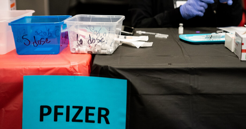
Research shows that the pandemic took a toll on our overall well-being and left many of us drained. Here are seven simple steps to get you thriving again.With vaccination rates on the rise, hope is in the air. But after a year of trauma, isolation and grief, how long will it take before life finally — finally — feels good?Post-pandemic, the answer to that question may be in your own hands. A growing body of research shows that there are simple steps you can take to recharge your emotional batteries and spark a sense of fulfillment, purpose and happiness. The psychology community calls this lofty combination of physical, mental and emotional fitness “flourishing.” It is the exact opposite of languishing, that sense of stagnation Adam Grant wrote about recently for The Times.“Flourishing really is what people are ultimately after,” said Tyler J. VanderWeele, an epidemiology and biostatistics professor and director of Harvard’s Human Flourishing Program. “It’s living the good life. We usually think about flourishing as living in a state in which all aspects of a person’s life are good — it’s really an all-encompassing notion.”The good news is that the scientific evidence related to flourishing is robust, and numerous studies show simple activities can lead to marked improvement in overall well-being. Here are some practical activities, backed by science, that can help you get started.Assess yourself.First, how do you know if you’re languishing, flourishing or somewhere in between? Simply asking someone is an effective diagnostic tool, said Laurie Santos, a psychology professor at Yale who teaches a free 10-week course called the “The Science of Well-Being.” Do you wake up ready to start your day or would you rather go back to sleep? Do you have a sense of purpose or do you find how you spend much of your day to be meaningless? “You are kind of the expert on your own sense of flourishing,” she said.Dr. VanderWeele uses a 10-question assessment in his program at Harvard, which you can try here. Participants rate five areas of their lives on a scale of one to 10, with questions focusing on happiness and life satisfaction, physical and mental health, meaning and purpose, character and virtue and close social relationships. Just taking the quiz and reflecting on the questions it asks can put you on a path to making positive changes, Dr. VanderWeele said.Savor and celebrate small things.After a year of Zoom birthday parties and virtual graduations, many of us want to revel in gathering together again. Celebrations help to create and cement relationships. “It’s really important that post-pandemic we embrace more and more celebrating,” Dr. VanderWeele said.But it’s not just the big occasions that should be marked. Acknowledging small moments is also important for well-being, research shows. Psychologists call it “savoring.” Savoring is about appreciating an event or activity in the moment, sharing tiny victories and noticing the good things around you.A 2012 study of college students found that taking part in a savoring activity called “mindful photography” resulted in overall improvements in mood and a significantly greater sense of appreciation for college life. The students were instructed to take at least five photos of their everyday lives — friends, their favorite view on campus, books they enjoyed — twice a week for two weeks. Reflecting on the photos, and the small moments that brought them joy, helped the students focus on the good in their lives.If snapping photos of your favorite things sounds like too much work, research shows you also benefit when you savor enjoyable experiences like luxuriating in a warm bath, spending the day with your best friend or taking an “awe” walk.Try “Sunday dinner gratitude.”Some people expressed gratitude more during the pandemic, whether it was clapping for health care workers or thanking a grocery checkout person. But creating a weekly gratitude ritual can cement the habit. Numerous studies show that taking time to reflect on what we’re grateful for improves our quality of life.In a 2003 study, researchers instructed college students to list, once a week, five things they were grateful for, both big and small. (Some wrote that they were grateful for waking up that morning; one included gratitude for the Rolling Stones.) Compared to a control group, the students assigned to the gratitude intervention for 10 weeks had better feelings about life as a whole and fewer physical complaints.A gratitude practice should not be a burden. Try to stack a new gratitude habit on a weekly ritual — like Sunday dinner with family; taking out the trash, or your weekly grocery run.Do five good deeds.Acts of kindness not only help others, they also can help you flourish. Research shows that performing five acts of kindness in a single day, once a week, can have a powerful effect. A 2004 study showed that when college students spent a day doing five acts of kindness — like donating blood, helping a friend with a paper or writing a thank you note to a former professor — they experienced more significant increases in well-being than those who spread out five kind things over the course of a week.Volunteer work can also improve well being. Dr. VanderWeele and other researchers looked at data from a cohort of nearly 13,000 older adults and found that participants who volunteered at least two hours a week during the study period experienced higher levels of happiness, optimism and purpose in life, compared to those who did not volunteer at all.To make it easy, Dr. Grant recommends starting off with a daily “five-minute favor,” like introducing two people who could benefit from knowing each other, or sending an article or podcast link to a friend, saying you were thinking of them.Look for communities and connection.Even a quick chat with a stranger or a momentary bond with someone new can foster a sense of fulfillment, particularly when what researchers call a high quality connection occurs. “They don’t have to be lasting relationships or long interactions,” Dr. Grant said. “Sometimes people feel an extra spring in their step when they talk to a stranger on a plane or a subway, or when somebody greets them at a restaurant.”Moments of being seen by other people, and being met with respect or even enthusiasm, can energize and invigorate us and help create bonds within our neighborhood or community.As you emerge from pandemic life, try to reconnect with a community you’ve missed. It might be going back to church or choir practice, a running group or yoga class or even just hanging out at your local coffee shop. And don’t be afraid to chat with a stranger, reconnect with your barista or strike up a conversation at the dog park.Find purpose in everyday routines.What things do you look forward to each day? What gives your life meaning? Research has found that flourishing comes from daily routines, like working on a new skill or reaching out to thank the people you value in your life, and small moments of mastery, connection and meaning.“There are lots of American adults that would meet the qualifications of feeling happy, but they don’t feel sense of purpose,” said Corey Keyes, a professor of sociology at Emory University. “Feeling good about life is not enough.”While work doesn’t have to be the main driver behind your sense of purpose, studies show that reframing how you think about your job can improve your sense of satisfaction. Deepening relationships with co-workers and reminding yourself how your job contributes to a greater good can change how you think about work. If you’re an insurance agent, for example, perceiving your job as a means of helping people get back on their feet after an accident, rather than focusing on a rote task like processing claims, can make your work more fulfilling“People think that in order to flourish, they need to do whatever their version of winning the Olympics is, or climbing a mountain, or having some epic experience,” Dr. Grant said.If you’re feeling down, choose a small project. It could be as simple as cleaning the kitchen or doing yard work, or even washing your pillow cases. Maybe you set a 10-minute timer and go for a short jog, or try a one-minute meditation. Completing a simple, impactful task can build toward a sense of accomplishment.Try something new.“Many of us think we need to change our circumstances, get a job where we earn tons more money, or switch our relationships, buy something new,” said Dr. Santos. “But what the research really shows is that flourishing comes from a different set of behaviors and habits.”And now that life is getting back closer to normal, there are more opportunities to branch out. You can join a book club or running group, take a pottery class, visit a museum or outdoor art exhibit, try a new recipe, explore a nearby trail or neighborhood or test out a free language learning app like Duolingo.Most important for overall well being, Dr. Keyes said, is being interested in life; a sense of satisfaction or happiness tends to follow that. The pandemic has challenged us because we haven’t been able to pursue many of our previous interests, he said. “The first key to feeling good about life is to seek out new interests,” he said.Dr. Grant also said learning a skill and then teaching it to someone, or taking on passion projects as hobbies, can lead to fulfillment. The end of the pandemic offers a new opportunity to reflect, he said, and to ask a new question: “How do I want to spend my time?”

Read more →

Testing tool can quickly distinguish between viral and bacterial infections

 When patients complain of coughing, runny nose, sneezing and fever, doctors are often stumped because they have no fundamental tool to identify the source of the respiratory symptoms and guide appropriate treatments.
That tool might finally be on its way. In a study proving feasibility, researchers at Duke Health showed that their testing technology can accurately distinguish between a viral and a bacterial infection for respiratory illness – a critical difference that determines whether antibiotics are warranted. And, importantly, the test provided results in under an hour.
“This is exciting progress,” said study lead Ephraim Tsalik, associate professor in the departments of Medicine and Molecular Genetics and Microbiology at Duke University School of Medicine.
“We’ve been working on this for over a decade,” Tsalik said. “We knew in 2016 that our test worked in the research setting, but it’s always been our goal to have a test that could produce results rapidly, while patients are at their doctor’s office. It’s important that the distinction can be made quickly to ensure that antibiotics are not inappropriately prescribed.”
Tsalik and colleagues published results of their study in the journal Critical Care Medicine, which confirm the test’s accuracy with results available in under an hour.
The researchers have developed a gene expression method that diverges from current diagnostic strategies, which focus on identifying specific pathogens. The current tests are time-consuming and can only identify a pathogen if it’s specifically targeted by the test in the first place.
Host gene expression, however, looks for a distinct immune signal that is unique to the type of infection the body is fighting. The immune system activates one set of genes when fighting bacterial infections and a different set of genes in response to a viral infection. After the team discovered these gene expression signatures for bacterial and viral infection, they collaborated with BioFire Diagnostics, a company that specializes in molecular diagnostics, to develop this first-of-its kind test.
In a multisite study of more than 600 patients presenting to hospital emergency departments with respiratory infections, the tests identified bacterial infections with 80% accuracy and viral infections with nearly 87% accuracy. The current standard tests have about 69-percent accuracy. Tests provided results in less than an hour, and their accuracy was confirmed retrospectively using two different methods.
“Acute respiratory illness is the most common reason that people visit a health care provider when feeling sick,” Tsalik said. “Patients with these symptoms are inappropriately treated with antibiotics far too often due to challenges in discriminating the cause of illness, fueling antibiotic resistance. Our study shows that a rapid test to distinguish between these two sources of illness is possible and could improve clinical care.”
Tsalik said additional studies are underway to validate this approach in additional groups of patients. The researchers are also working to adapt the technology to produce more specific information, including whether the virus causing illness is influenza or SARS-CoV-2.
Story Source:
Materials provided by Duke University Medical Center. Note: Content may be edited for style and length.

Read more →

Chronic exposure to low levels of blast may be associated with neurotrauma

Scientists at the Walter Reed Army Institute for Research demonstrated that biomarkers associated with traumatic brain injury were elevated among law enforcement and military personnel, particularly in active duty participants with longer duration of service. Most notably, these elevated biomarker levels were observed in individuals without a diagnosed brain injury or concussion.
Some law enforcement and military personnel are regularly exposed to low levels of blast, particularly during training, due to the use of explosive charges and high caliber weapons. Understanding effects from these occupational exposures is a military health care priority to improve diagnosis and mitigation of ill effects.
While repeated exposure to low level blast is not known to result in clinically diagnosed traumatic brain injury, exposures have been linked to a series of reported symptoms such as headaches, fatigue, dizziness, memory difficulties, and tinnitus (ringing in the ears) — collectively referred to as “breacher’s brain” among members of affected communities.
This study, published in the Journal of the American Medical Association, measured neurotrauma biomarker concentrations in blood samples from 106 military and law enforcement personnel who were not actively engaged in training or physical activity at the time of blood collection and compared those concentrations with commercially available samples from individuals who were similar in sex and age but unlikely to have been exposed to blast.
“We found that five biomarkers previously associated with TBI and brain diseases were elevated among personnel when compared to controls,” said Dr. Angela Boutte, lead author on the paper and a researcher at the WRAIR Brain Trauma Neuroprotection branch. “Given the difficulty of identifying and evaluating injury associated with repeated low level blast exposure, we hope these data are the first step in our collective goal to identify objective biomarkers as clinically relevant diagnostic tools.”
Dr. Bharani Thangavelu and Dr. Walter Carr, WRAIR brain health researchers and co-authors, emphasized the potential impact of blast exposure experienced by military personnel stating, “Low level blast exposure in routine military training should not be expected to result in acute, gross behavioral deficits for the majority of personnel. However, repeated exposure across years does correlate with symptomology, especially when a history of chronic exposure is exacerbated by new, large magnitude exposures.”
Efforts to identify and quantify the impact of blast and traumatic brain injury on Service Members have increased dramatically in recent years, including initiatives in response to Congressional mandates. Biomarkers of blast effects on brain health will be a useful tool in this effort, especially as tools that augment decision-making based on symptoms reported by personnel.
Story Source:
Materials provided by Walter Reed Army Institute of Research. Note: Content may be edited for style and length.

Read more →

Nanoplastics and other harmful pollutants found in disposable face masks

Swansea University scientists have uncovered potentially dangerous chemical pollutants that are released from disposable face masks when submerged in water.
The research reveals high levels of pollutants, including lead, antimony, and copper, within the silicon-based and plastic fibres of common disposable face masks.
The work is supported by the Institute for Innovative Materials, Processing and Numerical Technologies (IMPACT) and the SPECIFIC Innovation & Knowledge Centre
Project lead Dr Sarper Sarp of Swansea University College of Engineering said: “All of us need to keep wearing masks as they are essential in ending the pandemic. But we also urgently need more research and regulation on mask production, so we can reduce any risks to the environment and human health.”
Outlined in a recent paper, the tests carried out by the research team used a variety of masks — from standard plain face masks to novelty and festive masks for children with many currently being sold in UK retail outlets.
The rise in single-use masks, and the associated waste, due to the COVID-19 pandemic has been documented as a new cause of pollution. The study aimed to explore this direct link — with investigations to identify the level of toxic substances present.

Read more →