Clean water and toilets for healthy shelters

Regular, standardized assessments of evacuation shelters can help keep people healthy following natural disasters, according to research published by Tohoku University scientists and colleagues in the journal Heliyon. The study found that a clean tap water supply and hygienic toilets were especially important for protecting evacuees from the spread of infectious diseases.
“A clean water supply and maintaining hygiene are important for reducing environmental health risks among victims of natural disasters,” says Tadashi Ishii, who specializes in disaster medicine at Tohoku University. “But scientists have not yet established a strong evidence base that describes the relationship between damage in resource supplies and infrastructure on the one hand and disaster victims’ health status on the other.”
Ishii led the Ishinomaki Zone Joint Relief Team following the Great East Japan Earthquake of March 11, 2011. More than 15,000 people died and 2,500 went missing following the disaster, with some 500,000 evacuated to shelters across Japan. It took nearly a year before all shelters were shut down.
The team conducted regular visits to the shelters in order to assess resource availability, infrastructure, and the health status and needs of people residing in the shelters. Now, Ishii and his research team have analysed these 2011 records to evaluate the impacts of resource supply levels and infrastructure damage on the physical health of evacuees.
Their study included 28 mid- to large-sized shelters regularly assessed in the weeks following the earthquake. The study looked specifically at changes made to resources and infrastructure between days 14 and 25 after the earthquake.
The team found that inadequate clean tap water and toilets were insufficiently improved during the assessment period in about half the shelters. Clinical symptoms of common respiratory and gastrointestinal infections were more prevalent in shelters where these two resources had not improved. Shelters that were able to improve the supply of clean tap water and toilet hygiene witnessed significant reductions in the prevalence of gastrointestinal symptoms among evacuees.
“Our study demonstrated the difficulty of quickly collecting objective assessment data from evacuation shelters during the acute phase of a massive disaster,” says Ishii. “It also shows the validity of quick visual assessments of resources by trained staff. Importantly, the study reveals the importance of rapidly restoring clean water supply and toilet hygiene in shelters to reduce environmental health risks among evacuees.”
Ishii and his team next plan to develop easy, reliable and quick assessment tools for evaluating resource damage and health status in evacuation shelters. He also stresses the importance of collaborating with local governments to set up effective supply chains that can rapidly deploy clean water and hygienic rescue toilets in the aftermath of natural disasters.
Story Source:
Materials provided by Tohoku University. Note: Content may be edited for style and length.

Read more →

Diabetes vaccine gives promising results in a genetic subgroup

A clinical study led by Linköping University and financed by pharmaceuticals company Diamyd Medical has investigated whether immunotherapy against type 1 diabetes can preserve the body’s own production of insulin. The results suggest that injection of a protein, GAD, into lymph nodes can be effective in a subgroup of individuals. The results have been published in Diabetes Care.
In type 1 diabetes, the body’s immune system attacks the cells that produce insulin. When the insulin-producing cells have disappeared, the body can no longer regulate blood sugar level, and a person with type 1 diabetes must take exogenous insulin for the rest of his or her life.
A highly topical question in research into type 1 diabetes is whether, and if so how, the attack of the immune system can be slowed or even completely stopped. One possible strategy is based on altering the immune defence by injecting a protein that the cells of the immune system react to, in a form of vaccination. One of the proteins against which the immune system often forms antibodies in type 1 diabetes is known as GAD65 (glutamic acid decarboxylase). Professor Johnny Ludvigsson at Linköping University has studied for many years the possibility of vaccinating people who have newly diagnosed type 1 diabetes with GAD. It is hoped that the immune system will become more tolerant against the body’s own GAD, and stop damaging the insulin-producing cells, such that the body can continue to form some insulin.
“Studies have shown that even an extremely small production of insulin in the body is highly beneficial for patient health. People with diabetes who produce a certain amount of insulin naturally do not develop low blood sugar levels, hypoglycaemia, so easily. They have also a lower risk of developing the life-threatening condition ketoacidosis, which can arise when the insulin level is low,” says Johnny Ludvigsson, senior professor in the Department of Biomedical and Clinical Sciences at Linköping University.
Johnny Ludvigsson has led DIAGNODE-2, a clinical phase 2 study in which researchers investigated the effect of GAD-alum (Diamyd) injections into the lymph nodes of 109 young people with recently diagnosed type 1 diabetes. The natural insulin production of the participants was measured at the start of the study and again after 15 months. Several other outcome measures were also followed, such as change in long-term blood sugar levels (HbA1c), and how much supplementary insulin the patients needed to take every day.
Previous studies of immunotherapy in diabetes have suggested that genetic factors play a role in how patients respond to the treatment. This led the researchers in DIAGNODE-2 to look at several variants of what are known as “HLA genes.” These genes code for proteins located on the surface of some cells. They function as holders of proteins, and expose them to immune system cells passing by. If the protein fragment exposed in this way comes from, for example, bacteria, the immune system should form antibodies against the foreign protein. However, the immune system sometimes reacts against the body’s own substances, and certain types of HLA are associated with an increased risk of type 1 diabetes. The HLA variant HLA-DR3-DQ2 exposes the GAD65 protein to cells of the immune system, and patients with this variant often form antibodies against GAD65 at an early stage of the disease. Around half of the participants in the study had the HLA-DR3-DQ2 variant.

Read more →

Digital Twin technology a 'powerful tool' but requires significant investment, say experts

Healthcare and aerospace experts at King’s College London, The Alan Turing Institute, the University of Cambridge, and the Oden Institute for Computational Engineering and Sciences at UT Austin in Texas have said advances in digital twin technology make it a powerful tool for facilitating predictive and precision medicine and enhancing decision-making for aerospace systems. Their opinion piece was published today in Nature Computational Science.
When applied to healthcare, the digital twin, a virtual version of real-life objects that can be used to predict how that object will perform, could predict how a patient’s disease will develop and how patients are likely to respond to different therapies.
It is also of huge benefit in aerospace, where, for example, the technology will be needed to monitor and control thousands of drones, ensuring that they are maintained, have efficient and safe flight plans and can automatically adapt to changes in conditions, such as weather, without the need for human interaction.
However, current digital twins are largely the result of bespoke technical solutions that are difficult to scale.
The authors say that these use cases place new demands on the speed, robustness, validation, verification and uncertainty quantification in digital twin creation workflows.
Achieving digital twins at scale will require a drastic reduction in technical barriers to their adoption.

Read more →

Obesity protects against death in severe bacterial infection

For many diseases, overweight and obesity are risk factors. But now a study shows that a higher BMI may be linked to higher survival rates in patients hospitalized for severe bacterial infections.
Scientists at Sahlgrenska Academy, University of Gothenburg, and Skaraborg Hospital in Skövde carried out the research, and their study has now been published in the journal PLOS ONE. The data were collected before the COVID-19 pandemic.
The population-based study involved observations, over a nine-month period, of all 2,196 individual adults receiving care for suspected severe bacterial infection at Skaraborg Hospital in Skövde. The researchers followed the patients in this study population over time, during and after their hospital stay.
The results show that the raised chances of survival were associated with a higher body mass index (BMI) in both the short and long term, at 28 days and one year after hospitalization respectively. The differences in survival rates were clear. In the normal-weight group, 26 percent were dead within a year. The corresponding figures in the groups with higher BMI were 9-17 percent.
Occasional surveys of limited patient groups have previously shown similar results. The new findings clarify and confirm the “obesity survival paradox”: that overweight and obesity afford protection against severe bacterial infections.
Åsa Alsiö, adjunct senior lecturer in infectious diseases at Sahlgrenska Academy and senior consultant in infectious diseases at Skövde, is the study’s first and corresponding author.
“In the context of most other diseases, overweight and obesity are disadvantageous. This applies to several types of cancer, cardiovascular disease and, in particular, COVID-19, in which a higher BMI is associated with higher mortality. Paradoxically, it’s the other way round here.
“What we don’t know,” Alsiö continues, “is how being overweight can benefit the patient with a bacterial infection, or whether it’s connected with functions in the immune system and how they’re regulated. More knowledge is needed about how being overweight affects the immune system. One patient category it could be studied in is individuals undergoing bariatric surgery.”
Gunnar Jacobsson, Sahlgrenska Academy and senior consultant in infectious diseases at Skaraborg Hospital in Skövde, is the senior author of the study:
“The COVID-19 pandemic has highlighted vulnerable patient groups, and overweight people have been hit hard. Maybe experience and handling of care for patients with severe bacterial infections can be used to improve the prognosis of COVID-19 and overweight. Globally, obesity is increasing at an alarming rate. More knowledge is needed to shed light on how body weight affects the body’s defenses against infection, so that treatment can be individualized,” Jacobsson says.
The researchers think there is a need for more studies, at the population level, of how BMI affects treatment outcomes in various infectious diseases and what connections with regulation of the immune system may exist.
Story Source:
Materials provided by University of Gothenburg. Note: Content may be edited for style and length.

Read more →

Virus infection cycle revealed in dynamic detail

A critical process in the infection cycle of viruses has been revealed for the first time in dynamic detail using pioneering plant-based technology.
Evidence about the process of virus maturation revealed in the research could help us develop new methods for treating viral infections.
Maturation plays a critical role for all animal and bacterial viruses and is required to produce infectious virions or particles. Though the outlines of the process have been determined for many groups of viruses, detailed mechanistic studies have not been reported.
To provide the first detailed mechanistic study of maturation, Roger Castells-Graells, a rotation Ph.D. student working in Professor. George Lomonossoff’s laboratory at the John Innes Centre infiltrated genetic material of the insect virus Nudaurelia capensis omega virus (N?V) into dwarf tobacco plants N.benthamiana.
This transient expression technique uses Virus Like Particles (VLPs) which are mimics of the authentic virus. The capsid or protein coat of the virus is produced by plant cells and the research team then analyses the material purified from infiltrated leaves.
The research demonstrated that maturation of procapsids — immature viral structures — can occur within plant cells to yield fully functional mature capsids. This has not been observed previously in the absence of a natural infection and is a new application for the transient expression system pioneered by Professor Lomonossoff at the John Innes Centre.
Comparative cryo-EM analysis of the structures of the procapsids and mature capsids revealed the large structural rearrangements both inside and between the protein subunits of the capsid that accompany maturation. These shape changes enable the chemical reactions that are necessary for the virus to infect the host.
Professor Lomonossoff, a group leader at the John Innes Centre, said: “Most structural studies of virus particles to date have given a static picture of the particles. By isolating particles from plants that are undergoing the process of maturation, we have managed to obtain a picture of the dynamics of an essential part of a virus infection cycle.”
The present study, a collaboration involving scientists at the University of Leeds, in Brazil and the USA, as well as at the John Innes Centre, reveals details of the structures at the beginning and the end of the maturation process. What is now required is an analysis of intermediate steps to get a complete understanding of the dynamics.
This will enable the research team to determine the 3-D structures of intermediates in the maturation process to create a “movie.”
“We have shown that maturation occurs over time within plant cells and that means we have discovered a valuable tool for studying virus maturation. We hope it will be of interest to potential collaborators and industry,” said Professor Lomonossoff.
Story Source:
Materials provided by John Innes Centre. Note: Content may be edited for style and length.

Read more →

Mild COVID-19 induces lasting antibody protection, study finds

Months after recovering from mild cases of COVID-19, people still have immune cells in their body pumping out antibodies against the virus that causes COVID-19, according to a study from researchers at Washington University School of Medicine in St. Louis. Such cells could persist for a lifetime, churning out antibodies all the while.
The findings, published May 24 in the journal Nature, suggest that mild cases of COVID-19 leave those infected with lasting antibody protection and that repeated bouts of illness are likely to be uncommon.
“Last fall, there were reports that antibodies wane quickly after infection with the virus that causes COVID-19, and mainstream media interpreted that to mean that immunity was not long-lived,” said senior author Ali Ellebedy, PhD, an associate professor of pathology & immunology, of medicine and of molecular microbiology. “But that’s a misinterpretation of the data. It’s normal for antibody levels to go down after acute infection, but they don’t go down to zero; they plateau. Here, we found antibody-producing cells in people 11 months after first symptoms. These cells will live and produce antibodies for the rest of people’s lives. That’s strong evidence for long-lasting immunity.”
During a viral infection, antibody-producing immune cells rapidly multiply and circulate in the blood, driving antibody levels sky-high. Once the infection is resolved, most such cells die off, and blood antibody levels drop. A small population of antibody-producing cells, called long-lived plasma cells, migrate to the bone marrow and settle in, where they continually secrete low levels of antibodies into the bloodstream to help guard against another encounter with the virus.
The key to figuring out whether COVID-19 leads to long-lasting antibody protection, Ellebedy realized, lies in the bone marrow. To find out whether those who have recovered from mild cases of COVID-19 harbor long-lived plasma cells that produce antibodies specifically targeted to SARS-CoV-2, the virus that causes COVID-19, Ellebedy teamed up with co-author Iskra Pusic, MD, an associate professor of medicine. Ellebedy already was working with co-authors Rachel Presti, MD, PhD, an associate professor of medicine, and Jane O’Halloran, MD, PhD, an assistant professor of medicine, on a project to track antibody levels in blood samples from COVID-19 survivors.
The team already had enrolled 77 participants who were giving blood samples at three-month intervals starting about a month after initial infection. Most participants had had mild cases of COVID-19; only six had been hospitalized.
With Pusic’s help, Ellebedy and colleagues obtained bone marrow from 18 of the participants seven or eight months after their initial infections. Five of them came back four months later and provided a second bone marrow sample. For comparison, the scientists also obtained bone marrow from 11 people who had never had COVID-19.
As expected, antibody levels in the blood of the COVID-19 participants dropped quickly in the first few months after infection and then mostly leveled off, with some antibodies detectable even 11 months after infection. Further, 15 of the 19 bone marrow samples from people who had had COVID-19 contained antibody-producing cells specifically targeting the virus that causes COVID-19. Such cells could still be found four months later in the five people who came back to provide a second bone-marrow sample. None of the 11 people who had never had COVID-19 had such antibody-producing cells in their bone marrow.
“People with mild cases of COVID-19 clear the virus from their bodies two to three weeks after infection, so there would be no virus driving an active immune response seven or 11 months after infection,” Ellebedy said. “These cells are not dividing. They are quiescent, just sitting in the bone marrow and secreting antibodies. They have been doing that ever since the infection resolved, and they will continue doing that indefinitely.”
People who were infected and never had symptoms also may be left with long-lasting immunity, the researchers speculated. But it’s yet to be investigated whether those who endured more severe infection would be protected against a future bout of disease, they said.
“It could go either way,” said first author Jackson Turner, PhD, an instructor in pathology & immunology. “Inflammation plays a major role in severe COVID-19, and too much inflammation can lead to defective immune responses. But on the other hand, the reason why people get really sick is often because they have a lot of virus in their bodies, and having a lot of virus around can lead to a good immune response. So it’s not clear. We need to replicate the study in people with moderate to severe infections to understand whether they are likely to be protected from reinfection.”
Ellebedy and colleagues now are studying whether vaccination also induces long-lived antibody-producing cells.

Read more →

Researchers discover oligodendrocyte loss and subtype alteration in CTE brains

Since 2008, researchers at Boston University School of Medicine (BUSM) and VA Boston Healthcare System have studied Chronic Traumatic Encephalopathy (CTE), a progressive brain disease associated with repetitive head impacts that has been diagnosed after death in the brains of American football players and other contact sport athletes as well as members of the armed services. Clinically, impulsivity, explosivity, depression, memory impairment and executive dysfunction have been reported to occur in the disease.
While many of the scientific studies to date have focused on repetitive head trauma leading to the development of abnormal tau, a new study provides insights into white matter changes that may offer new targets for therapies.
“Research to date has focused on the deposition of abnormal tau in the gray matter in CTE. This study shows that the white matter undergoes important alterations as well. There is loss of oligodendrocytes and alteration of oligodendrocyte subtypes in CTE that might provide new targets for prevention and therapies,” said corresponding author Ann McKee, MD, chief of neuropathology at VA Boston Healthcare, director of the BU CTE Center.
McKee and her team isolated cellular nuclei from the postmortem dorsolateral frontal white matter in eight cases of CTE and eight matched controls. They conducted single-nucleus RNA-seq (snRNA-seq) with these nuclei, revealing transcriptomic, cell-type-specific differences between the CTE and control cases. What they discovered was that the white matter in CTE had fewer oligodendrocytes and the oligodendroglial subtypes were altered compared to control tissue.
These findings are of particular interest because previous studies have largely focused on the CTE-specific tau lesion located in the cortex in the brain. “In comparison, the cellular death process occurring in white matter oligodendrocytes in CTE appears to be separate from the accumulation of hyperphosphorylated tau,” she said. “We know that the behavioral and mood changes that occur in CTE are not explained by tau deposition. This study suggests that white matter alterations are also important features of the disease, and future studies will determine whether these white matter changes play a role in the production of behavioral or mood symptoms in CTE, such as explosivity, violence, impulsivity, and depression.”
These findings appear online in the journal Acta Neuropathologica.
This work was supported in part by the Harvard/MIT Joint Research Grants Program in Basic Neuroscience (K. Blake Chancellor, Joseph E. Duke-Cohan, Ann C. McKee) and the Concussion Legacy Foundation Young Investigator Fellowship (Sarah E. Chancellor). This work was supported by grant funding from: National Institute on Aging (AG057902, AG06234), The National Institute of Neurological Disorders and Stroke (U54NS115266, U01NS086659), National Institute of Aging Boston University Alzheimer’s Disease Center (P30AG13846); the Concussion Legacy Foundation, and the Nick and Lynn Buoniconti Foundation.
Story Source:
Materials provided by Boston University School of Medicine. Note: Content may be edited for style and length.

Read more →

Telomere length, a longevity measure, may be determined early in life

Telomeres are protective caps on DNA that shorten as we grow older. Now, one of the first studies to examine telomere length (TL) in childhood finds that the initial setting of TL during prenatal development and in the first years of life may determine one’s TL throughout childhood and potentially even into adulthood or older age. The study also finds that TL decreases most rapidly from birth to age 3, followed by a period of maintenance into the pre-puberty period, although it was sometimes seen to lengthen.
The study, which followed children from birth to age 9, was led by researchers at the Columbia Center for Children’s Environmental Health at Columbia University Mailman School of Public Health. Results appear in the journal Psychoneuroendocrinology.
The researchers discovered that a mother’s TL is predictive of newborn TL and tracks with her child’s TL through pre-adolescence. While all telomeres are expected to shorten with age, the reasons why some children have telomeres that shorten faster are unknown, one explanation may be that telomeres are susceptible to environmental pollutants. It is also unknown why some children had telomeres that lengthened across the study period though it is notable that this phenomenon has also been observed in other studies.
“Given the importance of telomere length in cellular health and aging, it is critical to understand the dynamics of telomeres in childhood,” says senior author Julie Herbstman, PhD, director of CCCEH and associate professor of environmental health science at Columbia Mailman School. “The rapid rate of telomere attrition between birth and age 3 years may render telomeres particularly susceptible to environmental influences during this developmental window, potentially influencing life-long health and longevity.”
In the new study, researchers used polymerase chain reaction to measure TL in white blood cells isolated from cord blood and blood collected at ages 3, 5, 7, and 9, from 224 children. They also measured maternal TL at delivery in a subset of mothers.
The researchers say more research is needed to understand the biological mechanisms driving variability in the rate of TL change during the first years of life, as well as modifiable environmental factors that contribute to shifts in the rate of attrition.
Story Source:
Materials provided by Columbia University’s Mailman School of Public Health. Note: Content may be edited for style and length.

Read more →

Blind Man's Sight Partially Restored with 'Optogenetics' Gene Therapy

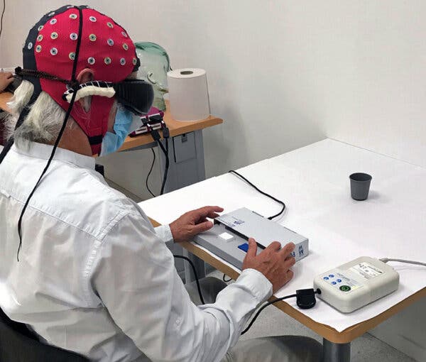
Using a technique called optogenetics, researchers added light-sensitive proteins to the man’s retina, giving him a blurry view of objects.A team of scientists announced Monday that they had partially restored the sight of a blind man by building light-catching proteins in one of his eyes. Their report, which appeared in the journal Nature Medicine, is the first published study to describe the successful use of this treatment.“Seeing for the first time that it did work — even if only in one patient and in one eye — is exciting,” said Ehud Isacoff, a neuroscientist at the University of California, Berkeley, who was not involved in the study.The procedure is a far cry from full vision. The volunteer, a 58-year-old man who lives in France, had to wear special goggles that gave him the ghostly perception of objects in a narrow field of view. But the authors of the report say that the trial — the result of 13 years of work — is a proof of concept for more effective treatments to come.“It’s obviously not the end of the road, but it’s a major milestone,” said Dr. José-Alain Sahel, an ophthalmologist who splits his time between the University of Pittsburgh and the Sorbonne in Paris.Dr. Sahel and other scientists have tried for decades to find a cure for inherited forms of blindness. These genetic disorders rob the eyes of essential proteins required for vision.When light enters the eye, it is captured by so-called photoreceptor cells. The photoreceptors then send an electrical signal to their neighbors, called ganglion cells, which can identify important features like motion. They then send signals of their own to the optic nerve, which delivers the information to the brain.In previous studies, researchers have been able to treat a genetic form of blindness called Leber congenital amaurosis, by fixing a faulty gene that would otherwise cause photoreceptors to gradually degenerate.But other forms of blindness can’t be treated so simply, because their victims lose their photoreceptors completely. “Once the cells are dead, you cannot repair the gene defect,” Dr. Sahel said.For these diseases, Dr. Sahel and other researchers have been experimenting with a more radical kind of repair. They are using gene therapy to turn ganglion cells into new photoreceptor cells, even though they don’t normally capture light.The scientists are taking advantage of proteins derived from algae and other microbes that can make any nerve cell sensitive to light.In the early 2000s, neuroscientists figured out how to install some of these proteins into the brain cells of mice and other lab animals by injecting viruses carrying their genes. The viruses infected certain types of brain cells, which then used the new gene to build light-sensitive channels.Originally, researchers developed this technique, called optogenetics, as a way to probe the workings of the brain. By inserting a tiny light into the animal’s brain, they could switch a certain type of brain cell on or off with the flick of a switch. The method has enabled them to discover the circuitry underlying many kinds of behavior.Dr. Sahel and other researchers wondered if they could use optogenetics to add light-sensitive proteins to cells in the retina. After all, they reasoned, retinal cells are nerves as well — an extension of the brain, in other words.For Ed Boyden, a neuroscientist at M.I.T. who helped pioneer the field of optogenetics, the quest to use these proteins to cure blindness took him by surprise. “So far, I’ve thought of optogenetics as a tool for scientists primarily, since it’s being used by thousands of people to study the brain,” he said. “But if optogenetics proves itself in the clinic, that would be extremely exciting.”Dr. Sahel and his colleagues recognized that the optogenetic proteins created by Dr. Boyden and others were not sensitive enough to produce an image from ordinary light entering the eye. But the scientists could not beam amplified light into the eye, because the glare would destroy the delicate tissue of the retina.So the scientists chose an optogenetic protein that’s sensitive only to amber light, which is easier on the eye than other colors, and used viruses to deliver these amber proteins to the ganglion cells in the retina.The experimental set-up, where the volunteer was asked to say whether or not the cup was on the white table. Behavioral responses and brain activity were recorded simultaneously during the test.Sahel, et al.; Nature MedicineNext, the researchers invented a special device to transform visual information from the external world into amber light that could be recognized by the ganglion cells. They created goggles that scan their field of view thousands of times a second and register any pixels in which the light changes. The goggles then send a pulse of amber light from that pixel into the eye.The researchers reasoned that this strategy might be able to create images in the brain. Our eyes naturally dart around in tiny movements many times a second. With each jump, many pixels would change light levels.Still, it was an open question whether blind people could learn to use this information to recognize objects. “The brain has to learn a new language,” said Botond Roska, an ophthalmologist at the University of Basel and a co-author of the new study.After testing their gene therapy and goggles on monkeys, Dr. Roska, Dr. Sahel and their colleagues were ready to try it out on people. Their plan was to inject gene-bearing viruses into one eye of each blind volunteer, then wait several months for the ganglion cells to grow optogenetic proteins. They would then train the volunteers to use the goggles.Unfortunately, they only managed to train one volunteer before the coronavirus pandemic shut down the project. After years of preparation for the study, it was now stuck in limbo.But then the one volunteer they had managed to train got in touch. For seven months, he had been wearing the goggles at home and on walks. One day he realized he could see the stripes of a crosswalk.When the pandemic subsided in France over the summer, the scientists managed to bring him into their lab for more training and tests. They discovered that he could reach out and touch a notebook sitting on a table, but had less luck with a smaller box of staples. When the scientists set out either two or three tumblers in front of the volunteer, he managed to count them correctly 12 out of 19 times.During some of the trials, the volunteer wore a cap with electrodes that could detect brain activity through his scalp. When the goggle sent signals to his retina, it activated parts of the brain involved in vision.“It is a major achievement from a scientific point of view, and most importantly for blind people,” said Lucie Pellissier, a neuroscientist at the University of Tours in France who was not involved in the study.Dr. Sahel and his colleagues founded a company called GenSight to move their technique through clinical trials with the hopes of getting it approved by regulators. They’re not alone. Dr. Isacoff and his colleagues have founded a similar company called Vedere Bio that was acquired last October by Novartis.It will take many more positive results from clinical trials before optogenetics can become a standard treatment for some forms of blindness. For now, Dr. Sahel and his colleagues are bringing in the other volunteers for training, as well as testing higher doses of the virus and upgrading their goggles to thin spectacles that would be more comfortable while also delivering more information to the retina.Dr. Isacoff and his colleagues have carried out experiments of their own that raise the possibility that other optogenetic proteins could make retinal cells sensitive enough to detect light without the help of goggles. “I think it’s going to perform quite well,” he said.For all the time that Dr. Sahel has put into his own system, he hesitated to guess how far it could improve. “Until you have a patient tell you what they are seeing, you really can’t predict anything,” he said.

Read more →

What Convinced Me to Start Wearing Sun Protection

I’ve failed to practice what I preached about limiting sun exposure, but a new report has prompted me to reform.Pick your favorite cliché: Do as I say, not as I do; an ounce of prevention is worth a pound of cure; better safe than sorry; forewarned is forearmed.Mea culpa. All the above relate to my failure to follow the well-established health advice about sun exposure that I’ve offered repeatedly to my readers: Routinely protect your skin from the cancer-causing and aging effects of the sun’s ultraviolet rays.For decades I’ve failed to practice what I preached (OK to wince) and am now paying for my negligence with unsightly splotches, bumps and bruises and at least one cancerous lesion on my sun-damaged skin. My litany of excuses has included: hats mess up my hair, long sleeves and pants are too hot in summer and exercising while coated with sunscreen is suffocating.Annually vowing to do better, every summer I dutifully purchase the latest dermatology-recommended sunscreen that, alas, spends the summer unopened on a bathroom shelf. I hereby pledge to do better this year, albeit late in the game.A new report from a dermatology team at Kaiser Permanente health care centers in California has prompted me to reform. The team, headed by the epidemiologist Lisa Herrinton in Oakland, followed nearly half a million patients seen at the centers for up to 10 years. Half had already developed one or more actinic keratosis, a precancerous rough, scaly skin lesion caused by years of unprotected sun exposure.As you might expect, these lesions most often form on the face, ears, back of the hands, forearms, scalp and neck and are — or should be — routinely removed when found by dermatologists to prevent progression to cancer. The lesions are markers of sun damage and can serve as an early warning system for people at risk of developing cancer somewhere on sun-exposed skin.While the hazard is greatest for people with light skin, blue eyes, freckles or red hair, having a dark complexion is not a free pass. Tanning, not just burning, is a form of sun damage.Among patients in the Kaiser Permanente study who were younger than 50, those with a diagnosis of actinic keratosis were nearly seven times more likely to develop a skin cancer called squamous cell carcinoma during the decade-long follow-up. The cancer risk was eight times higher among patients older than 50 who had one or more actinic keratosis removed, and the more such lesions these patients had, the more likely they were to develop skin cancer during the follow-up.Furthermore, the older the patient, the sooner cancer was diagnosed after actinic keratosis was found and presumably treated. It took seven to eight years for 10 percent of patients in their 50s with an actinic keratosis to receive a diagnosis of skin cancer, but it took only three to four years for patients in their 70s and one to two years for those in their 80s.Alas, those of us in the upper decades of life knew little in our younger years about the risks of sun damage beyond the need to avoid a bad sunburn. Many youngsters like me swam, hiked, biked and played sports minimally clothed while the sun tanned or burned our skin. We sunbathed coated in baby oil in a misguided effort to acquire a rich tan. And many of us, myself included, failed to reach adulthood with sun-protective habits that could have prevented the skin damage now woefully apparent.Given that the risk of ultraviolet light to healthy skin has since been widely publicized, I’m astonished at how many people today visit tanning salons or use tanning beds at home, damaging the wholesome cutaneous barrier nature gave us.Happily, the new study suggests that more people now have a greater understanding and respect for the sun’s effects on skin and can look forward to a healthier future, said Dr. Sangeeta Marwaha, a dermatologist in Sacramento and co-author of the study. Among people who entered the study in 2018, the risk of developing skin cancer was two-thirds that of study entrants in 2008 who were followed for an equal number of years.“There’s been an increase in sun-protective habits and a resulting decrease in the development of skin cancer,” Dr. Marwaha said in an interview. “Parents today are more likely to protect their children from undue sun exposure, and the use of sunscreen is now more mainstream.”But there’s still a long way to go. Fostering a healthy respect for sun protection in young children is especially important because experts estimate that 80 percent of a person’s lifetime sun exposure is acquired before age 18.Repeated exposure to the sun’s ultraviolet radiation causes most of the skin changes — wrinkles, age spots and tiny broken blood vessels — generally considered a normal result of aging. Yes, aging plays a role, but these effects occur much earlier in life on sun-exposed skin. UV light damages the elastin fibers in skin, causing it to stretch, sag and wrinkle. It also damages surface blood vessels, rendering them more fragile and easily bruised.And Zachary W. Lipsky, a biomedical engineer at Binghamton University, found that UV radiation weakens the bonds that help the cells in the top layer of skin stick together, damaging the skin’s structural integrity and leaving it more vulnerable to infection.But while some of these effects can be masked by cosmetic treatments and plastic surgery, the most serious damage done by UV radiation — to the DNA of skin cells — is permanent and irreversible and can result in cancer-causing mutations.Furthermore, preventing sun damage is easier and cheaper than reversing it and less likely to result in premature wrinkles and scars. Try to schedule your outdoor activities early or late in the day, avoiding the peak sun hours of 10 a.m. to 4 p.m.Routinely apply a broad-spectrum sunscreen with an SPF of 30 or higher to exposed skin year-round even on cloudy days, using at least a quarter teaspoon on your face alone. Apply sunscreen half an hour before going out and reapply it every two hours and after being in the water. Modern sunscreens are not greasy or pasty, but they lose effectiveness with time so be sure to check the expiration date. Even if you sit under an umbrella at the beach or in the park, the sun’s reflected rays will hit your skin.Wear a hat with a wide brim, especially important for men who are balding. If you have the means, invest in top-quality sunglasses and clothing, including swimwear, with built-in SPF protection. The darker and heavier the fabric, the better. “A plain white T-shirt has an SPF of 4, whereas dark blue denim jeans could have an SPF of 2000,” Dr. Marwaha said.This summer I plan to apply sunscreen daily and wear a long-sleeved sun-protective shirt when I walk, cycle and garden, even on cloudy days, a habit I wish I’d cultivated decades ago.

Read more →