Virus Variants Can Infect Mice, Scientists Report

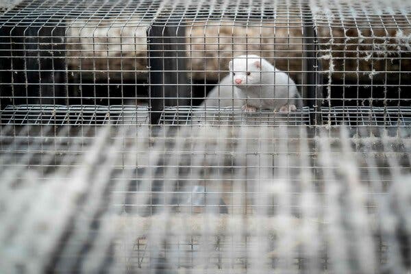
Infected rodents pose no immediate danger to humans, but the research suggests that mutations are helping the coronavirus expand its range of potential hosts.Bats, humans, monkeys, minks, big cats and big apes — the coronavirus can make a home in many different animals. But now the list of potential hosts has expanded to include mice, according to an unnerving new study.Infected rodents pose no immediate risk to people, even in cities like London and New York, where they are ubiquitous and unwelcome occupants of subway stations, basements and backyards.Still, the finding is worrying. Along with previous work, it suggests that new mutations are giving the virus the ability to replicate in a wider array of animal species, experts said.“The virus is changing, and unfortunately it’s changing pretty fast,” said Timothy Sheahan, a virologist at the University of North Carolina at Chapel Hill, who was not involved in the new study.In the study, the researchers introduced the virus into the nasal passages of laboratory mice. The form of the virus first identified in Wuhan, China, cannot infect laboratory mice, nor can B.1.1.7, a variant that has been spreading across much of Europe, the researchers found.But B.1.351 and P1, the variants discovered in South Africa and Brazil, can replicate in rodents, said Dr. Xavier Montagutelli, a veterinarian and mouse geneticist at the Pasteur Institute in Paris, who led the study. The research, posted online earlier this month, has not yet been reviewed for publication in a scientific journal.The results indicate only that infection in mice is possible, Dr. Montagutelli said. Mice caught in the wild have not been found to be infected with the coronavirus, and so far, the virus does not seem to be able to jump from humans to mice, from mice to humans, or from mice to mice.“What our results emphasize is that it is necessary to regularly assess the range of species that the virus can infect, especially with the emergence of new variants,” Dr. Montagutelli said.The coronavirus is thought to have emerged from bats, with perhaps another animal acting as an intermediate host, and scientists worry that the virus may return to what they describe as an animal “reservoir.”Apart from potentially devastating those animal populations, a coronavirus spreading in another species may then acquire dangerous mutations, returning to humans in a form the current vaccines weren’t designed to fend off.A mink looks out from its cage at a farm in Denmark, where mink populations were hit hard by the coronavirus.Mads Claus Rasmussen/Agence France-Presse — Getty ImagesMinks are the only animals known to be able to catch the coronavirus from humans and pass it back. In early November, Denmark culled 17 million farmed mink to prevent the virus from evolving into dangerous new variants in the animals.More recently, researchers found that B.1.1.7 infections in domesticated cats and dogs can cause the pets to develop heart problems similar to those seen in people with Covid-19.To establish a successful infection, the coronavirus must bind to a protein on the surface of animal cells, gain entry into the cells, and exploit their machinery to make copies of itself. The virus must also evade the immune system’s early attempts at thwarting the infection.Given all those requirements, it is “quite extraordinary” that the coronavirus can infect so many species, said Vincent Munster, a virologist at the National Institute of Allergy and Infectious Diseases. “Typically, viruses have a more curtailed host range.”Mice are a known reservoir for hantavirus, which causes a rare and deadly disease in people. Even though the coronavirus variants don’t seem to be able to jump from mice to people, there is potential for them to spread among rodents, evolve into new variants, and then infect people again, Dr. Munster said.The variants may also threaten endangered species like black-footed ferrets. “This virus seems to be able to surprise us more than anything else, or any other previous virus,” Dr. Munster said. “We have to err on the side of caution.”Dr. Sheahan said he was more concerned about transmission to people from farm animals and pets than from mice.“You’re not catching wild mice in your house and snuggling — getting all up in their face and sharing the same airspace, like maybe with your cat or your dog,” he said. “I’d be more worried about wild or domestic animals with which we have a more intimate relationship.”But he and other experts said the results emphasized the need to closely monitor the rapid changes in the virus.“It’s like a moving target — it’s crazy,” he added. “There’s nothing we can do about it, other than try and get people vaccinated really fast.”

Read more →

Estimating lifetime microplastic exposure

Every day, people are exposed to microplastics from food, water, beverages and air. But it’s unclear just how many of these particles accumulate in the human body, and whether they pose health risks. Now, researchers reporting in ACS’ Environmental Science & Technology have developed a lifetime microplastic exposure model that accounts for variable levels from different sources and in different populations. The new model indicates a lower average mass of microplastic accumulation than previous estimates.
Microplastics, which are tiny pieces of plastic ranging in size from 1 µm to 5 mm (about the width of a pencil eraser), are ingested from a variety of sources, such as bottled water, salt and seafood. Their fate and transport in the human body are largely unknown, although the particles have been detected in human stool. In addition to possibly causing tissue damage and inflammation, microplastics could be a source of carcinogens and other harmful compounds that leach from plastic into the body. Previous studies have tried to estimate human exposure to the particles and their leached chemicals, but they have limitations, including discrepancies in the databases used, a failure to consider the entire microplastic size range and the use of average exposure rates that do not reflect global intakes. Nur Hazimah Mohamed Nor, Albert Koelmans and colleagues wanted to develop a comprehensive model to estimate the lifetime exposure of adults and children to microplastics and their associated chemicals.
To make their model, the researchers identified 134 studies that reported microplastic concentrations in fish, mollusks, crustaceans, tap or bottled water, beer, milk, salt and air. They performed corrections to the data so that they could be accurately compared among the different studies. Then, the team used data on food consumption in different countries for various age groups to estimate ranges of microplastic ingestion. This information, combined with rates of microplastic absorption from the gastrointestinal tract and excretion by the liver, was used to estimate microplastic distribution in the gut and tissues. The model predicted that, by the age of 18, children could accumulate an average of 8,300 particles (6.4 ng) of microplastics in their tissues, whereas by the age of 70, adults could accrue an average of 50,100 microplastic particles (40.7 ng). The estimated amounts of four chemicals leaching from the plastics were small compared with a person’s total intake of these compounds, the researchers concluded. These data suggest that prior studies might have overestimated microplastic exposure and possible health risks, but it will be important to assess the contributions of other food types to ingestion and accumulation, the researchers say.
Story Source:
Materials provided by American Chemical Society. Note: Content may be edited for style and length.

Read more →

Vitamin A for nerve cells

Neuroscientists agree that a person’s brain is constantly changing, rewiring itself and adapting to environmental stimuli. This is how humans learn new things and create memories. This adaptability and malleability is called plasticity. “Physicians have long suspected that remodeling processes also take place in humans at the contact points between nerve cells, i.e. directly at the synapses. Until now, however, such a coordinated adaptation of structure and function could only be demonstrated in animal experiments,” says Prof. Dr. Andreas Vlachos from the Institute of Anatomy and Cell Biology at the University of Freiburg. But now Vlachos, together with Prof. Dr. Jürgen Beck, head of the Department of Neurosurgery at the University Medical Center Freiburg, has provided experimental evidence for synaptic plasticity in humans. In addition to Vlachos and Beck, the research team consists of Dr. Maximilian Lenz, Pia Kruse and Amelie Eichler from the University of Freiburg, Dr. Jakob Strähle from the University Medical Center Freiburg and colleagues from Goethe University Frankfurt. The results were presented in the scientific journal eLife.

advertisement

Read more →

Sugar not so nice for your child's brain development

Sugar practically screams from the shelves of your grocery store, especially those products marketed to kids.
Children are the highest consumers of added sugar, even as high-sugar diets have been linked to health effects like obesity and heart disease and even impaired memory function.
However, less is known about how high sugar consumption during childhood affects the development of the brain, specifically a region known to be critically important for learning and memory called the hippocampus.
New research led by a University of Georgia faculty member in collaboration with a University of Southern California research group has shown in a rodent model that daily consumption of sugar-sweetened beverages during adolescence impairs performance on a learning and memory task during adulthood. The group further showed that changes in the bacteria in the gut may be the key to the sugar-induced memory impairment.
Supporting this possibility, they found that similar memory deficits were observed even when the bacteria, called Parabacteroides, were experimentally enriched in the guts of animals that had never consumed sugar.
“Early life sugar increased Parabacteroides levels, and the higher the levels of Parabacteroides, the worse the animals did in the task,” said Emily Noble, assistant professor in the UGA College of Family and Consumer Sciences who served as first author on the paper. “We found that the bacteria alone was sufficient to impair memory in the same way as sugar, but it also impaired other types of memory functions as well.”
Guidelines recommend limiting sugar

Read more →

Masks make little difference to facial identification

But the study also shows for the first time that performance may be improved by using super recognisers — people who are very skilled at recognising faces. It also reveals that masks do make recognising someone’s emotions more difficult.
There are many questions surrounding face masks and the impact that masks will have on face identification. Can we recognise the faces of people who we know well if they are wearing a mask? And, relevant to policing and security scenarios or a supermarket ID check, can an unfamiliar face be recognized across images if it is masked? And how do masks impact our ability to recognize a person’s emotions?
Dr Noyes is Senior Lecturer in Cognitive Psychology and conducted the study, published by the Royal Society, in collaboration with researchers at the University of Greenwich, University of Reading, and University of Lincoln. A leading expert in the field, Dr Noyes was intrigued what the enforced wearing of masks due to COVID-19 would have on facial recognition.
The study consisted of three experiments which tested recognition of familiar faces, recognition of an unfamiliar face (comparing images, aka face matching), and emotion recognition. The researchers compared face recognition and emotion recognition for faces with no concealment, faces in masks, and faces in sunglasses — something far more commonplace than masks and often a matter of choice rather than necessity.
In the first experiment, participants were presented with pairs of famous faces, and were asked to decide if the images were of the same person or two different people.
“People are typically very good at identifying the faces of people they know well,” says Dr Noyes. “However, we found that face masks reduced accuracy on this task. There was no difference in accuracy for faces in masks compared to faces in sunglasses. Accuracy on the familiar face recognition task remained high — around 90% — even for faces in masks.
“Face comparisons are much more difficult if the faces are unknown to the identifier, but it is this task which mimics what can happen in many security scenarios. In the unfamiliar face comparison task, both masks and sunglasses reduced identification accuracy. Masks impaired performance the most, but only a little more than sunglasses.” This difference in recognition was at only around 3%.
A group of people who were known to be ‘super recognisers’ also took part in the task. Super recognisers have an exceptional natural ability for recognising a face, an ability that only 2% of the population have.
Super recognisers outperformed typical observers for unconcealed faces, faces in masks, and faces in sunglasses, showing that they still outperform typical observers even when looking at concealed faces. This study is the first to test the performance of super recognisers for faces in masks.
What about the recognition of a person’s emotional expressions? Participants in the study viewed face images and were asked to decide which emotion had been displayed (see Fig 2).
“The effect of masks on emotion categorization was more complex than the results for the recognition task,” Dr Noyes explains. “The emotions ‘happiness’, ‘disgust’ and ‘surprise’ were particularly difficult to recognise when the faces were in masks, but the recognition of the emotions ‘anger’ and ‘fear’ were impaired by both masks and sunglasses.”
Dr Noyes continues, “The results of the study show that the lower half of the face is important for face identification and emotion recognition. It’s not all in the eyes!”
Story Source:
Materials provided by University of Huddersfield. Note: Content may be edited for style and length.

Read more →

Why SARS-CoV-2 replicates better in the upper respiratory tract

A team of researchers from the Institute for Infectious Diseases (IFIK) at the University of Bern and the Federal Institute of Virology and Immunology (IVI) have assessed virus growth and activation of the cellular defense mechanisms in the respiratory tract. They have shown that natural temperature differences that exist in the upper and lower respiratory tract have a profound influence on SARS-CoV-2 replication and subsequent innate immune activation in human cells. The findings can help to develop antiviral drugs and preventive measures.
“SARS-CoV-2 and SARS-CoV are highly similar genetically, generate a homologous repertoire of viral proteins, and use the same receptor to infect human cells. However, despite these similarities, there are also important differences between the two viruses,” says Ronald Dijkman from the Institute for Infectious Diseases (IFIK) at the University of Bern. For example, SARS-CoV infection is characterized by severe disease and inflammation in the lower respiratory tract and infected individuals are only contagious after the onset of symptoms, making it easier to identify and interrupt infection chains.
In contrast, SARS-CoV-2 preferentially replicates in the upper airways (nasal cavity, pharynx, trachea) and can be efficiently transmitted from one individual to another before the appearance of disease symptoms. Moreover, the outcome of SARS-CoV-2 infection varies widely from person to person, and can manifest as asymptomatic, mild, or severe disease. Older people as well as individuals with certain underlying medical conditions (heart conditions, diabetes, cancer) are at greater risk of developing severe illness, which is often associated with infection of lower respiratory tissues, high levels of inflammation, and lung failure.
Temperature is key
To better understand why infections with SARS-CoV and SARS-CoV-2 result in such different clinical outcomes, researchers from the University of Bern used specialized human airway cell cultures to investigate the impact of respiratory tract temperatures on SARS-CoV and SARS-CoV-2 replication. The cells originate from human samples and mimic the complexity of the cells found in the respiratory tract. They grow in special containers, are nourished from the bottom side and are exposed to air on the top side, just like the cells in the human trachea. The cultures also make mucus and have cilia that beat very quickly. “Because the organization of these cells greatly resembles the cells found in human tissues, they are a relevant system that can be used in a laboratory to study respiratory viruses,” Dijkman explains.
The researchers have now used this existing model for the first time to study the effects of respiratory temperatures on SARS-CoV and SARS-CoV-2 replication. They found that temperature plays an important role as SARS-CoV-2 preferred to replicate at temperatures typically found in the upper airways (33°C). Colder incubation temperatures allowed the virus to replicate faster and to a higher extent than when infections were carried out at 37°C to mimic the lower lung environment. Unlike SARS-CoV-2, replication of SARS-CoV was not impacted by different incubation temperatures. The experiments were conducted both in the high security laboratory of the IVI in Mittelhäusern and in the biosafety laboratory of the Institute for Infectious Diseases (IFIK) at the University of Bern in the building of sitem-insel, the Swiss Institute for Translational Medicine and Entrepreneurship.

Read more →

Temperature sensor could help safeguard mRNA vaccines

Scientists have developed vaccines for COVID-19 with record speed. The first two vaccines widely distributed in the U.S. are mRNA-based and require ultracold storage (-70 C for one and -20 C for the other). Now, researchers reporting in ACS Omega have developed a tamper-proof temperature indicator that can alert health care workers when a vial of vaccine reaches an unsafe temperature for a certain period, which could help ensure distribution of effective mRNA vaccines.
The two COVID mRNA vaccines contain instructions for building harmless pieces of the SARS-CoV-2 spike protein. Once the vaccine is injected into the body, human cells use the mRNA instructions to make the spike protein, which they temporarily display on their surface, triggering an immune response. But mRNA is highly unstable, requiring ultracold storage and transport conditions for the vaccines to remain effective. Sung Yeon Hwang, Dongyeop Oh, Jeyoung Park and colleagues wanted to develop a time-temperature indicator (TTI) to identify mRNA vaccines that are exposed to undesirable temperatures during storage or transport, so that they could be discarded.
To make their TTI, the researchers added a mixture of ethylene glycol (antifreeze), water and blue dye to a small tube and froze it in liquid nitrogen. Then, they added a white cellulose absorbent to the top of the frozen coolant, turned the tube upside down, and adhered it to a larger glass vial containing simulated vaccine at -70 C. At temperatures above -60 C, the antifreeze mixture melted, and the dye diffused into the white absorbent, turning it light blue. The color change happened about 2 minutes after the simulated vaccine was exposed to a higher temperature. Importantly, exposures of less than 2 minutes — which are unlikely to impair vaccine efficacy — did not turn the TTI blue. The color change persisted if the tube was refrozen at -70 C, making the system tamper-proof. By changing the coolants or their mixing ratio, or by using different absorbents, the TTI could be tailored to monitor the ideal storage conditions of different mRNA vaccines, the researchers say.
The authors acknowledge funding from the Korea Research Institute of Chemical Technology.
Story Source:
Materials provided by American Chemical Society. Note: Content may be edited for style and length.

Read more →

Can drinking cocoa protect your heart when you're stressed?

Increased consumption of flavanols — a group of molecules occurring naturally in fruit and vegetables — could protect people from mental stress-induced cardiovascular events such as stroke, heart disease and thrombosis, according to new research.
Researchers have discovered that blood vessels were able to function better during mental stress when people were given a cocoa drink containing high levels of flavanols than when drinking a non-flavanol enriched drink.
A thin membrane of cells lining the heart and blood vessels, when functioning efficiently the endothelium helps to reduce the risk of peripheral vascular disease, stroke, heart disease, diabetes, kidney failure, tumour growth, thrombosis, and severe viral infectious diseases. We know that mental stress can have a negative effect on blood vessel function.
A UK research team from the University of Birmingham examined the effects of cocoa flavanols on stress-induced changes on vascular function — publishing their findings in Nutrients.
Lead author, Dr. Catarina Rendeiro, of the University of Birmingham’s School of Sport, Exercise and Rehabilitation Sciences, explains: “We found that drinking flavanol-rich cocoa can be an effective dietary strategy to reduce temporary impairments in endothelial function following mental stress and also improve blood flow during stressful episodes.”
“Flavanols are extremely common in a wide range of fruit and vegetables. By utilizing the known cardiovascular benefits of these compounds during periods of acute vascular vulnerability (such as stress) we can offer improved guidance to people about how to make the most of their dietary choices during stressful periods.”
In a randomized study, conducted by postgraduate student Rosalind Baynham, a group of healthy men drank a high-flavanol cocoa beverage 90 minutes before completing an eight-minute mental stress task.
The researchers measured forearm blood flow and cardiovascular activity at rest and during stress and assessed functioning of the blood vessels up to 90 min post stress — discovering that blood vessel function was less impaired when the participants drank high-flavanol cocoa. The researchers also discovered that flavanols improve blood flow during stress.
Stress is highly prevalent in today’s society and has been linked with both psychological and physical health. Mental stress induces immediate increases in heart rate and blood pressure (BP) in healthy adults and also results in temporary impairments in the function of arteries even after the episode of stress has ceased.
Single episodes of stress have been shown to increase the risk of acute cardiovascular events and the impact of stress on the blood vessels has been suggested to contribute to these stress-induced cardiovascular events. Indeed, previous research by Dr Jet Veldhuijzen van Zanten, co-investigator on this study, has shown that people at risk for cardiovascular disease show poorer vascular responses to acute stress.
“Our findings are significant for everyday diet, given that the daily dosage administered could be achieved by consuming a variety of foods rich in flavanols — particularly apples, black grapes, blackberries, cherries, raspberries, pears, pulses, green tea and unprocessed cocoa. This has important implications for measures to protect the blood vessels of those individuals who are more vulnerable to the effects of mental stress,” commented Dr. Rendeiro.
Story Source:
Materials provided by University of Birmingham. Note: Content may be edited for style and length.

Read more →

Tadpole nerve regeneration capacity provides clue to treating spinal cord injury

Nagoya University researchers have identified a gene that plays a crucial role in regenerating neurons of African clawed frog tadpoles, which has an unusually high capacity for nerve regeneration. Their study, recently published in the journal iScience, showed that introducing the gene into mice with spinal cord injury (SCI) led to a partial recovery of their lost motor functions. These findings could contribute to the development of a new therapy for SCI, which often causes a person to experience permanent and severe physical and neurological disabilities.
Repairing spinal cord injuries in humans and other mammals is difficult, partly because of their limited ability to repair and regenerate neural tissues in the spinal cord. In contrast, there are animals with a high capacity for nerve regeneration, such as the African clawed frog. “As a tadpole, it is fully capable of functional recovery after a spinal cord injury,” said Drs. Dasfne Lee-Liu and Juan Larrain from the P. Universidad Catolica de Chile in their study, “Genome-wide expression profile of the response to spinal cord injury in Xenopus laevis reveals extensive differences between regenerative and non-regenerative stages,” published in 2014.
In this context, the Nagoya University research team conducted a collaborative study with Drs. Lee-Liu and Larrain to identify transcription factors that regulate nerve regeneration in the African clawed frog tadpole, with the aim of inducing regenerative effects in mammals. The team comprehensively analyzed the gene expression profiles of tadpoles in response to SCI, and found that a gene called Neurod4 was expressed predominantly during nerve regeneration. The team thus hypothesized that this gene is a key factor in the regeneration of neural tissues after an injury.
In this study, the team also focused on the fact that in mammals, neural stem cells (known as self-renewing cells) derived from the ependymal cells lining the central canal of the spinal cord are activated and proliferate in the early stage of SCI, although these types of neural stem cells eventually transform into astrocytes — a type of cell that forms rigid glial scars.
“Taking these things together, we thought that introducing Neurod4 into activated neural stem cells may help regenerate neurons,” said Associate Professor Atsushi Natsume of the Nagoya University Graduate School of Medicine, the corresponding author of the study.
To that end, the team conducted experiments in which the Neurod4 gene was introduced to activated neural stem cells in mice just after SCI. The researchers observed that the neural stem cells were successfully converted into neurons and, interestingly, the mice occasionally moved their paralyzed hind legs. Dr. Natsume explained, “Neurod4 introduced into activated neural stem cells facilitates the production of relay neurons, which project to motor neurons of the hind legs. As a secondary effect, glial scar formation was suppressed after the subacute phase of spinal cord injury. This effect allows an environment that was conducive for axons to elongate beyond the injury site and form synapses, thereby improving the motor function of the hind legs.”
“Our method is to introduce a neuro regenerative gene directly into neural stem cells that are already present in the spinal cord. This could lessen the problems of rejection and tumor formation, which often occur in conventional stem cell transplantation methods. We believe this study will contribute to the development of new therapeutic approaches to spinal cord injury,” he added.
Story Source:
Materials provided by Nagoya University. Note: Content may be edited for style and length.

Read more →

B.1.1.7 variant of COVID-19 spreading rapidly in United States

The faster-spreading B.1.1.7 variant of SARS-CoV-2 first detected in the United Kingdom, the coronavirus that causes COVID-19, is quickly on its way to becoming the dominant variant of the virus in the United States, according to a study from scientists at Scripps Research and the COVID-19 test maker Helix.
The findings, which appear today in Cell, suggest that future COVID-19 case numbers and mortality rates in the United States will be higher than would have been otherwise. The analysis suggests that the variant, which has been detectable in an increasing proportion of SARS-CoV-2 samples, is 40-50 percent more transmissible than SARS-CoV-2 lineages that were previously dominant. Other studies have found evidence that the B.1.1.7 variant may be about 50 percent more likely to cause fatal COVID-19.
“B.1.1.7 rapidly became the dominant SARS-CoV-2 variant in the U.K. and other countries after its emergence late last year, and the U.S. is now on a similar trajectory,” says study co-senior author Kristian Andersen, PhD, a professor in the Department of Immunology and Microbiology at Scripps Research and director of Infectious Disease Genomics at the Scripps Research Translational Research Institute. “We need immediate and decisive action to minimize COVID-19 morbidity and mortality.”
In addition to Andersen, the other senior author was William Lee, PhD, vice president of science at Helix, which provides PCR-based COVID-19 testing to organizations across the United States. The study was also authored by Nicole Washington, PhD, associate director of research at Helix, and Karthik Gangavarapu of the Andersen Lab.
“B.1.1.7 has a doubling rate of a little over a week and an increased transmission rate of 40-50 percent, which means it could have a meaningful impact on public health,” says Lee. “It is critical that we continue to monitor the spread of this and other emerging variants, but our current level of surveillance is inadequate and lags behind that of other countries. We need a more comprehensive national SARS-CoV-2 genomics surveillance program to address this.”
The B.1.1.7 variant emerged in southern England last year and has since become the dominant variant in the U.K. In December, Andersen’s team at Scripps Research with colleagues at University of California, San Diego confirmed the first evidence of the variant in California.

Read more →