The Maldives Lured Tourists Back. Now It Needs Nurses.

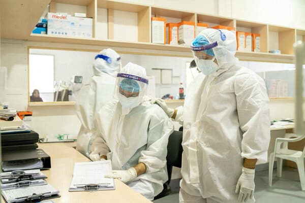
The island nation kept Covid cases low, and its resorts open, for much of the pandemic. But a recent surge exposed its overreliance on expatriate health workers.MALÉ, Maldives — The largest Covid-19 treatment facility in the Maldives has nearly 300 beds and a steady supply of oxygen. But as the country reported some of the world’s highest caseloads per capita last month, Covid wards ran low on another essential resource: employees.“At worst, we had one nurse to attend to 20 patients in the general wards,” said Mariya Saeed, the head of the Hulhumalé Medical Facility in Malé, the capital. “We needed human resources to provide proper care to the many bedridden elderly, but the nurses were exhausted.”The pandemic has triggered shortages of health workers around the world, forcing governments to scramble. Spain, for instance, launched an emergency plan last year to recruit medical students and retired doctors for Covid duty. And in India last month, Prime Minister Narendra Modi asked local officials to start recruiting final year medical students.But the Maldives, an archipelago of about 1,200 islands in the Indian Ocean, faces unique challenges. It can’t easily call up masses of students because it has only one university with a school of medicine. And it can’t rely just on its citizens, because its health care system depends largely on foreign workers. Many of those doctors and nurses come from India, a country that is facing its own gargantuan outbreak.The Hulhumalé Medical Facility, in Malé, shown in another image from the government, is turning to volunteers to help care for patients.Health Emergency Operations CenterOne result is that the Maldives, which has otherwise tackled the pandemic with meticulous attention to detail, isn’t sure how to staff its hospitals for the next crisis.“We have spoken to countries like Bangladesh and India” about recruiting their doctors and nurses, President Ibrahim Mohamed Solih told reporters last month. “But they are unable to provide any assistance due to their own Covid situations.”The Maldives, a Muslim-majority nation with a population of about 540,000, has styled itself a model of pandemic response for small countries. Using aggressive contact tracing, and relying on dispersed island geography to slow outbreaks, the government kept its Covid caseload low enough to lift curbs on domestic movements and lure international tourists back to its luxury resorts, a mainstay of the economy. In April it allowed Ramadan feasts and nationwide council elections to go ahead as usual.“You never know what will happen tomorrow,” Thoyyib Mohamed, the managing director of the country’s official public relations agency, told The New York Times in February. “But for the time being, I must say: This is a really good case study for the entire world, especially tropical destinations.”Though 59 percent of the population has received at least one dose of a Covid-19 vaccine, the recent surge took a heavy toll. Nearly half of the country’s 200 Covid-19 deaths during the entire pandemic were reported in May.Healthcare workers taking swab samples in Malé. The country’s health care system depends heavily on foreign doctors and nurses. Ahmed Shurau/Agence France-Presse — Getty ImagesMany people in Malé now have someone in their extended families who has died, said Marjan Montazemi, the Unicef representative to the Maldives. “Because the numbers are not the same as in other countries, it doesn’t attract that much attention,” she said. “But for the country it has been quite difficult.”Officials in the Maldives have not confirmed how variants may have influenced the latest outbreak, but local doctors say the Delta variant, first detected in neighboring India, likely played a role.As cases surged to more than 1,500 a day last month, hundreds of Covid-19 patients descended on the Hulhumalé Medical Facility. . Despite being built last year for the purpose of treating Covid patients, the facility — staffed with 16 doctors and 89 nurses — was not ready.“We were always prepared for a possible surge, but a wave this sudden and massive was just unexpected,” said Nazla Musthafa, a health adviser to the government.To compensate for a shortage of doctors and nurses, Maldives National University’s medical school — which opened in 2019 and has a total of 115 students — sent dozens of medical and nursing students to help work in Malé’s Covid wards. The government also called nurses out of retirement and drafted volunteers with no medical experience.Ms. Saeed, the head of the Hulhumalé Medical Facility, said that volunteers mostly helped patients go to the toilet, roll over in bed, maneuver wheelchairs and oxygen cylinders, and perform other basic functions. She said volunteers wore protective gear but that there was no time to screen them for Covid-19.President Solih, middle, shown in an image provided by his office, visited the Hulhumalé facility, where the government plans to build a ward with 270 more beds. The President’s Office Republic of MaldivesOne volunteer, Rizna Zareer, 35, said she mainly provided moral support to patients who were not allowed to receive visitors.“We were their family, and I saw them that way, too,” she said.The shortage of medical workers is so bad that lab technicians involved in contact tracing must work around the clock, a team of World Bank experts said in a statement.The bottleneck highlights a dependence on foreign health workers that the government knew was a problem even before the pandemic hit.In 2018, expatriates made up all but a fifth of the Maldives’s 900 or so doctors and more than half of its nearly 3,000 nurses, leading to high turnover that affects the quality of health care, a government report said.Other countries, including Ireland, Israel and New Zealand, also rely heavily on expatriates to work in health care. But unlike them, the Maldives isn’t rich. That means it can’t compete as aggressively to lure foreign doctors and nurses, especially during a pandemic that has left virtually every nation’s health work force short-handed.The President’s Office Republic Of MaldivesS. Irudaya Rajan, the chairman of the International Institute of Migration and Development, a research organization based in southern India, said that he expected countries that send large numbers of health workers abroad, including India and the Philippines, to tweak policies to keep more workers at home.The Maldives needs a better strategy for securing a steadier supply of expatriate doctors and nurses, Mr. Rajan said. One option would be to sponsor Indian medical students in India and require them to work in the Maldives for a few years after graduating, he said.“One lesson every country should learn from Covid-19 is: Don’t exploit poor countries like India and the Philippines,” Mr. Rajan said. “Invest in them and their people, and they can benefit you.”A spokesman for President Solih of the Maldives did not respond to requests for comment.The daily average of new cases in the Maldives is now about 260, or less than a quarter what it was during the peak last month. But as of Friday, the country still had about 21,000 active cases, and a 12-hour curfew implemented in Malé last month remained in effect. The call to prayer still rings out five times a day from the city’s mosques, but worshipers are allowed inside only in small numbers.The government recently announced a plan to build an additional 270-bed ward at the Hulhumalé Medical Facility to deal with future outbreaks and boost the country’s total bed capacity for Covid patients from 460 to 730. The main challenge for the new ward may be figuring out how to staff it.Mr. Solih told reporters last month that his health minister, Ahmed Naseem, hoped to recruit 40 doctors and 100 nurses from India and Bangladesh by the end of June. But at the same news conference, Mr. Naseem tried to lower expectations.“Currently it is difficult to employ people from India, Bangladesh and Sri Lanka,” he said. “Sri Lanka, especially, is near impossible. I have been trying for many days.”An Air India flight carrying doses of the Covishield vaccine, made in India, is seen after its arrival in Malé in January.Agence France-Presse — Getty ImagesMaahil Mohamed reported from Malé, the Maldives, and Mike Ives from Hong Kong.

Read more →

Parents and caregivers reported mental health issues more often than others during the pandemic, a C.D.C. study says.

Parents and unpaid caregivers of adults in the United States reported far higher rates of mental health issues during the coronavirus pandemic than people who held neither of those roles, federal researchers reported on Thursday.About 70 percent of parents and adult caregivers — such as those tending to older people, for example — and about 85 percent of people who were both reported adverse mental health symptoms during the pandemic, versus about a third of people who did not hold those responsibilities, according to new research by the Centers for Disease Control and Prevention.The study also found that people who were both parent and caregivers were eight times more likely to have seriously considered suicide than people who held neither role.“These findings highlight that parents and caregivers, especially those balancing roles both as parents and caregivers, experienced higher levels of adverse mental health symptoms during the Covid-19 pandemic than adults without these responsibilities,” the authors said.“Caregivers who had someone to rely on for support had lower odds of experiencing any adverse mental health symptoms,” they said.The report follows innumerable anecdotes and several studies suggesting spikes in mental health problems among parents and caregivers during the pandemic. But the new C.D.C. report noted that “without prepandemic mental health data in this sample, whether adverse mental health symptoms were caused by or worsened by the pandemic is unknown.”The study is based on data from online English-language surveys administered to panels of U.S. residents run by Qualtrics, a company that conducts commercial surveys, for the Covid-19 Outbreak Public Evaluation Initiative, an effort to track American attitudes and behaviors during the pandemic. The data was gathered from Dec. 6 to 27 last year, and from Feb. 16 to March 8 of this year, and relied on 10,444 respondents, weighted to match U.S. demographic data, 42 percent of whom identified as parents or adult caregivers.The study noted that the results might not fully represent the U.S. population, because of factors like the surveys only being presented online and in English.The surveys included screening items for depression, anxiety, Covid-19 trauma and stress-related disorders, and asked respondents whether they had experienced suicidal thinking in the past month. About half of the parent-caregivers who responded said that they had recently had suicidal thoughts.Elizabeth A. Rohan, a health scientist at the C.D.C. and one of the study’s authors, said in an interview that what set this research apart was a large sample size and a broad definition of caregiver, which allowed for a more inclusive picture of people in that role.“Our net captured more people than other surveys,” Dr. Rohan said.Dr. Rohan said that the study reinforced the need to destigmatize mental health issues among caregivers and for better support systems. Communication is key, she said, and “it doesn’t have to be professional help.”She added, “We cannot underestimate the importance of staying connected to one another,” which is helpful whether the person is “a trusted friend, a family member or a professional.”If you are having thoughts of suicide, call the National Suicide Prevention Lifeline at 1-800-273-8255 (TALK). You can find a list of additional resources at SpeakingOfSuicide.com/resources.

Read more →

Blood cancer patients with COVID-19 fare better with convalescent plasma

A large, retrospective, multicenter study involving Washington University School of Medicine in St. Louis indicates that convalescent plasma from recovered COVID-19 patients can dramatically improve likelihood of survival among blood cancer patients hospitalized with the virus.
The therapy involves transfusing plasma — the pale yellow liquid in blood that is rich in antibodies — from people who have recovered from COVID-19 into patients who have leukemia, lymphoma or other blood cancers and are hospitalized with the viral infection. The goal is to accelerate their disease-fighting response. Cancer patients may be at a higher risk of death related to COVID-19 because of their weakened immune systems.
The data, collected as part of a national registry, indicate that patients who received convalescent plasma from donors who had recovered from COVID-19 had a death rate of 13.3% compared with 24.8% for those who did not receive it.
The difference was especially striking among severely ill patients admitted to intensive care units (ICUs). Such patients treated with convalescent plasma had a death rate of 15.8% compared with 46.9% for those who didn’t receive the treatment.
“These results suggest that convalescent plasma may not only help COVID-19 patients with blood cancers whose immune systems are compromised, it may also help patients with other illnesses who have weakened antibody responses to this virus or to the vaccines,” said Jeffrey P. Henderson, MD, PhD, an associate professor of medicine and of molecular microbiology at Washington University. “The data also emphasize the value of an antibody therapy such as convalescent plasma as a virus-directed treatment option for hospitalized COVID-19 patients.”
The research is published June 17 in the journal JAMA Oncology.

Read more →

Comprehensive RNA-Atlas

By cleverly combining complementary sequencing techniques, researchers have deepened our understanding of the function of known RNA molecules and discovered thousands of new RNAs. A better understanding of our transcriptome is essential to better understand disease processes and uncover novel genes that may serve as therapeutic targets or biomarkers.
The article ‘The RNA Atlas expands the catalog of human non-coding RNAs’, published today in Nature Biotechnology, is the result of more than five years of hard work to further unravel the complexity of the human transcriptome. Never before such a comprehensive effort was undertaken to characterize all RNA-molecules in human cells and tissues.
RNAs in all shapes and sizes
Our transcriptome is — analogous to our genome — the sum of all RNA molecules that are transcribed from the DNA strands that make up our genome. However, there’s no 1-on-1 relationship with the latter. Firstly, each cell and tissue hasve a unique transcriptomes, with varying RNA production and compositions, including tissue-specific RNAs. Secondly, not all RNAs are transcribed from typical — protein coding — genes that eventually produce proteins. Many of our RNA molecules are not used as a template to build proteins, but originate from what once was called junk DNA: long sequences of DNA with unknown functions.
These non-coding RNAs (ncRNAs) come in all kinds of shapes and sizes: short, long, and even circular RNAs. Many of them even lack the tail of adenine-molecules that is typical for protein-coding RNAs.
300 human cell and tissue types and three sequencing methods
“There have been other projects to catalogue our transcriptome but the RNA-Atlas project is unique because of the applied sequencing methods,” says prof. Pieter Mestdagh from the Center for Medical Genetics at Ghent University. “Not only did we look at the transcriptome of as many as 300 human cell and tissue types, but most importantly, we did so with three complementary sequencing technologies, one aimed at small RNAs, one aimed at polyadenylated (polyA) RNAs and a technique called total RNA sequencing.”
This last sequencing technology led to the discovery of thousands of novel non-coding RNA genes, including a novel class of non-polyadenylated single exon genes and many new circular RNAs. By combining and comparing the results of the different sequencing methods the researchers were able to define for every measured RNA transcript, the abundance in the different cells and tissues, whether it has a polyA-tail or not (it appears that for some genes this can differ from cell type to cell type), and whether it is linear of circular. Moreover, the consortium searched and found important clues in determining the function of some of the ncRNAs. By looking at the abundancy of different RNA’s in different cell types they found correlations that indicate regulatory functions, and could determine whether this regulation happens on the transcription level (by preventing or stimulating transcription of protein coding genes) or post-transcriptional (e.g. by breaking down RNAs).
An invaluable resource for biomedical science
All data, analyses and results (equivalent to a few libraries of information) are available for download and interrogation in the R2 web portal, enabling the community to implement this resource as a tool for exploration of non-coding RNA biology and function.
Prof. Pavel Sumazin of the Baylor College of Medicine: “By combining all data in one comprehensive catalogue, we have created a new valuable resource for biomedical scientists around the world studying disease processes. A better understanding of the complexity of the transcriptome is indeed essential to better understand disease processes and uncover novel genes that may serve as therapeutic targets or biomarkers. The age of RNA therapeutics is swiftly rising — we’ve all witnessed the impressive creation of RNA vaccines, and already the first medicines that target RNA are used in the clinic. I’m sure we’ll see lots more of these therapies in the next years and decades.”
Story Source:
Materials provided by Ghent University. Note: Content may be edited for style and length.

Read more →

Beneficial bacteria can be restored to C-section babies at birth

Babies born by cesarean section don’t have the same healthy bacteria as those born vaginally, but a Rutgers-led study for the first time finds that these natural bacteria can be restored.
The study appears in the journal Med.
The human microbiota consists of trillions of bacteria, viruses, fungi and other microorganisms — some beneficial, some harmful — that live in and on our bodies. Women naturally provide these pioneer colonizers to their babies’ sterile bodies during labor and birth, helping their immune system to develop. But antibiotics and C-sections disturb this passing of microbes and are related to increased risks of obesity, asthma and metabolic diseases.
The researchers followed 177 babies from four countries over the first year of their lives — 98 were born vaginally and 79 were born by C-section, 30 of which were swabbed with a maternal vaginal gauze right after birth.
Lab analysis showed that the microbiota of the C-section babies swabbed with their mother’s vaginal fluids was close to that of vaginally born babies. Also, the mother’s vaginal microbiomes on the day of birth were similar to other areas of their bodies (gut, mouth and skin), showing that maternal vaginal fluids help to colonize bacteria across their babies’ bodies.
This was the first large observational study to show that restoring a C-section baby’s natural exposure to maternal vaginal microbes at birth normalizes the microbiome development during their first year of life. The researchers said the next step is conducting randomized clinical trials to determine if the microbiota normalization translates into disease protection.
“Further research is needed to determine which bacteria protect against obesity, asthma and allergies, diseases with underlying inflammation,” said senior author Maria Gloria Dominguez Bello, a professor in the Department of Biochemistry and Microbiology in the School of Environmental and Biological Sciences at Rutgers University-New Brunswick. “Our results support the hypothesis that acquiring maternal vaginal microbes normalizes microbiome development in the babies.”
According to the World Health Organization, C-section is needed in about 15 percent of births to avoid risking the life of the mother or child, but in many countries such as in Brazil, the Dominican Republic, Iran and China, C-section is performed in more than 70% of urban births.
Story Source:
Materials provided by Rutgers University. Note: Content may be edited for style and length.

Read more →

Thin, stretchable biosensors could make surgery safer

A research team from Los Alamos National Laboratory and Purdue University have developed bio-inks for biosensors that could help localize critical regions in tissues and organs during surgical operations.
“The ink used in the biosensors is biocompatible and provides a user-friendly design with excellent workable time frames of more than one day,” said Kwan-Soo Lee, of Los Alamos’ Chemical Diagnostics and Engineering group.
The new biosensors allow for simultaneous recording and imaging of tissues and organs during surgical procedures.
“Simultaneous recording and imaging could be useful during heart surgery in localizing critical regions and guiding surgical interventions such as a procedure for restoring normal heart rhythms,” said Chi Hwan Lee, the Leslie A. Geddes Assistant Professor of Biomedical Engineering and Assistant Professor of Mechanical Engineering and, by courtesy, of Materials Engineering at Purdue University.
Los Alamos was responsible for formulating and synthesizing the bio-inks, with the goal of creating create an ultra-soft, thin and stretchable material for biosensors that is capable of seamlessly interfacing with the surface of organs. They did this using 3D-printing techniques.
“Silicone materials are liquid and flow like honey, which is why it is very challenging to 3D-print without sagging and flowing issues during printing,” Kwan-Soo Lee said. “It is very exciting to have found a way to create printed inks that do not have any shape deformation during the curing process.”
The bio-inks are softer than tissue, stretch without experiencing sensor degradation, and have reliable natural adhesion to the wet surface of organs without needing additional adhesives.
Craig Goergen, the Leslie A. Geddes Associate Professor of Biomedical Engineering at Purdue University, aided with the in vivo assessment of the patch via testing in both mice and pigs. The results showed the biosensor was able to reliably measure electrical signal while not impairing cardiac function.
The research was published today in Nature Communications. It was funded by Science Campaign 2.
Story Source:
Materials provided by DOE/Los Alamos National Laboratory. Note: Content may be edited for style and length.

Read more →

'Nanodecoy' therapy binds and neutralizes SARS-CoV-2 virus

Nanodecoys made from human lung spheroid cells (LSCs) can bind to and neutralize SARS-CoV-2, promoting viral clearance and reducing lung injury in a macaque model of COVID-19. By mimicking the receptor that the virus binds to rather than targeting the virus itself, nanodecoy therapy could remain effective against emerging variants of the virus.
SARS-CoV-2 enters a cell when its spike protein binds to the angiotensin-converting enzyme 2 (ACE2) receptor on the cell’s surface. LSCs — a natural mixture of lung epithelial stem cells and mesenchymal cells — also express ACE2, making them a perfect vehicle for tricking the virus.
“If you think of the spike protein as a key and the cell’s ACE2 receptor as a lock, then what we are doing with the nanodecoys is overwhelming the virus with fake locks so that it cannot find the ones that let it enter lung cells,” says Ke Cheng, corresponding author of the research. “The fake locks bind and trap the virus, preventing it from infecting cells and replicating, and the body’s immune system takes care of the rest.”
Cheng is the Randall B. Terry Jr. Distinguished Professor in Regenerative Medicine at North Carolina State University and a professor in the NC State/UNC-Chapel Hill Joint Department of Biomedical Engineering.
Cheng and colleagues from NC State and UNC-CH converted individual LSCs into nanovesicles, or tiny cell membrane bubbles with ACE2 receptors and other lung cell-specific proteins on the surface.
They confirmed that the spike protein did bind to the ACE2 receptors on the decoys in vitro, then used a fabricated SARS-Co-V-2 mimic virus for in vivo testing in a mouse model. The decoys were delivered via inhalation therapy. In mice, the nanodecoys remained in the lungs for 72 hours after one dose and accelerated clearance of the mimic virus.
Finally, a contract research organization conducted a pilot study in a macaque model and found that inhalation therapy with the nanodecoys accelerated viral clearance, and reduced inflammation and fibrosis in the lungs. Although no toxicity was noted in either the mouse or macaque study, further study will be necessary to translate this therapy for human testing and determine exactly how the nanodecoys are cleared by the body.
“These nanodecoys are essentially cell ‘ghosts,’ and one LSC can generate around 11,000 of them,” Cheng says. “Deploying millions of these decoys exponentially increases the surface area of fake binding sites for trapping the virus, and their small size basically turns them into little bite-sized snacks for macrophages, so they are cleared very efficiently.”
The researchers point out three other benefits of the LSC nanodecoys. First, they can be delivered non-invasively to the lungs via inhalation therapy. Second, since the nanodecoys are acellular — there’s nothing living inside — they can be easily preserved and remain stable longer, enabling off-the-shelf use. Finally, LSCs are already in use in other clinical trials, so there is an increased likelihood of being able to use them in the near future.
“By focusing on the body’s defenses rather than a virus that will keep mutating we have the potential to create a therapy that will be useful long-term,” Cheng says. “As long as the virus needs to enter the lung cell, we can keep tricking it.”
The research appears in Nature Nanotechnology and was supported by the National Institutes of Health and the American Heart Association. Dr. Jason Lobo, pulmonologist at UNC-CH, is co-author of the paper.
Story Source:
Materials provided by North Carolina State University. Original written by Tracey Peake. Note: Content may be edited for style and length.

Read more →

Unraveling the origin of Alzheimer's disease

Case Western Reserve University researchers studying prions — misfolded proteins that cause lethal incurable diseases — have identified for the first time surface features of human prions responsible for their replication in the brain.
The ultimate goal of the research is to help design a strategy to stop prion disease in humans — and, ultimately, to translate new approaches to work on Alzheimer’s and other neurodegenerative diseases.
Scientists have yet to discover the exact cause of Alzheimer’s disease, but largely agree that protein issues play a role in its emergence and progression. Alzheimer’s disease afflicts more than 6 million people in the U.S., and the Alzheimer’s Association estimates that their care will cost an estimated $355 billion this year.
Research was done at the Safar Laboratory in the Department of Pathology and the Center for Proteomics and Bioinformatics at Case Western Reserve University School of Medicine, and at Case Western Reserve’s Center for Synchrotron Bioscience at Brookhaven Laboratories in New York. Jiri Safar, professor of pathology, neurology and neurosciences at the Case Western Reserve School of Medicine, leads the work. The report, “Structurally distinct external domains drive replication of major human prions,” was published in the June 17 issue of PLOS Pathogens.
Prions were first discovered in the late 1980s as a protein-containing biological agent that could replicate itself in living cells without nucleic acid. The public health impact of medically transmitted human prion diseases — and also animal transmissions of bovine spongiform encephalopathy (BSE, “mad cow disease”) prions — dramatically accelerated the development of a new scientific concept of self-replicating protein.
Human prions can bind to neighboring normal proteins in the brain, and cause microscopic holes. In essence, they turn brains into sponge-like structures and lead to dementia and death. These discoveries led to the ongoing scientific debate on whether prion-like mechanisms may be involved in the origin and spread of other neurodegenerative disorders in humans.
“Human prion diseases are conceivably the most heterogenous neurodegenerative disorders, and a growing body of research indicates that they are caused by distinct strains of human prions,” Safar said. “However, the structural studies of human prions have lagged behind the recent progress in rodent laboratory prions, in part because of their complex molecular characteristics and prohibitive biosafety requirements necessary for investigating disease which is invariably fatal and has no treatment.”
The researchers developed a new three-step process to study human prions: Human brain-derived prions were first exposed to a high-intensity synchrotron X-ray beam. That beam created hydroxyl radical species which, with short bursts of light, selectively and progressively changed the prion’s surface chemical composition. The unique properties of this type of light source include its enormous intensity; it can be millions of times brighter than light from the sun to the Earth. The rapid chemical modifications of prions by short bursts of light were monitored with anti-prion antibodies. The antibodies recognize the prion surface features, and mass spectrometry that identifies exact sites of prion-specific, strain-based differences, providing an even more precise description of the prion’s defects. Illuminated prions were then allowed to replicate in a test tube. The progressive loss of their replication activity as the synchrotron modifies them helped identify key structural elements responsible for prions’ replication and propagation in the brain.”The work is a critical first step for identifying sites of structural importance that reflect differences between prions of different diagnosis and aggressiveness,” said Mark Chance, vice dean for research at the School of Medicine and a co-investigator on the work. “Thus, we can now envision designing small molecules to bind to these sites of nucleation and replication and block progression of human prion disease in patients.”
This structural approach, Chance said, also provides a template for how to identify structurally important sites on misfolded proteins in other diseases such as Alzheimer’s, which involves protein propagation from cell to cell in a similar way to prions.
Story Source:
Materials provided by Case Western Reserve University. Note: Content may be edited for style and length.

Read more →

Yellow fever mosquitoes evolve different strategies to resist pesticides

The Yellow fever mosquito (scientific name, Aedes aegypti) spreads multiple untreatable viruses in humans and is primarily controlled using a pesticide called permethrin. However, many mosquitoes are evolving resistance to the pesticide. A new study by Karla Saavedra-Rodriguez of Colorado State University and colleagues, published in the journal PLOS Genetics, identifies mutations linked to different permethrin resistance strategies, which threaten our ability to control disease outbreaks.
When treated mosquitoes encounter permethrin in the wild, they will do one of the following: immediately die, be knocked out but recover, or be unaffected. Saavedra-Rodriguez and her colleagues decided to investigate the genetic variations that lead to these three responses to the pesticide. The team collected mosquitoes from a permethrin-resistant colony in Tapachula, Mexico, exposed them to the pesticide in the lab and then sequenced the genomes from each group.
When they compared the genomes from the three types of mosquitos, they found that unaffected mosquitoes primarily had mutations in VGSC, a gene for a cellular sodium channel known to be involved in pesticide resistance. To a lesser extent, these mosquitoes also carried mutations in detoxification genes and cuticle protein genes. The cuticle makes up the insects’ tough outer shell and can slow the pesticide as it enters the body. Insects that recovered from exposure had mutations in cuticle protein genes and in a different group of detoxification genes. The results show that there are distinct genetic changes behind these two types of permethrin resistance.
The results of the new study will help scientists to track mutations linked to resistance in populations of the Yellow fever mosquito from Southeastern Mexico. This knowledge can help scientists understand how mosquitoes have evolved resistance and when a population can no longer be controlled with permethrin. This understanding will be necessary to develop tools to support future insecticide management strategies.
Story Source:
Materials provided by PLOS. Note: Content may be edited for style and length.

Read more →

First months decisive for immune system development

Many diseases caused by a dysregulated immune system, such as allergies, asthma and autoimmunity, can be traced back to events in the first few months after birth. To date, the mechanisms behind the development of the immune system have not been fully understood. Now, researchers at Karolinska Institutet show a connection between breast milk, beneficial gut bacteria and the development of the immune system. The study is published in Cell.
“A possible application of our results is a preventative method for reducing the risk of allergies, asthma and autoimmune disease later in life by helping the immune system to establish its regulatory mechanisms,” says the paper’s last author Petter Brodin, paediatrician and researcher at the Department of Women’s and Children’s Health, Karolinska Institutet. “We also believe that certain mechanisms that the study identifies can eventually lead to other types of treatment for such diseases, not just a prophylactic.”
The incidence of autoimmune diseases such as asthma, type 1 diabetes and Crohn’s disease is increasing in children and adolescents in parts of the world. These diseases are debilitating, but not as common in low-income countries as they are in Europe and the USA.
It has long been known that the risk of developing these diseases is largely determined by early life events; for instance, there is a correlation between the early use of antibiotics and a higher risk of asthma. It is also known that breastfeeding protects against most of these disorders.
There is a link between specific, protective bacteria on the skin and in the airways and gut and a lower risk of immunological diseases. However, there is still much to learn about how these bacteria form the immune system.
Researchers at Karolinska Institutet, Evolve Biosystems, Inc, the University of California Davis, University of Nebraska, Lincoln, and University of Nevada, Reno studied how the neonatal immune system adapts to and is shaped by the many bacteria, viruses, nutrients and other environmental factors to which the baby is exposed during the first few months of life.

Read more →