Stephen Karanja: Kenyan anti-vaccine doctor dies from Covid-19

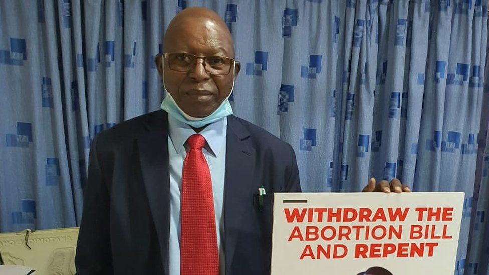
SharecloseShare pageCopy linkAbout sharingimage copyrightCitizenGOA Kenyan doctor who became a vociferous opponent of Covid-19 vaccines has succumbed to the virus, weeks after saying the jabs were “totally unnecessary”. Dr Stephen Karanja, chairman of the Kenya Catholic Doctors Association, advocated steam inhalation and hydroxychloroquine tablets. He clashed with the Catholic church over the safety of Covid jabs.Health authorities and the World Health Organization (WHO) rejected his claims. “[The vaccine] being distributed in Kenya, has been reviewed and found safe not only by the WHO rigorous process but also by several stringent regulatory authorities,” the WHO said in March.Africa Live: Latest updates from across the continentKenyan nurse: ‘I was shunned over coronavirus fears’The women fighting South Africa’s ‘infodemic’The Kenya Conference of Catholic Bishops also distanced itself from Dr Karanja’s view on Covid-19 vaccines, saying the vaccines were “licit and ethically acceptable.” Kenya received just over a million vaccine doses from the global Covax initiative, most of which have been administered. The country has confirmed more than 160,000 cases and 2,707 deaths. In March, the government imposed another lockdown restricting movement in five counties after a surge in new infections.What did Dr Karanja say about Covid vaccines?In a letter dated 3 March Dr Karanja said that “there are drugs that have been repurposed and used effectively to treat Covid-19,” adding that “we also know that vaccination for this disease is totally unnecessary making the motivation suspect.”He went on in different forums to advocate alternative treatments, including steam inhalation and a cocktail of drugs – including hydroxychloroquine and Ivermectin, which have not been approved by the WHO to treat Covid-19.Covid-19 and hydroxychloroquine: What do we know?The South African debate over IvermectinDr Karanja, who was an obstetrician and gynaecologist, died on Thursday a week after he was admitted to hospital suffering from complications caused by a Covid-19 infection.What else has Dr Karanja said?Before falling out with the Catholic church in Kenya about the safety and efficacy of the Covid-19 vaccine, Dr Karanja often allied with the religious leaders to oppose mass vaccination campaigns. In 2019 he led opposition against vaccination of schoolgirls against cervical cancer, saying the jab against Human Papilloma Virus (HPV) was unnecessary because it affected those “whose lifestyle involves irresponsible sexual behaviours”. In 2014, his association opposed the government’s rollout of a tetanus vaccine targeting women, claiming it was a sterilisation campaign, despite local health authorities, the WHO, and the UN children’s agency Unicef saying the vaccine was safe.In both instances the government carried on with its plans, but officials reported that they encountered vaccine hesitancy as a result of the objections raised by Dr Karanja.He was also a prominent anti-abortion campaigner and appeared in court in 2018 as an expert witness in a case in which the government was sued for withdrawing guidelines on abortion. The high court ruled that the government decision was unlawful and illegal. Though shunned by a majority of health professionals in Kenya, the Catholic church recognised his association, but often hastened to add that Dr Karanja did not speak for the Catholic church.”The mandate of the church is to speak on matters of morality and faith. The mandate of the doctors is to speak on their understanding of their scientific practice. We are not at variance,” Father Ferdinand Lugonzo, the Kenya Conference of Catholic Bishops spokesperson, told the BBC.A SIMPLE GUIDE: How do I protect myself?IMPACT: What the virus does to the bodyRECOVERY: How long does it take?

Read more →

Engineering T cells to attack cancer broadly

Through T cell engineering, researchers at Virginia Commonwealth University Massey Cancer Center show that it’s possible to arrest tumor growth for a variety of cancers and squash the spread of cancer to other tissues. This research will be published in tomorrow’s print edition of Cancer Research.
The paper builds on decades of research by study co-senior author Paul B. Fisher, M.Ph., Ph.D., a member of Massey’s Cancer Biology research program, who discovered a protein called IL-24 that attacks a variety of cancers in several different ways.
In this latest study, Fisher teamed up with his colleague Xiang-Yang (Shawn) Wang, Ph.D., who co-leads the Developmental Therapeutics research program at Massey, to deliver the gene coding for IL-24, which is called MDA-7, to solid tumors using T cells.
“I think the beauty of what we’ve been involved in is that it expands the scope of immunotherapy,” said Fisher, professor and chair of the Department of Human and Molecular Genetics at the VCU School of Medicine, director of the VCU Institute of Molecular Medicine (VIMM) and Thelma Newmeyer Corman Endowed Chair in Oncology Research. “Our approach is less dependent on cancer cells expressing something specific to target.”
After all, this isn’t the first time T cells have been engineered for cancer immunotherapy. FDA-approved chimeric antigen receptor T (CAR-T) cell therapy — which is designed to destroy cancer cells expressing specific surface molecules — has shown tremendous success for treating advanced cancers of the blood and lymphatic systems.
But CAR-T has made limited progress on solid tumors, such as prostate cancer or melanoma, because the cells that make up those tumors aren’t all the same, which blocks the engineered T cells from recognizing and attacking.

Read more →

Move over CRISPR, the Retrons are coming

Researchers have created a new gene editing tool called Retron Library Recombineering (RLR) that can generate up to millions of mutations simultaneously, and ‘barcodes’ mutant bacterial cells so that the entire pool can be screened at once. It can be used in contexts where CRISPR is toxic or not feasible, and results in better editing rates.
While the CRISPR-Cas9 gene editing system has become the poster child for innovation in synthetic biology, it has some major limitations. CRISPR-Cas9 can be programmed to find and cut specific pieces of DNA, but editing the DNA to create desired mutations requires tricking the cell into using a new piece of DNA to repair the break. This bait-and-switch can be complicated to orchestrate, and can even be toxic to cells because Cas9 often cuts unintended, off-target sites as well.
Alternative gene editing techniques called recombineering instead perform this bait-and-switch by introducing an alternate piece of DNA while a cell is replicating its genome, efficiently creating genetic mutations without breaking DNA. These methods are simple enough that they can be used in many cells at once to create complex pools of mutations for researchers to study. Figuring out what the effects of those mutations are, however, requires that each mutant be isolated, sequenced, and characterized: a time-consuming and impractical task.
Researchers at the Wyss Institute for Biologically Inspired Engineering at Harvard University and Harvard Medical School (HMS) have created a new gene editing tool called Retron Library Recombineering (RLR) that makes this task easier. RLR generates up to millions of mutations simultaneously, and “barcodes” mutant cells so that the entire pool can be screened at once, enabling massive amounts of data to be easily generated and analyzed. The achievement, which has been accomplished in bacterial cells, is described in a recent paper in PNAS.
“RLR enabled us to do something that’s impossible to do with CRISPR: we randomly chopped up a bacterial genome, turned those genetic fragments into single-stranded DNA in situ, and used them to screen millions of sequences simultaneously,” said co-first author Max Schubert, Ph.D., a postdoc in the lab of Wyss Core Faculty member George Church, Ph.D. “RLR is a simpler, more flexible gene editing tool that can be used for highly multiplexed experiments, which eliminates the toxicity often observed with CRISPR and improves researchers’ ability to explore mutations at the genome level.”
Retrons: from enigma to engineering tool
Retrons are segments of bacterial DNA that undergo reverse transcription to produce fragments of single-stranded DNA (ssDNA). Retrons’ existence has been known for decades, but the function of the ssDNA they produce flummoxed scientists from the 1980s until June 2020, when a team finally figured out that retron ssDNA detects whether a virus has infected the cell, forming part of the bacterial immune system.

Read more →

Novel coronavirus' spike protein plays additional key role in illness

Scientists have known for a while that SARS-CoV-2’s distinctive “spike” proteins help the virus infect its host by latching on to healthy cells. Now, a major new study shows that they also play a key role in the disease itself.
The paper, published on April 30, 2021, in Circulation Research, also shows conclusively that COVID-19 is a vascular disease, demonstrating exactly how the SARS-CoV-2 virus damages and attacks the vascular system on a cellular level. The findings help explain COVID-19’s wide variety of seemingly unconnected complications, and could open the door for new research into more effective therapies.
“A lot of people think of it as a respiratory disease, but it’s really a vascular disease,” says Assistant Research Professor Uri Manor, who is co-senior author of the study. “That could explain why some people have strokes, and why some people have issues in other parts of the body. The commonality between them is that they all have vascular underpinnings.”
Salk researchers collaborated with scientists at the University of California San Diego on the paper, including co-first author Jiao Zhang and co-senior author John Shyy, among others.
While the findings themselves aren’t entirely a surprise, the paper provides clear confirmation and a detailed explanation of the mechanism through which the protein damages vascular cells for the first time. There’s been a growing consensus that SARS-CoV-2 affects the vascular system, but exactly how it did so was not understood. Similarly, scientists studying other coronaviruses have long suspected that the spike protein contributed to damaging vascular endothelial cells, but this is the first time the process has been documented.
In the new study, the researchers created a “pseudovirus” that was surrounded by SARS-CoV-2 classic crown of spike proteins, but did not contain any actual virus. Exposure to this pseudovirus resulted in damage to the lungs and arteries of an animal model — proving that the spike protein alone was enough to cause disease. Tissue samples showed inflammation in endothelial cells lining the pulmonary artery walls.
The team then replicated this process in the lab, exposing healthy endothelial cells (which line arteries) to the spike protein. They showed that the spike protein damaged the cells by binding ACE2. This binding disrupted ACE2’s molecular signaling to mitochondria (organelles that generate energy for cells), causing the mitochondria to become damaged and fragmented.
Previous studies have shown a similar effect when cells were exposed to the SARS-CoV-2 virus, but this is the first study to show that the damage occurs when cells are exposed to the spike protein on its own.
“If you remove the replicating capabilities of the virus, it still has a major damaging effect on the vascular cells, simply by virtue of its ability to bind to this ACE2 receptor, the S protein receptor, now famous thanks to COVID,” Manor explains. “Further studies with mutant spike proteins will also provide new insight towards the infectivity and severity of mutant SARS CoV-2 viruses.”
The researchers next hope to take a closer look at the mechanism by which the disrupted ACE2 protein damages mitochondria and causes them to change shape.
Other authors on the study are Yuyang Lei and Zu-Yi Yuan of Jiaotong University in Xi’an, China; Cara R. Schiavon, Leonardo Andrade, and Gerald S. Shadel of Salk; Ming He, Hui Shen, Yichi Zhang, Yoshitake Cho, Mark Hepokoski, Jason X.-J. Yuan, Atul Malhotra, Jin Zhang of the University of California San Diego; Lili Chen, Qian Yin, Ting Lei, Hongliang Wang and Shengpeng Wang of Xi’an Jiatong University Health Science Center in Xi’an, China.
The research was supported by the National Institutes of Health, the National Natural Science Foundation of China, the Shaanxi Natural Science Fund, the National Key Research and Development Program, the First Affiliated Hospital of Xi’an Jiaotong University; and Xi’an Jiaotong University.

Read more →

Fiber-optic ultrasonic imaging probe for future nanoscale disease diagnostics

Scientists at the University of Nottingham have developed an ultrasonic imaging system, which can be deployed on the tip of a hair-thin optical fibre, and will be insertable into the human body to visualise cell abnormalities in 3D.
The new technology produces microscopic and nanoscopic resolution images that will one day help clinicians to examine cells inhabiting hard-to-reach parts of the body, such as the gastrointestinal tract, and offer more effective diagnoses for diseases ranging from gastric cancer to bacterial meningitis.
The high level of performance the technology delivers is currently only possible in state-of-the-art research labs with large, scientific instruments — whereas this compact system has the potential to bring it into clinical settings to improve patient care.
The Engineering and Physical Sciences Research Council (EPSRC)-funded innovation also reduces the need for conventional fluorescent labels — chemicals used to examine cell biology under a microscope — which can be harmful to human cells in large doses.
The findings are being reported in a new paper, entitled ‘Phonon imaging in 3D with a fibre probe’ published in the Nature journal, Light: Science & Applications.
Paper author, Salvatore La Cavera, an EPSRC Doctoral Prize Fellow from the University of Nottingham Optics and Photonics Research Group, said of the ultrasonic imaging system: “We believe its ability to measure the stiffness of a specimen, its bio-compatibility, and its endoscopic-potential, all while accessing the nanoscale, are what set it apart. These features set the technology up for future measurements inside the body; towards the ultimate goal of minimally invasive point-of-care diagnostics.”
Currently at prototype stage, the non-invasive imaging tool, described by the researchers as a “phonon probe,” is capable of being inserted into a standard optical endoscope, which is a thin tube with a powerful light and camera at the end that is navigated into the body to find, analyse, and operate on cancerous lesions, among many other diseases. Combining optical and phonon technologies could be advantageous; speeding up the clinical workflow process and reducing the number of invasive test procedures for patients.

Read more →

Shortage of DNA building blocks in the cell releases mitochondrial DNA

Mitochondria are the energy suppliers of our body cells. These tiny cell components have their own genetic material, which triggers an inflammatory response when released into the interior of the cell. The reasons for the release are not yet known, but some cardiac and neurodegenerative diseases as well as the ageing process are linked to the mitochondrial genome. Researchers at the Max Planck Institute for Biology of Ageing and the CECAD Cluster of Excellence in Ageing research have investigated the reasons for the release of mitochondrial genetic material and found a direct link to cellular metabolism: when the cell’s DNA building blocks are in short supply, mitochondria release their genetic material and trigger inflammation. The researchers hope to find new therapeutic approaches by influencing this metabolic pathway.
Our body needs energy — for every metabolic process, every movement and for breathing. This energy is produced in tiny components of our body cells, the so-called mitochondria. Unlike other cell components, mitochondria have their own genetic material, mitochondrial DNA. However, in certain situations, mitochondria release their DNA into the interior of the cell, causing a reaction from the cell’s own immune system and being associated with various diseases as well as the ageing process. The reasons for the release of mitochondrial DNA are not yet known.
Shortage of DNA building blocks triggers inflammatory reaction
To answer the question of when mitochondria release their DNA, researchers at the Max Planck Institute for Biology of Ageing have focused on the mitochondrial protein YME1L, which owes its name to yeast mutants that release their mitochondrial DNA — yeast mitochondrial escape 1. “In cells lacking YME1L, we observed the release of mitochondrial DNA into the cell interior and a related immune response in the cells,” said Thomas MacVicar, one of the study’s two first authors. Closer examination revealed a direct link to the building blocks of DNA. “If the cells lack YME1L, there is a deficiency of DNA building blocks inside the cell,” Thomas MacVicar describes. “This deficiency triggers the release of mitochondrial DNA, which in turn causes an inflammatory response in the cell: the cell stimulates similar inflammatory reactions as it does during a bacterial or viral infection. If we add DNA building blocks to the cells from the outside, that also stops the inflammation.”
New therapeutic approaches based on the metabolism of DNA building blocks
The discovered link between the cellular inflammatory response and the metabolism of DNA building blocks could have far-reaching consequences, explains Thomas MacVicar: “Some viral inhibitors stop the production of certain DNA building blocks, thereby triggering an inflammatory response. The release of mitochondrial DNA could be a crucial factor in this, contributing to the effect of these inhibitors.” Several ageing-associated inflammatory diseases, including cardiac and neurodegenerative diseases, as well as obesity and cancer, are linked to mitochondrial DNA. The authors hope that modulating the metabolism of DNA building blocks will offer new therapeutic opportunities in such diseases.
Story Source:
Materials provided by Max-Planck-Gesellschaft. Note: Content may be edited for style and length.

Read more →

'Pokemonas': Bacteria related to lung parasites discovered, named after Pokémon

‘Pokemonas’ live in round amoebae, similar to Pokémon, which are caught inside balls in the popular video game.  A research team at the University of Cologne has discovered previously undescribed bacteria in amoebae that are related to Legionella and may even cause disease. The researchers from Professor Dr Michael Bonkowski’s working group at the Institute of Zoology have named one of the newly discovered bacteria ‘Pokemonas’ because they live in spherical amoebae, comparable to Pokémon in the game, which are caught in balls. The results of their research have been published in the journal Frontiers in Cellular and Infection Microbiology.
Bacteria of the order Legionellales have long been of scientific interest because some of these bacteria are known to cause lung disease in humans and animals — such as ‘Legionnaires’ disease’, which is caused by the species Legionella pneumophila and can sometimes be fatal. Legionellales bacteria live and multiply as intracellular parasites in the cells of organisms as hosts. In particular, the hosts of Legionellales are amoebae. The term ‘amoeba’ is used to describe a variety of microorganisms that are not closely related, but share a variable shape and crawling locomotion by means of pseudopods. ‘We wanted to screen amoebae for Legionellales and chose a group of amoebae for our research that had no close relationship to the hosts that were previously studied. The choice fell on the amoeba group Thecofilosea, which is often overlooked by researchers,’ explains Marcel Dominik Solbach.
And indeed, the spherical Thecofilosea serve as host organisms for Legionellales. In Thecofilosea amoebae from environmental samples, the scientists were able to detect various Legionellales species, including two previously undescribed genera and one undescribed species from the genus Legionella. ‘The results show that the range of known host organisms of these bacteria is considerably wider than previously thought. In addition, these findings suggest that many more amoebae may serve as hosts for Legionellales — and thus potentially as vectors of disease. To investigate this further, we are now sequencing the complete genome of these bacteria,’ said Dr Kenneth Dumack, who led the project.
In the future, these new findings should help to better understand how Legionellales bacteria are related amongst each other, and clarify their interactions with their hosts as well as the routes of infection in order to prevent outbreaks of the diseases in humans.
The researchers named one of the genera of bacteria they discovered ‘Pokemonas.’ The genus name ‘Pokemonas’ is a play on words based on the video game franchise ‘Pokémon,’ which celebrates its 25th anniversary this year and which most schoolchildren, students, and their parents should be familiar with. The name alludes to the intracellular lifestyle of the bacteria in the ball-shaped Thecofilosea amoebae, because in the ‘Pokémon’ series games, little monsters are caught in balls, much like ‘Pokemonas’ in the Thecofilosea.
Story Source:
Materials provided by University of Cologne. Note: Content may be edited for style and length.

Read more →

Prior SARS-CoV-2 infection boosts response to variants after first vaccine dose

A single dose of vaccine boosts protection against SARS-CoV-2 coronavirus variants, but only in those with previous COVID-19, a study has found.
In those who have not previously been infected and have so far only received one dose of vaccine the immune response to variants of concern may be insufficient.
The findings, published today in the journal Science and led by researchers at Imperial College London, Queen Mary University of London and University College London, looked at immune responses in UK healthcare workers at Barts and Royal Free hospitals following their first dose of the Pfizer/BioNTech vaccine.
They found that people who had previously had mild or asymptomatic infection had significantly enhanced protection against the Kent and South Africa variants, after a single dose of the mRNA vaccine. In those without prior COVID-19, the immune response was less strong after a first dose, potentially leaving them at risk from variants.
Professor Rosemary Boyton, Professor of Immunology and Respiratory Medicine at Imperial College London, who led the research, said: “Our findings show that people who have had their first dose of vaccine, and who have not previously been infected with SARS-CoV-2, are not fully protected against the circulating variants of concern. This study highlights the importance of getting second doses of the vaccine rolled out to protect the population.”
Blood samples were analysed for the presence and levels of immunity against the original strain of SARS-CoV-2, as well as the Kent (B.1.1.7) and South Africa (B.1.351) variants of concern. Along with antibodies — the Y-shaped proteins which stick to the virus and help block or neutralize the threat — the researchers also focused on two types of white blood cell: B-cells, which ‘remember’ the virus; and T cells, which help B cell memory and recognise and destroy cells infected with coronavirus.

Read more →

New genetic target for blood cancer treatment

Targeting a pathway that is essential for the survival of certain types of acute myeloid leukaemia could provide a new therapy avenue for patients, the latest research has found.
Researchers from the Wellcome Sanger Institute found that a specific genetic mutation, which is linked with poor prognosis in blood cancer, is involved in the development of the disease when combined with other mutations in mice and human cell lines.
The study, published today (30th April) in Nature Communications, provides a greater understanding of how the loss-of-function mutation in the CUX1 gene leads to the development and survival of acute myeloid leukaemia. The findings suggest that targeting a pathway that is essential for these cancer cells to continue growing could lead to new targeted therapies for some patients.
Acute myeloid leukaemia (AML) is an aggressive blood cancer that affects people of all ages, often requiring months of intensive chemotherapy and prolonged hospital admissions. It typically develops in cells within the bone marrow to crowd out the healthy cells, in turn leading to life-threatening infections and bleeding. Mainstream AML treatments have remained unchanged for decades and fewer than one in three people survive the cancer.
Previously through large-scale DNA sequencing analysis, researchers at the Wellcome Sanger Institute found that loss-of-function mutations in the CUX1 gene on chromosome 7q were seen in several types of cancer, including AML, where it is associated with poor prognosis. However, the role of this gene in AML development is unclear.
In this new study, the team used CRISPR/Cas9 gene-editing technology to show that lack of functioning CUX1 leads to expansion of certain types of blood stem cells, which are defective in a type of regulated cell death known as apoptosis. They found that the loss of CUX1 causes increased expression of the CFLAR gene — which encodes a protein that restrains apoptosis — potentially providing a means for mutated cancer cells to evade cell death and propagate. The researchers showed that targeting CFLAR, or apoptosis evasion pathways in general, could be a possible treatment for those living with this type of AML that is linked to poor prognosis. Currently, there are no clinically approved drugs that target CFLAR.
Dr Saskia Rudat, co-first author and Postdoctoral Fellow at the Wellcome Sanger Institute, said: “By investigating the role of CUX1 further, we now have new insight into how this gene, and the lack of it when mutated, plays a key role in the survival of blood cancer cells. While this mutation doesn’t seem to cause the development of malignant disease on its own, focusing on the pathways involved with CUX1 is a good target for further research.”
Dr Emmanuelle Supper, co-first author and Postdoctoral Fellow at the Wellcome Sanger Institute, said: “By building on our previous analysis, this research has allowed us to gain crucial information about the development of this disease, and would not have been possible without the new and exciting CRISPR/Cas9 and genome sequencing technologies that enable us to investigate genetic weaknesses in cancer. Understanding more about the genetic basis of disease, and how multiple mutations come together to cause blood cancer is vital if we hope to save lives in the future.”
Dr Chi Wong, senior author and Wellcome Clinical Fellow at the Wellcome Sanger Institute and Honorary Consultant Haematologist at Addenbrooke’s Hospital, said: “Acute myeloid leukaemia is a devastating disease, which is currently difficult to treat, especially in cases characterised by genetic lesions such as loss of CUX1 and chromosome 7q deletions. This new study provides evidence that could be used to help develop new targeted treatment for some people living with acute myeloid leukaemia, offering hope for this group of patients who unfortunately are more likely to have a poor prognosis.”
Story Source:
Materials provided by Wellcome Trust Sanger Institute. Note: Content may be edited for style and length.

Read more →