
Dr. J. Allan Hobson, Who Studied the Dreaming Brain, Dies at 88
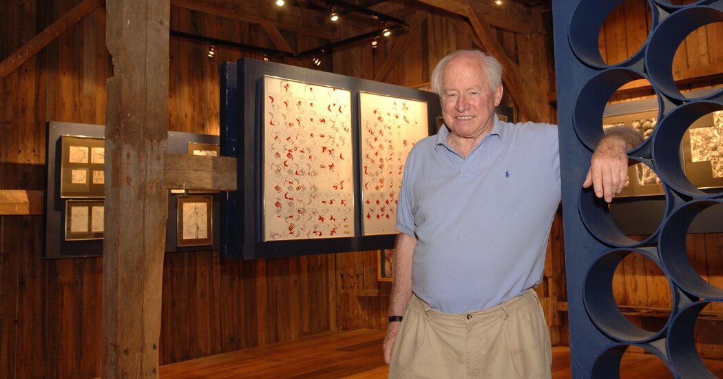
He disputed the Freudian view that dreams held encrypted codes of meaning, believing instead that they resulted from random firings of neurons in the brain.Dr. J. Allan Hobson, a psychotherapist and pioneering sleep researcher who disputed Freud’s view that dreams held hidden psychological meaning, died on July 7 at his home in East Burke, Vt. He was 88. The cause was kidney failure resulting from diabetes, said his daughter, Julia Hobson Haggerty.For some time, sleep was not taken seriously as an academic pursuit. Even Dr. Hobson, who was a professor of psychiatry at Harvard Medical School and director of the Laboratory of Neurophysiology at the Massachusetts Mental Health Center, joked that the only known function of sleep was to cure sleepiness.But over a career that spanned more than four decades, his own research and that of others showed that sleep is crucial to normal cognitive and emotional function, including learning and memory.In more than 20 books — among them “The Dreaming Brain” (1988); “Dreaming as Delirium: How the Brain Goes Out of its Mind” (1999), and “Dream Self” (2021), a memoir — he popularized his research and that of others, including the findings that sleep begins in utero and is essential for tissue growth and repair throughout life.“He showed that sleep isn’t a nothing state,” Ralph Lydic, who conducted research with Dr. Hobson in the 1980s and is a professor of neuroscience at the University of Tennessee, said in a phone interview.“He demonstrated that the brain is as active during R.E.M. sleep as it is during wakefulness,” he added, referring to sleep characterized by rapid eye movement. “We know as much about sleep as we do in part because of him.”One of his most influential contributions to dream research came in 1977, when Dr. Hobson and a colleague, Robert McCarley, produced a cellular and mathematical model that they believed showed how dreams occur. Dreams, they said, are not mysterious codes sent by the subconscious but rather the brain’s attempt to attribute meaning to random firings of neurons in the brain.This view, that dreams are the byproduct of chemical reactions, was a departure from psychological orthodoxy and heresy to Freudians, and it remains in dispute.But to Dr. Hobson, the content of dreams was not as important as the electrical activity of the brain during the dream state.His work became foundational for many other sleep researchers, including Carlos H. Schenck, whose team in Minnesota found a link between behavioral disorders during R.E.M. sleep — punching one’s bed partner, for example, or even jumping out of a window — and the likelihood in some of those people of developing Parkinson’s disease.“Allan Hobson helped us understand the dream abnormalities of R.B.D. right from the beginning in 1986,” Dr. Schenck said in an email, referring to R.E.M. sleep behavioral disorders.Dr. Hobson wrote about dream research in more than 20 books. “The Dreaming Brain” was published in 1988.Basic BooksDr. Hobson thrived on controversy, and it was no surprise to many that he challenged his own profession of psychoanalysis and its founding father. Even as a child, he constantly questioned the status quo. At 4, he took measurements and concluded that Santa Claus could not fit down the chimney.“I’m skeptical about any absolute set of rules, scientific rules, moral rules, behavioral rules,” he said in a 2011 interview with The Boston Globe. “That’s one reason why I don’t feel bad taking on Sigmund Freud. I think Sigmund Freud has become politically correct. Psychoanalysis has become the bible, and I think that’s crazy.”.css-1xzcza9{list-style-type:disc;padding-inline-start:1em;}.css-rqynmc{font-family:nyt-franklin,helvetica,arial,sans-serif;font-size:0.9375rem;line-height:1.25rem;color:#333;margin-bottom:0.78125rem;}@media (min-width:740px){.css-rqynmc{font-size:1.0625rem;line-height:1.5rem;margin-bottom:0.9375rem;}}.css-rqynmc strong{font-weight:600;}.css-rqynmc em{font-style:italic;}.css-yoay6m{margin:0 auto 5px;font-family:nyt-franklin,helvetica,arial,sans-serif;font-weight:700;font-size:1.125rem;line-height:1.3125rem;color:#121212;}@media (min-width:740px){.css-yoay6m{font-size:1.25rem;line-height:1.4375rem;}}.css-1dg6kl4{margin-top:5px;margin-bottom:15px;}#masthead-bar-one{display:none;}#masthead-bar-one{display:none;}.css-1pxllx6 header h4{font-family:nyt-cheltenham,georgia,’times new roman’,times,serif;font-weight:500;font-size:1.25rem;line-height:1.5625rem;margin-bottom:5px;}@media (min-width:740px){.css-1pxllx6 header h4{font-size:1.5625rem;line-height:1.875rem;}}.css-1pd7fgo{background-color:white;border:1px solid #e2e2e2;width:calc(100% – 40px);max-width:600px;margin:1.5rem auto 1.9rem;padding:15px;box-sizing:border-box;}@media (min-width:740px){.css-1pd7fgo{padding:20px;width:100%;}}.css-1pd7fgo:focus{outline:1px solid #e2e2e2;}#NYT_BELOW_MAIN_CONTENT_REGION .css-1pd7fgo{border:none;padding:20px 0 0;border-top:1px solid #121212;}.css-1pd7fgo[data-truncated] .css-rdoyk0{-webkit-transform:rotate(0deg);-ms-transform:rotate(0deg);transform:rotate(0deg);}.css-1pd7fgo[data-truncated] .css-eb027h{max-height:300px;overflow:hidden;-webkit-transition:none;transition:none;}.css-1pd7fgo[data-truncated] .css-5gimkt:after{content:’See more’;}.css-1pd7fgo[data-truncated] .css-6mllg9{opacity:1;}.css-1rh1sk1{margin:0 auto;overflow:hidden;}.css-1rh1sk1 strong{font-weight:700;}.css-1rh1sk1 em{font-style:italic;}.css-1rh1sk1 a{color:#326891;-webkit-text-decoration:underline;text-decoration:underline;text-underline-offset:1px;-webkit-text-decoration-thickness:1px;text-decoration-thickness:1px;-webkit-text-decoration-color:#ccd9e3;text-decoration-color:#ccd9e3;}.css-1rh1sk1 a:visited{color:#333;-webkit-text-decoration-color:#ccc;text-decoration-color:#ccc;}.css-1rh1sk1 a:hover{-webkit-text-decoration:none;text-decoration:none;}In one of his books, “Out of Its Mind: Psychiatry in Crisis — A Call for Reform” (2002, with Jonathan A. Leonard), he called for an overhaul of the profession.“I think people became disillusioned with psychoanalysis because it was, ultimately, a strange way of caring for people,” he told The New York Times in 2002.“There was this tendency in the psychoanalytic world to imply that everything was psychodynamic,” he added, noting that some doctors reflexively blamed mothers for their children’s behavior.But Dr. Hobson softened his views in his later years.“He came to believe that psychoanalysis could be useful for treating mental disorders,” Dr. Lydic said, “but he did not believe in a rigid symbolism in the interpretation of dreams.”For the most part, Dr. Hobson still believed, as the saying goes, that a cigar was just a cigar.John Allan Hobson was born on June 3, 1933, in Hartford, Conn. His mother, Ann (Cotter) Hobson, was a homemaker. His father, John Robert Hobson, was a lawyer.John attended the Loomis School, now the Loomis Chaffee School, in Windsor, Conn., graduating in 1951. He spent a year abroad, then returned to study at Wesleyan University in Connecticut, where he majored in English, graduating in 1955. He received his medical degree from Harvard Medical School in 1959.He married Joan Harlowe in 1956; they divorced in 1992. He married Dr. Rosalia Silvestri in the mid-1990s, and she survives him.In addition to his wife and daughter, Dr. Hobson is survived by four sons, Ian, Christopher, Andrew and Matthew; his brother, Bruce; and four grandchildren.After medical school, Dr. Hobson interned for two years at Bellevue Hospital in Manhattan. In lieu of military service, he served in the Public Health Service of the National Institutes of Health.He was influenced by Michel Jouvet, a neurophysiologist who discovered the region of the brain that controls rapid eye movement and who helped steer Dr. Hobson to study sleep and dreams.Apart from his research, Dr. Hobson was most passionate about his farm in Vermont, which he acquired in 1965 and had since been its steward.He converted part of one barn on the property into a small, interactive sleep museum and classroom for local students, basing it on his multimedia exhibit “Dreamstage,” which celebrated the art and science of sleep and toured science museums across the United States in the late 1970s. His museum featured, among other things, a preserved brain as well as artwork of brains.The farm was a gathering place for family and friends. Dr. Hobson’s children said that the dining room table was often the scene of celebratory recitations of poetry and song. Afterward, the kitchen would fill with the sound of Big Band favorites and become a dance floor.Dr. Hobson wrote in his memoir that he spent 10 years reading all of Marcel Proust — twice. He read 10 pages a day.“I simply admire his persistent and revealing self-analysis and his description of mental life in and at the edges of sleep,” Dr. Hobson told The Globe. “His self-observation is much more careful than that of Freud.”
Read more →Scientists advance breast, ovarian cancer research with cryo-electron microscopy

Using advanced imaging technology, Mayo Clinic scientists have provided an unprecedented understanding of the BRCA1-BARD1 protein complex, which is often mutated in patients with breast or ovarian cancer. Their paper, published in Nature, identifies aspects of how BRCA1-BARD1 functions, supporting future translational research, cancer prevention efforts and drug development.
“BRCA1-BARD1 is important for DNA repair. It has direct relevance to cancer because hundreds of mutations in the BRCA1 and BARD1 genes have been identified in cancer patients,” says Georges Mer, Ph.D., a Mayo Clinic structural biologist and biochemist who is the lead author of the paper. “But no one knows if these mutations, or variants of unknown significance, are cancer-predisposing or not because we do not know whether the variants are located in a region of BRCA1-BARD1 that is important for function. Now because we can see how BRCA1-BARD1 works, we have a good idea of what regions of BRCA1-BARD1 are important for function.”
In a cell, the complex of DNA and histone proteins are complexed into what’s called chromatin, and packaged into bundles called nucleosomes. DNA damage response proteins need to access chromatin to repair damaged DNA. BRCA1-BARD1 contributes to fixing broken DNA strands, which helps in the maintenance and survival of cells. But it is also a function that could possibly be blocked or inactivated if this is a strategy a cancer cell uses to survive chemotherapy.
Cryo-electron microscopy and nuclear magnetic resonance spectroscopy
“We used two techniques — cryo-electron microscopy and nuclear magnetic resonance spectroscopy — to understand at near-atomic resolution how BRCA1-BARD1 associates with the nucleosome, the repeating unit of chromatin, and how BRCA1-BARD1 modifies chromatin,” explains Dr. Mer.
In cryo-electron microscopy, purified BRCA1-BARD1 bound to nucleosomes, together referred to as macromolecules, are flash-frozen then imaged using an electron microscope. The macromolecules are oriented in various ways within the sample so a computer program evaluates all the orientation data to create a 3D structure. Dr. Mer and his team also examined BRCA1-BARD1 nucleosome complexes with nuclear magnetic resonance spectroscopy, which uses a strong magnet to probe the relative positions of atoms within macromolecules. Using these imaging tools, the scientists could visualize BRCA1-BARD1 in action and uncover a new function of the complex.
“We showed how BRCA1-BARD1 attaches ubiquitin to the nucleosome, but we also determined that BRCA1-BARD1 recognizes ubiquitin already attached to the nucleosome, which serves as a signal for broken DNA,” says Dr. Mer. “We discovered an unexpected cross-talk by which ubiquitin recognition by BRCA1-BARD1 enhances its ubiquitin attachment activity, and this helps us better understand how BRCA1-BARD1 performs its function.”
The researchers created a video from the cryo-electron microscopy data to show where the protein complex interacts with the nucleosome [see link below].
From discovery science to patient care
Dr. Mer and his team expect that high-resolution images of BRCA1-BARD1 can help guide patient care and future treatment of cancer in two ways: classifying variants of unknown significance and directing drug development with more accuracy.
“With these 3D structures, we should be able to convert several variants of unknown significance to likely cancer-predisposing variants,” says Dr. Mer. “This work is also expected to have an impact on drug development in the long term because the 3D structures of BRCA1-BARD1 in complex with the nucleosome we generated may help in the design of small molecules that could, for example, inactivate BRCA1-BARD1.”
In addition to Dr. Mer, other authors on the paper are Qi Hu, Ph.D.; Maria Victoria Botuyan, Ph.D.; Debiao Zhao, Ph.D.; Gaofeng Cui, Ph.D.; and Elie Mer. This research was funded by the National Institutes of Health, Mayo Clinic Cancer Center, Mayo Clinic Center for Biomedical Discovery, and the Ovarian Cancer Research Alliance, and was made possible through cryo-electron microscopy and nuclear magnetic resonance instrumentation at the Pacific Northwest Center for Cryo-EM and Mayo Clinic, respectively.
Story Source:
Materials provided by Mayo Clinic. Original written by Sara Tiner. Note: Content may be edited for style and length.
Cats Are So Not Appreciated. Think Again.

Researchers who work on the genomes of domestic and wild cats say their DNA holds clues to human as well as feline health.Leslie Lyons is a veterinarian and specialist in cat genetics. She is also a cat owner and general cat partisan who has been known to tease her colleagues who study dog genetics with the well-worn adage that “Cats rule. Dogs drool.”That has not been the case with research money and attention to the genetics of disease in cats and dogs, partly because the number of dog breeds offers variety in terms of genetic ailments and perhaps because of a general bias in favor of dogs. But Dr. Lyons, a professor at the University of Missouri, says there are many reasons cats and their diseases are invaluable models for human diseases. She took up the cause of cat science this week in an article in Trends in Genetics.“People tend to either love them or hate them, and cats are often underappreciated by the scientific community,” she writes. But, she says, in some ways the organization of the cat genome is much like the human genome, and cat genomics could help in the understanding of the vast amount of mammalian DNA that does not constitute genes, and is poorly understood.Among the advances in veterinary medicine that have benefited humans, she pointed out that remdesivir, an important drug in combating Covid, was first successfully used against a cat disease caused by another coronavirus.She is the director of the 99 Lives Cat Genome Sequencing Initiative and as part of that project, she and a group of colleagues, including Wes Warren at the University of Missouri and William Murphy at Texas A&M University, recently produced the most detailed genome of the cat to date, which surpasses the dog genome.“For the moment,” Dr. Lyons said.I spoke last week with Dr. Lyons, Dr. Warren and Dr. Murphy, who refer to themselves as Team Feline. Dr. Lyons was visiting Texas, and with two of her colleagues she talked about why the genomes of cats are important to medical knowledge.I report on animal science, and over the years, I admitted to the members of Team Feline, I seem to have written more about dogs than cats. The dog-cat rivalry in genomic science is mostly a good-natured rivalry, but just to assess what I was getting myself into I first asked about the scientists’ nonscientific approach to cats and dogs.The conversation has been edited for length and clarity.First, their personal preferences:Dr. William Murphy: I do have cats and dogs as pets, but I prefer cats.Dr. Wes Warren: I’m a dog owner. Unfortunately I’m allergic to cats.Dr. Leslie Lyons: He has a very expensive dog that keeps having problems.Why were you moved to write the article promoting the cause of cat science?Dr. Lyons: Throughout my career, I’ve been trying to get people to recognize that our everyday pets have the same diseases as us and can really provide important information if we can understand what makes them tick a little bit better, how their genomes are constructed.A Pallas’s cat, or manul. Wildscotphotos/AlamyYou have high quality genomes of several species of cats beyond the domestic cat?Dr. Lyons: We already have the lions and tigers, the Asian leopard cat, Geoffroy’s cat, a half-dozen species with really, really good genomes that are even better than the dog genomes at this point in time.Dr. Murphy: By far. It was actually better quality than the human reference genome until very recently. The goal is to have the complete encyclopedia of the cat’s DNA, so we can actually fully understand the genetic basis for all traits in the cat.Dr. Lyons: For example the allergy gene that Wes is allergic to. We completely understand that gene now. We can maybe even knock it out of the cat to produce cats that are more hypoallergenic or at least understand what elicits the immune response better.How are cat diseases a good model for human diseases?Dr. Lyons: What we’re discovering is different species have different health problems. We should really be picking the right species.Dr. Warren: We know that dogs get cancer more frequently, similar to ourselves. Cats don’t get cancer very often. And that’s a fascinating story of evolution. So are there signals or clues in the genome of the cat that allows us to zero in better on why cats get certain types of cancers and understand the differences among dogs, cats and humans.How about the cats that are subjects of the research?Dr. Lyons: Genomic research is fantastic because all we need is maybe a blood sample. And so once we have the blood sample, we don’t have to do experimentation on an animal. We’re actually observing what animals already have. We’re working with the diseases that are already there.What about wild species?Dr. Murphy: High quality genomes for wild cats can aid in their species survival plans and their recovery in the wild.Dr. Lyons: We see half a dozen health problems in wild felids. We have a study of transitional cell carcinoma in fishing cats, inherited blindness in black-footed cats, polycystic kidney disease in Pallas’s cats. Snow leopards have terrible eye problems, probably because of inbreeding in zoos. So understanding their genomes can help us to stop those problems in the zoo populations, and that will help humans with the same conditions as well.How about ancient DNA and cats? There’s been a lot of work on that in dogs. How is that progressing in cats?Dr. Lyons: A couple of groups are moving forward with ancient DNA. I worked on some mummy cats and we showed that the mitochondrial DNA types that we found in the mummified cats are present more commonly in Egyptian cats today than they are anywhere else. So the cats of the pharaohs are the cats of present day Egyptians.To switch gears: I’ve always been a dog person but I’ve been thinking about getting a cat. Any tips?Dr. Lyons: Get two. They’ll be buddies. And give them something to scratch. Otherwise it is going to be your couch.
Read more →Seeking a unique treatment for lobular breast cancer
Though the two main histological types of breast cancer — lobular and ductal — are treated with the same hormonal therapies, women with lobular breast cancer often have recurrence or metastasis of the disease several years after their initial treatment.
In an attempt to find out why the long-term outcomes are poorer for patients with lobular breast cancer — which affects some 40,000 women a year — University of Colorado Cancer Center member Matthew Sikora, PhD, began looking at the role of the protein MDC1 in tumor cells.
“This is a protein that’s normally involved in DNA repair, but it seems to have some new function in lobular cancer cells,” Sikora says. “It’s now required for estrogen receptor activity.”
MDC1 and estrogen
Ductal cancer and lobular cancer cells both use the hormone estrogen to grow, Sikora explains, and the antiestrogen drug tamoxifen typically blocks estrogen in the tumor cell, thwarting that growth. In a paper published in May in the journal Molecular Cancer Research, however, Sikora and his fellow researchers from the CU School of Medicine examined how in lobular cancer cells, the MDC1 protein allows cells to use tamoxifen as a weak estrogen, causing them to keep growing, albeit at a lesser rate.
“We think this MDC1 protein may be what influences how lobular cells respond to estrogen in the first place,” Sikora says. “Back in the 1980s, when women got hormone replacement therapy and there was an uptick in breast cancer risk, most of that was lobular. It’s this idea that these cells are just seeing anything that is estrogen-like differently. Not only is MDC1 possibly promoting this resistance to tamoxifen because of how it changes the way estrogen receptors work, but it may be playing a role in how estrogen works in the tumor cell.”
Looking for new treatments
Because lobular tumors often metastasize to the abdomen, GI tract, and ovaries, they can be harder to detect, Sikora says. This makes it all the more imperative to figure out a novel way to treat this type of tumor. Based on his initial research, Sikora is now looking at what other proteins work with MDC1 to promote tumor growth — and novel ways to stop that growth from happening.
“The way MDC1 normally works in DNA repair is like a scaffold. It sits on damaged sites and then recruits in other repair proteins,” he says. “It’s plausible that it would do something similar for estrogen receptor — instead of repair proteins, it might bring in partners that open and close DNA to let genes turn on and off. We have to figure out if there are other partners involved, if there’s a bigger complex that makes that process possible.”
With funding from the American Cancer Society, Sikora and his research partners will spend the next few years identifying those partners and how to combat them. They also plan to explore how the role of MDC1 in DNA repair changes in lobular cancer cells, and how that might reveal other vulnerabilities. Ultimately, he hopes the research will lead to better treatment for lobular cancer patients — treatment that reduces the risk of metastases and recurrences years down the road.
“Right now, even though patients are differentially diagnosed with either lobular or ductal breast cancer, there are few different therapy decisions, despite what we’re learning about how outcomes are different,” Sikora says. “For a patient with lobular cancer, ideally we could identify, using gene-expression signatures, whether this estrogen receptor-MDC1 partnership is active in the tumor. Then we can treat them accordingly. That’s the long-term goal.”
Story Source:
Materials provided by University of Colorado Anschutz Medical Campus. Original written by Greg Glasgow. Note: Content may be edited for style and length.
First 3-D view of TB granulomas alters paradigm of their shape and formation
For 70 years, clinicians thought they knew the shape of tuberculosis granulomas in the lungs of patients. Histology — the study of microscopic structures in thin slices of lung tissue in the 1940s and 1950s — showed round features, and researchers intuitively assumed that meant the granulomas were spherical or ovoid.
That long-lived paradigm is now shown to be wrong, in a study by researchers at the University of Alabama at Birmingham and the Africa Health Research Institute, or AHRI, Durban, KwaZulu-Natal, South Africa. The historical histology, essentially, was a two-dimensional look at structures, similar to cutting a very thin slice through a tree branch, a slice that would look round or oval.
The new research has created a three-dimensional view of diseased lung tissue from tuberculosis patients, using micro-computed tomography, or microCT. This revealed that the larger granulomas were anything but round — rather they had complex, branched shapes. One granuloma looked somewhat like a ginger root, another like a cluster of early buds on a cherry tree, before the blossoms appear. The tuberculosis granulomas showed marked heterogeneity in shape, volume and number in the lung sections. (See link to study below for videos of the three-dimensional views.)
A granuloma is an aggregation of immune cells summoned in response to chronic inflammation. In tuberculosis, the granulomas are often necrotic, meaning they are a mass of formless dead debris.
Researchers led by Adrie Steyn, Ph.D., a UAB professor of microbiology and a member of the AHRI, used microCT, histology and immunohistochemistry to construct three-dimensional views of necrotic granulomas, and also vasculature and airways. This yielded an unanticipated insight into the spatial organization of tuberculosis granulomas in relation to airways and vasculature.
“Unlike depictions of granulomas as simple spherical structures,” Steyn said, “human necrotic granulomas exhibit complex, cylindrical, branched morphologies that are connected to the airways and shaped by the bronchi.”
Steyn says these visualizations have three impacts. They highlight the likelihood that a single structurally complex lesion could be mistakenly viewed as multiple independent lesions when evaluated in two dimensions. Second, the lack of vascularization within obstructed bronchi establishes a paradigm for anti-mycobacterial drug tolerance, since the microbes are protected from a full delivery of blood-borne bactericidal drugs. Third, the results suggest that a bronchogenic spread of the Mycobacterium tuberculosis pathogen re-seeds the lung.
Neanderthal and Denisovan blood groups deciphered
Blood group analyses for three Neanderthals and one Denisovan by a team from the Anthropologie Bio-Culturelle, Droit, Éthique et Santé research unit (CNRS / Aix-Marseille University / EFS) confirm hypotheses concerning their African origin, Eurasian dispersal, and interbreeding with early Homo sapiens. The researchers also found further evidence of low genetic diversity and possible demographic fragility. Their findings are published in PLOS ONE (28 July 2021).
The extinct hominin lineages of the Neanderthals and Denisovans were present throughout Eurasia from 300,000 to 40,000 years ago. Despite prior sequencing of about 15 Neanderthal and Denisovan individuals, the study of the genes underlying blood groups had hitherto been neglected. Yet blood group systems were the first markers used by anthropologists to reconstruct the origins of hominin populations, their migrations, and their interbreeding.
In a new study, scientists from the CNRS, Aix-Marseille University, and the French Blood Establishment (EFS) have examined the previously sequenced genomes of one Denisovan and three Neanderthal females who lived 100,000 to 40,000 years ago, in order to identify their blood groups and consider what they may reveal about human’s evolutionary history. Of the 40-some known blood group systems, the team concentrated on the seven usually considered for blood transfusion purposes, the most common of which are the ABO (determining the A, B, AB, and O blood types) and Rh systems.
The findings bolster previous hypotheses but also offer new surprises. While it was long thought that Neanderthals were all type O — just as chimpanzees are all type A and gorillas all type B — the researchers demonstrated that these ancient hominins already displayed the full range of ABO variability observed in modern humans. Extensive analysis covering other blood group systems turned up alleles that argue in favour of African origins for Neanderthals and Denisovans.
Especially surprising is the discovery that the Neanderthals harboured a unique Rh allele absent in modern humans — with the notable exceptions of one Aboriginal Australian and one Papuan. Do these two individuals bear testimony to interbreeding of Neanderthals and modern humans before the migration of the latter into Southeast Asia?
Finally, this study sheds light on Neanderthal demographics. It confirms that these ancient hominins exhibited very little genetic diversity, and that they may have been susceptible to haemolytic disease of the fetus and newborn (erythroblastosis fetalis) — due to maternofetal Rh incompatibility — in cases where Neanderthal mothers were carrying the children of Homo sapiens or Denisovan mates. These clues strengthen the hypothesis that low genetic diversity together with low reproductive success contributed to the disappearance of Neanderthals.
Story Source:
Materials provided by CNRS. Note: Content may be edited for style and length.
Vaccinated People Should Be Tested After Exposure Even Without Symptoms, C.D.C. Says

In addition to revising its mask guidance on Tuesday, the Centers for Disease Control and Prevention also quietly updated its testing recommendations for people who are fully vaccinated against the coronavirus.The agency now advises that vaccinated people be tested for the virus if they come into contact with someone with Covid-19, even if they have no symptoms. Previously, the health agency had said that fully vaccinated people did not need to be tested after exposure to the virus unless they were experiencing symptoms.“Our updated guidance recommends vaccinated people get tested upon exposure regardless of symptoms,” Dr. Rochelle P. Walensky, the agency’s director, said in an email to The New York Times. “Testing is widely available.”Fully vaccinated people should wear a mask in public indoor spaces after exposure, the agency said. Three to five days later, they should be tested for the virus.If the results come back negative, they can stop wearing masks indoors. If results are positive, the infected should isolate at home for 10 days.The new recommendation came on the same day that the agency recommended that fully vaccinated people return to wearing masks indoors under some circumstances. When levels of community transmission are high, everyone, regardless of vaccination status, should wear masks indoors when they are in public, the agency now says.The agency also recommended that vaccinated people in close contact with unvaccinated people, including children under age 12, consider wearing masks in public indoor spaces whatever the transmission rates in the local community. In a shift, the agency also recommended universal masking in schools.For months, the C.D.C. had resisted recommending masks for vaccinated people, even as the highly contagious Delta variant spread and the World Health Organization recommended continued mask wearing.The change was prompted by new data suggesting that even vaccinated people who are infected by Delta may carry large amounts of the virus and transmit it to others, Dr. Walensky said at a news briefing on Tuesday.The vaccines provide strong protection against the worst outcomes, however, including severe disease, hospitalization and death.Apoorva Mandavilli contributed reporting.
Read more →Pfizer-BioNTech’s vaccine gets slightly weaker over time, company data shows, but remains strong in preventing severe disease.
With cases rising in the U.S., the findings may influence the Biden administration’s deliberations about delivering a booster shot.
Read more →Biomedical scientists tie improved learning processes to reduced symptoms of depression
Virginia Tech scientists with the Fralin Biomedical Research Institute at VTC have identified neural learning processes to be associated with symptoms of depression and linked improvements in these processes to improved symptoms in research participants being treated for depression.
The findings, described in a study published July 28, 2021 in the Journal of the American Medical Association (JAMA) Psychiatry, suggest distinct paths to depression symptoms and new mathematically guided approaches for treating clinical depression.
Major depression is one of the most common mental disorders in the United States and can cause severe impairment, according to the National Institute of Mental Health. An estimated 7.1% of all U.S. adults have had at least one major depressive episode.
“Current medications and behavioral therapies are helpful, but for many people struggling with depression, existing treatments don’t work well,” said Pearl Chiu, an associate professor at the Fralin Biomedical Research Institute Computational Psychiatry Unit and the study’s corresponding author. “We need to consider other possible paths to depression. These paths, or mechanisms, could point to new treatment targets to explore.”
The scientists used computational models of brain functioning as a new way to consider mechanisms of depression. In a key discovery, the researchers found that the symptom improvements that followed cognitive behavioral therapy were related to improvements in reinforcement learning components that were disrupted prior to therapy.
“Depression is a very serious illness and a leading cause of disability in the world. We hope that our work can be a bridge between behavioral clinicians and computational scientists to more precisely identify what causes depression and new ways to treat the illness,” said first author Vanessa Brown, a former doctoral student with Chiu in Virginia Tech’s Department of Psychology and who is now an assistant professor of psychiatry at the University of Pittsburgh.