Home Care Is Broken. Can Congress Fix It?

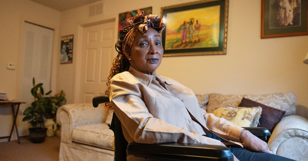
The latest Democratic proposal would funnel $150 billion toward subsidized home and community-based care. Experts worry that may not be enough to ease the severe shortage of workers.Spinal surgery four years ago left Alene Shaheed in a wheelchair and dependent on short daily visits from home health aides to help her get around.But her support system fell apart this year during the prolonged coronavirus pandemic, and her routine care became unpredictable. Four agencies serving her hometown, Jacksonville, Fla., failed to provide aides regularly, due to severe shortages of the low-wage workers.“If no one comes for three days, I don’t get a bath for three days,” said the 76-year-old. “I don’t have anyone to fix meals, so I’m eating ramen noodles until someone gets there.”About 800,000 people are on waiting lists to receive subsidized home care. For millions of Americans, finding reliable and affordable assistance to stay at home — instead of moving into a nursing home, where Covid-19 killed tens of thousands of people — has never seemed more urgent.Expanding home and community-based services is part of the legislative package that President Biden and Democrats have proposed. At this stage of negotiations in Congress, the amount for such programs under Medicaid — partly to increase the historically low wages of home care workers — has been reduced to $150 billion from $400 billion over eight years.“We’re going to expand services for seniors so families can get help from well-trained, well-paid professionals to help them take care of their parents at home — to cook a meal for them, to get their groceries for them, to help them get around, to help them live in their own home with the dignity they deserve to be afforded,” President Biden said on Thursday.”Will the amount in the current plan be enough? Supporters say the new health care money would shift Medicaid’s decades-long bias away from nursing home care. Many experts doubt promises that this round of funding can fix a system as broken as home care, especially as the growing retirement of the boomer generation requires more assistance to stay independent and strains health care funding.“You have to be very realistic about the amount of need you have in the system right now,” said David Grabowski, a professor of health care policy at Harvard Medical School. The $150 billion does represent a significant influx of funds, but there are limits, he said: “Once you start to do the math, the dollars don’t go as far as you’d like.”States are required to use Medicaid funds to cover nursing home care, but states have considerable leeway under federal regulations to decide how much should be allocated to provide home and community-based services.People who need help with tasks like feeding themselves, getting dressed or taking medication must often qualify for a Medicaid waiver to get home care. Medicaid, a federal-state program that is the primary source of coverage for long-term care, spends about $114 billion a year on these home and community-based services, representing well over half of the overall spending on long-term care. About 2.5 million people received waivers in 2018, according to the latest data available in a report by the Kaiser Family Foundation.Medicare, the federal insurance program for older and disabled adults, does not cover long-term care and it limits the kind of home care people can receive.It’s well known that demand far outstrips supply for home care for those who want to live independently. Some people may have private insurance or pay for the care themselves.Under Medicaid, the waiting lists for older and disabled Americans wanting home care keep growing because states cap enrollment. Most people on the lists live in states that did not expand Medicaid, according to a Kaiser analysis.Benefits for home care also vary widely from state to state. For example, someone in Pennsylvania is eligible for about $50,000 a year under Medicaid for home or community services, while someone in Iowa may get only $21,000.The lack of funding “really forces older adults into institutions,” said Amber Christ, an attorney with Justice in Aging, a nonprofit group. The new congressional package, she said, means that “we have an opportunity to flip the script.”She and other advocates plan to push for additional money. “We’re going to keep working to increase funding because more is needed to ensure all aging adults and people with disabilities have the option to receive the care they need at home,” she said in an email.Expanding home and community-based services is part of the legislative package that President Biden and Democrats have proposed.Stefani Reynolds for The New York TimesIncreasing wages for home health workers has been a contentious provision for Republicans, who see it as a giveaway to unions and would limit states’ flexibility in spending new funds.Calculating how the proposed $150 billion in the Democrats’ proposal is still guesswork, without detailed legislative language.Jonathan Gruber, a health economist at M.I.T., said the lower figure would provide home services for perhaps one million more people and create about 400,000 new jobs. That could include jobs for caretakers like family members who are unpaid but unable to go to work.But if the amount gets cut further — and negotiations on the bill are far from over — supporters warn that states may be less willing to expand services.“We need a big investment,” said Nicole T. Jorwic, senior director of public policy at The Arc, an advocacy group for people with physical and developmental disabilities. The bill has to provide at least $150 billion so “states will see the value and worth of taking it up,” she said.Even that level of funding might not eliminate the waiting lists, but “it will help take people off,” she said.Under the American Rescue Plan Act passed by Congress this year, all states made use of temporary funds allocated to shore up home and community-based services, Ms. Jorwic said.Still, the issue of wages in a pandemic economy where people are shunning lower-rung jobs bodes ill for the home health industry, whose workers have long been paid far less than others in service industries. Some businesses now pay $15 an hour or more, luring away those in underpaying jobs and leaving the vulnerable without reliable help.About 70 percent of long-term care workers earn less than $30,000 a year, according to Kaiser, and they are more likely to live in poverty. “It’s the same person who is aging into poverty and who is going to get pushed into a nursing home,” said Ms. Christ of Justice in Aging.While details are sparse, the proposed legislation would require states to prove that the funds were funneled toward higher wages. “This would be the first time that there was a large federal investment to increase wages,” Ms. Jorwic said.Higher wages are critical to finding more aides for those like Ms. Shaheed in Florida. “They’re no longer able to find anyone willing to come in for the low wages they are paying,” she said. “Nobody is going to come and help me for $10 an hour.”For those who have had to wait to qualify for financial aid for home care, the difference is palpable.People like Stephen Grammer, who has cerebral palsy, were warned since childhood that they faced institutionalization if home care could not be routinely provided.In his 20s, Mr. Grammer spent nearly a decade in a nursing home after his mother became ill. He chafed at the restrictions imposed on him while he was living with older adults with Alzheimer’s. If he left the premises, he had to be back by midnight or it would count against the 18 nights a year he was allowed to be outside.“When I had to use the restroom, I would press the call button, and many times the workers would come and turn off my call light and would walk away even though I had to go to the bathroom,” he said by email.Mr. Grammer, who uses an electric wheelchair and advocates for disabled individuals these days, eventually qualified for a Medicaid waiver and another state program that provides housing. Now at 41, he lives on his own in Roanoke, Va., and someone comes in 16 hours a day from 6 a.m. to 2 p.m. and from 4 p.m. to midnight.“I have the freedom to come and go as I please,” Mr. Grammer said.

Read more →

Muriel Lezak, Leading Authority on Brain Injuries, Dies at 94

A neuropsychologist, she wrote a textbook that became an essential guide to describing and evaluating brain damage and dysfunction.Muriel Lezak, a neuropsychologist who wrote a landmark textbook in the early days of her discipline that became an essential guide to the description and evaluation of brain injuries and disorders, died on Oct. 6 in a memory care facility in Portland, Ore. She was 94.Her death was confirmed by her nephew Stephen Lezak.Dr. Lezak began working as a clinical psychologist in the late 1940s. Two decades later, at the Veterans Administration Hospital in Portland, she brought her abiding curiosity about the connection between the brain and behavior to her treatment of soldiers who had suffered neurological damage in World War I, World War II and the Vietnam War.“I was the psychologist for neurology, neurosurgery and rehab,” she said in an oral history interview with Oregon Health & Science University in 2016. “It was like pig heaven, you know?”Dr. Lezak became keenly interested in patients with frontal lobe damage, which affects creativity, reasoning and the ability to relate to people and to plan and organize. As she dealt with interns and other medical personnel, she realized that there was no book in her evolving field that comprehensively reviewed the major disorders caused by brain dysfunction and injury, or the techniques, tests and procedures to evaluate patients.Her book “Neuropsychological Assessment,” published in 1976, filled that gap. It also added tests that she developed to evaluate brain dysfunction, like seeing how a patient drew a bicycle, that could provide insights into motor control and perception. She emphasized a flexible approach, adapting procedures to suit an individual patient’s problems, a departure from the standardized tests that were then common.“There had been nothing at the time that focused on the nuts and bolts of evaluation, and she did a really nice job of looking at the pattern of a broad range of assessments before making a conclusion about a diagnosis,” Kathleen Haaland, a neuropsychologist and professor of psychiatry and behavioral sciences at the University of New Mexico School of Medicine, said in a phone interview.Ida Sue Baron, a neuropsychologist and clinical professor emeritus of pediatrics at George Washington University School of Medicine in Washington, said in an email: “The publication of this book brought the field together coherently for the first time, by integrating the methods and the science for those of us who had no other references, and even for those not in our profession who wished to understand what neuropsychology was really all about.”Dr. Lezak was the sole author of two subsequent editions of the book and one of the writers of the fourth and fifth editions; a sixth edition, likely to be published in 2023, will be renamed “Lezak’s Neuropsychological Assessment.”At the V.A. hospital, where Dr. Lezak worked until 1985, she started a support group to help military wives cope with the altered behavior of their brain-injured husbands.“The people they were married to were no longer there; it was somebody else who was similar, looked pretty much the same, but was no longer the person they were able to love and interact with comfortably,” she said in the oral history.Muriel Elaine Deutsch was born on Aug. 26, 1927, in Chicago, where her father, Lester, was a furrier and her mother, Sylvia (Friedman) Deutsch, was a homemaker who helped with the fur business’s bookkeeping.Muriel’s fascination with medicine began when she wondered why her grandmother, who was in a wheelchair, couldn’t walk. But instead of going to a medical school — which she didn’t think would admit a woman with young children — she pursued psychology.She graduated from the University of Chicago with a bachelor’s degree in general studies in 1947 and a master’s in human development in 1949. That same year she married Sidney Lezak, a lawyer; they soon moved to Portland, where he would serve as the United States attorney in Oregon from 1961 to 1982.She received a doctorate in clinical psychology at the University of Portland in 1960. She later said that she could not have studied for that degree or worked simultaneously as a clinical psychologist without Mr. Lezak.“For me, he was ‘Sweetie,’” she said in an interview in 2016 with Dr. Haaland for the International Neuropsychological Society. “He was supportive from the get-go at a time when many husbands thought the little lady should be home baking cookies and going to the P.T.A. meetings and being nice.”She added, “As my career developed, it was fun; he wore me like a rose in his buttonhole.”Dr. Lezak worked at clinics and taught psychology at Portland State College (now University) and the University of Portland from 1949 until she began her 19-year tenure at the V.A. hospital in 1966. In 1985, she left to teach at the Oregon Health & Science University, where she was a professor of neurology, neurosurgery and psychiatry until 2005. She long had a private practice, and she continued to see patients until a few years ago.As early as 1982, Dr. Lezak sounded an alarm about the impact of head injuries incurred by athletes; in 1999 and 2001, she was an author and researcher of studies that found cognitive impairment in amateur and professional soccer players caused by repeatedly using their heads to hit the ball. She and Erik Matser, a co-author of both studies, warned of second-impact syndrome in which a seemingly harmless blow to the head can cause a serious injury.“I’d say that anybody under the age of 18 should not be heading,’‘ she told The New York Times in 2001. ‘’I think there’s some risks you just don’t take, because if you do have damage to the brain, there will be some residuals, and they won’t go away.”She was also an expert witness in various legal cases, including one in 2011 in which she concluded that Gary Haugen, a twice-convicted murderer who was sitting on death row in Oregon and wanted to be executed, had a “delusional disorder that makes him incompetent to be executed.” Mr. Haugen said he hadn’t given his permission to use the results of Dr. Lezak’s examination as part of his defense lawyers’ effort to block his execution.Dr. Lezak is survived by her daughters, Anne and Miriam Lezak, and nine grandchildren. Her son, David, died in 2014. Her husband died in 2006.In her interview with Dr. Haaland, Dr. Lezak recalled that before her textbook was published, patients with brain disorders and dysfunction were given a battery of standard tests by technicians, who gave the results to a psychologist.“God forbid the psychologist ever really saw the patient!” she said. “My book emphasized focusing on the patient and doing what was appropriate for the patient, not the test purveyor.”

Read more →

Covid-19 deaths pass five million worldwide

SharecloseShare pageCopy linkAbout sharingMore than five million people are known to have died of Covid-19 worldwide, 19 months since the pandemic began, according to Johns Hopkins University.Vaccines have slowed the death rate, but some health experts say the true toll could be far higher.The milestone comes amid warnings from health officials that cases and deaths in some places are rising for the first time in months.Nearly 250 million cases of the virus have been recorded worldwide.The World Health Organization (WHO) estimates the pandemic’s real global death toll could be two to three times higher than official records.In the US, more than 745,800 people have died, making it the country with the highest number of recorded deaths.COVID MAP: Where are cases the highest? COVID VACCINES: How fast is worldwide progress?BEHIND THE STORY: The Indian newsroom counting the uncounted deathsIt is followed by Brazil, with 607,824 recorded deaths, and India, with 458,437. But health experts believe these numbers are under reported, partly because of deaths at home and those in rural communities.It has taken the world longer to reach the latest one million deaths than the previous two. It took over 110 days to go from four million deaths to five million. That is compared to just under 90 days to rise from three million to four million.While vaccines have helped reduce the fatality rate, the WHO warned last week that the pandemic was “far from over”.Its director general Tedros Adhanom Ghebreyesus pointed to a rise in cases in Europe, where countries with low vaccination rates are seeing soaring infections and deaths.Last week, Russia recorded its highest number of daily cases and deaths since the start of the pandemic. Russia accounts for 10% of the last million deaths recorded globally.

mapped

Confirmed cases around the world

Zoom to

Show

246,743,962 cases

5,000,425 deaths

Read more →

New type of nerve cell discovered in the retina

Scientists at the John A. Moran Eye Center at the University of Utah have discovered a new type of nerve cell, or neuron, in the retina.
In the central nervous system a complex circuitry of neurons communicate with each other to relay sensory and motor information; so-called interneurons serve as intermediaries in the chain of communication. Publishing in the Proceedings of the National Academy of Sciences of the United States of America, a research team led by Ning Tian, PhD, identifies a previously unknown type of interneuron in the mammalian retina.
The discovery marks a notable development for the field as scientists work toward a better understanding of the central nervous system by identifying all classes of neurons and their connections.
“Based on its morphology, physiology, and genetic properties, this cell doesn’t fit into the five classes of retinal neurons first identified more than 100 years ago,” said Tian. “We propose they might belong to a new retinal neuron class by themselves.”
The research team named their discovery the Campana cell after its shape, which resembles a hand bell. Campana cells relay visual signals from both types of light-sensing rod and cone photoreceptors in the retina, but their precise purpose is the subject of ongoing research. Experiments showed Campana cells remain activated for an unusually long time — as long as 30 seconds — in response to a 10 millisecond light flash stimulation.
“In the brain, persistent firing cells are believed to be involved in memory and learning,” said Tian. “Since Campana cells have a similar behavior, we theorize they could play a role in prompting a temporal ‘memory’ of a recent stimulation.”
The published research study is: “An uncommon neuronal class conveys visual signals from rods and cones to retinal ganglion cells.” Authors are: Brent K. Young, Charu Ramakrishnan, Tushar Ganjawala, Ping Wang, Karl Deisseroth, and Ning Tian.
Funding for the research came from National Institutes of Health grants R01EY012345, R01EY031699, T32EY024234, HHMI. This work was supported by NIH Core Grant (EY014800), and an Unrestricted Grant from Research to Prevent Blindness, New York, NY, to the Department of Ophthalmology & Visual Sciences, University of Utah, and the Department of Ophthalmology of Wayne State University School of Medicine. The work was also supported by Ligon Research Center of Vision, Kresge Eye Institute, and the Dryer Foundation.
Story Source:
Materials provided by University of Utah Health. Note: Content may be edited for style and length.

Read more →

The 5:2 diet: A good choice for gestational diabetes

Weight loss after gestational diabetes can prevent the onset of type 2 diabetes. Yet finding the most effective way to lose weight and keep it off can be a challenge, especially for mothers with a new baby.
Now, new research from the University of South Australia suggests that the popular 5:2 or intermittent fasting diet is just as effective as a conventional energy-restricting diet, enabling women greater choice and flexibility when it comes to weight loss.
The 5:2 diet allows five days of normal eating each week while substantially restricting calories over two days a week, as opposed to a typical diet that requires moderate energy restrictions daily.
Globally, one in five pregnancies are affected by gestational diabetes, with these women having a ten-fold risk of developing type 2 diabetes later in life. Women who have had gestational diabetes and are also overweight are at an even higher risk. Type 2 diabetes has lifelong consequences and can lead to other chronic diseases such as heart disease and cancer.
Lead researcher, UniSA’s Dr Kristy Gray, says the finding will be greatly welcomed by women looking to lose weight.
“Gestational diabetes is the fastest growing type of diabetes in Australia, affecting 15 per cent of pregnancies,” Dr Gray says.
“Healthy eating and regular physical activity are recommended to manage gestational diabetes, with continuous energy restriction diets — or diets that cut calories by 25-30 per cent — being the most common strategy for weight loss and diabetes prevention.
“The trouble is, however, that new mums often put themselves last — they’re struggling with fatigue and juggling family responsibilities — so when it comes to weight loss, many find it hard to stick to a low-calorie diet.
“The 5:2 diet may provide a less overwhelming option. As it only cuts calories over two days, some women may find it easier to adopt and adhere to, as opposed to a consistently low-calorie diet requiring constant management.
“Our research shows that the 5:2 diet is just as effective at achieving weight loss as a continuous energy-restricted diet in women who have had gestational diabetes, which is great, because it provides women with greater choice and control.
“Of course, women should seek advice from a health professional before commencing this type of diet, to make sure that it is suitable for them.”
The research investigated the effects of both the 5:2 diet (five days of normal eating and two days of 500 calories) and a continuous energy-restricted diet (1500 calories per day) on weight loss and diabetes risk markers in women with a previous diagnosis of gestational diabetes. Both diets restricted energy by approximately 25 percent each week.
Story Source:
Materials provided by University of South Australia. Note: Content may be edited for style and length.

Read more →

Children, adults equally vulnerable to coronavirus infection, but children less likely to become sick, research finds

New research addresses the misconception that children are less susceptible to infection with the new coronavirus. According to a recent report in JAMA Pediatrics, children and adults have similar risks of becoming infected with SARS-CoV-2, but a much larger proportion of infected children do not show symptoms of COVID-19. When one household member is infected, there is a 52% chance they will transmit it to at least one other person with whom they live.
The findings are based on the Coronavirus Household Evaluation and Respiratory Testing (C-HEaRT) study led by the Centers for Disease Control and Prevention (CDC) in collaboration with investigators at University of Utah Health, Columbia University, Marshfield Virology Laboratory, and Abt Associates.
“Often, it seemed like children weren’t sick because they didn’t have any symptoms,” says Christina Porucznik, Ph.D., professor of public health at U of U Health, who led investigation of 189 families in Utah. “But some were actually infected, and they could still spread COVID-19.”
Early in the pandemic, reports indicated that children accounted for the minority of COVID-19 cases. However, the observation was not able to distinguish between two scenarios. One was that children were less susceptible to infection. Another was that reported case rates in children were artificially low because they did not show symptoms, and therefore were not tested.
To better understand infection dynamics, the C-HEaRT study followed 310 households with one or more children aged 0 to 17 years in Utah and New York City. More than 1,236 study participants submitted samples for weekly molecular testing (PCR) for SARS-CoV-2 infections and completed weekly questionnaires about symptoms. On average, each person was observed for 17 weeks, and the report included a total of 21,465 person-weeks of surveillance time. The results were from September 2020 through April 2021, before the Delta variant emerged in the U.S.
The study showed that: Children and adults 18 years and older had similar rates of infection. Children in different age groups (birth to 4 years; 5 to 11 years; 12 to 17 years) also had similar rates of infection. Infection rates in each group were between 4.4 to 6.3/1,000 person-weeks. About half of the cases in children were symptomatic, compared with 88% of adult cases. In households with one or more infected individuals, the overall average household infection risk was 52%. The mean household infection risk was 40% in Utah and 80% in New York City.More research will need to be done to investigate whether differences in housing density, the timing of emergence of the Delta variant, or other factors contributed to differences in household transmission rates in Utah and New York. Additionally, infection rates and household infection risk may be higher in the general population since study participants could be more likely to carry out COVID-19 prevention behaviors.
This study’s results highlight that many infections in children go undetected, underscoring the need for surveillance testing and for children to continue public health safety measures to protect the people around them, Porucznik says. “We know that until kids can be vaccinated, it’s still important for them to wear masks when they’re in groups and to keep them apart,” she says. “And most of all, when they are sick, keep them home.”
The study published as “Incidence Rates, Household Infection Risk, and Clinical Characteristics of SARS-CoV-2 Infection Among Children and Adults in Utah and New York City, New York” and was funded by the U.S. Centers for Disease Control and Prevention.
Additional co-authors from U of U Health include Joseph Stanford, M.D., professor of family and preventive medicine, and Emily Hacker, a graduate student in public health.
Story Source:
Materials provided by University of Utah Health. Note: Content may be edited for style and length.

Read more →

Noninvasive imaging strategy detects dangerous blood clots in the body

Atrial fibrillation — an irregular and often rapid heart rate — is a common condition that can cause clots to form in the heart that may then dislodge and flow to the brain, potentially leading to a stroke. The standard way to detect these clots requires patients to be sedated and to have a fairly large tube inserted down the throat and esophagus for a transesophageal ultrasound. Investigators at Massachusetts General Hospital (MGH) have now developed and tested a targeted contrast agent to detect and image these clots noninvasively. They verified the potential of this strategy in a study published in JACC: Cardiovascular Imaging.
The agent has a strong affinity for fibrin, a component of blood clots, and is detected with a radioactive copper tag. “The idea behind the technology is that the agent will find and bind to blood clots anywhere in the body — not just in the heart — and make the clots detectable like a bright star in the night sky,” says senior author David Sosnovik, MD, FACC, director of the Program in Cardiovascular Imaging within MGH’s Martinos Center for Biomedical Imaging and an associate professor of Medicine at Harvard Medical School. “In some ways this is analogous to doing a smart search with a search engine such as Google, where the search terms one uses guide the search. We inject the agent into a small peripheral vein and it circulates throughout the human body on its search for clots.” If it doesn’t find any clots, then it’s rapidly excreted from the body; however, if it finds a clot and binds to it, clinicians can detect it with an imaging technique known as positron emission tomography.
Sosnovik and his colleagues first examined how the agent reacts (specifically, its metabolism and pharmacokinetics) in eight healthy volunteers. After injection, the agent was initially stable within the body and then was cleared from tissues within several hours, suggesting that it was safe. Next, the team administered the agent to patients with atrial fibrillation, some with clots in the heart and some without. Imaging tests of the heart revealed bright signals within the clots that were not seen in patients without clots.
“Obviously much more work and many more studies will need to be done before this changes routine clinical practice, but this first-in-human study is an important step,” says Sosnovik. “Importantly, this smart or molecularly targeted agent can be used to detect clots anywhere in the body.”
Sosnovik stressed that the multidisciplinary nature of this project was critical to its success, with vital roles played by diverse scientists, including Peter Caravan, PhD, who invented and developed the study’s agent and is the co-director of MGH’s Institute for Innovation in Imaging. “This probe was invented and optimized in my laboratory by a dedicated team of chemists and biologists through the support of the National Heart Lung and Blood Institute of the National Institutes of Health,” says Caravan. “It is extremely gratifying to see these years of effort come to fruition with a fibrin-specific PET probe with the potential to make a real impact on human health.”
Others playing a major role in the study included David Izquierdo-Garcia, PhD, an assistant in Biomedical Engineering at MGH, and Ciprian Catana, MD, PhD, director of Integrated MR-PET Imaging at MGH’s Martinos Center for Biomedical Imaging. Izquierdo-Garcia, the lead author on the paper, and Catana both stressed the innovative nature of the imaging platform used in the study. “Not only did we use a novel molecular imaging probe in humans for the first time, but also, this is one of the first studies to fully explore the synergies and advantages of integrated PET-MRI scanners,” says Izquierdo-Garcia.
“It is a privilege to work at the Martinos Center for Biomedical Imaging and be part of multidisciplinary teams that collaborate to develop and apply cutting-edge imaging technologies,” adds Catana. “We were the first site in the U.S. to install a fully integrated PET-MRI scanner and have played a major role in the advancement and clinical translation of this technology.”
Additional co-authors include Pauline Désogère, PhD, Anne L. Philip, MPH, Choukri Mekkaoui, PhD, Rory B. Weiner, MD, Onofrio A. Catalano, MD, Yin-Ching Iris Chen, PhD, Doreen DeFaria Yeh, MD, and Moussa Mansour, MD.
Funding for the study was provided by the National Institutes of Health.

Read more →

Study IDs strategies to help recovering alcoholics

A recent qualitative study has identified six strategies that recovering alcoholics use to negotiate social situations and remain sober, depending on how they feel about stigmas associated with drinking and alcoholism.
“There is a stigma in the United States associated with not drinking socially,” says Lynsey Romo, corresponding author of the study and an associate professor of communication at North Carolina State University. “There is also a stigma associated with problem drinking. We did this study because we wanted to understand how people negotiate this double-stigma socially in order to stay sober.
“There really is no clear guidance for people in recovery on how to deal with stigmas associated with drinking and alcohol abuse, and a lot of people in recovery grapple with shame and other issues associated with these stigmas. We’re optimistic that outlining these strategies can serve as something of a tool kit for helping people in recovery navigate these issues.”
For the study, researchers conducted in-depth interviews with 22 U.S. adults who have been sober for at least 10 years.
The researchers identified six strategies for managing stigma. Specifically, the researchers found that the strategies study participants chose depended both on whether they believed there was a societal stigma against alcoholism and whether they felt such a stigma applied to them. In other words, did they feel society was judging them? And did they feel ashamed about being alcoholics, even though they were in recovery?
The six strategies were: Accepting the stigma: In this strategy, participants felt there was a societal stigma and that it applied to them, essentially incorporating the stigma into their identity. Coping behaviors here might include using self-deprecating humor about the subject. Evading responsibility for the stigma: This strategy involved participants accepting that the stigma applied to them, but minimizing their personal responsibility. That means, for example, blaming it on hereditary factors or other factors beyond their control. Reducing offensiveness of stigma: This involved accepting that a stigma applied to them, but focusing on the value and importance of recovery, as well as how they have changed for the better since entering recovery. Avoiding the stigma: In this strategy, participants accepted that the public stigmatizes alcoholism, but did not think the stigma applied to them. This distances the individual from the stigma because they don’t identify with the label of alcoholism. Denying the stigma: This strategy challenges both the public understanding of stigma and whether it applies to them. Essentially, participants utilizing this strategy believe that nobody is perfect and other people don’t have the right to judge them. They also focus on the fact that they’re in recovery, which is an accomplishment in itself. Ignoring/displaying the stigma: This strategy also challenges both the public understanding of stigma and whether it applies to them. Participants using this strategy are open about their experiences and engage in advocacy to educate others and combat stereotypes about alcoholism and recovery.This study is part of a larger body of work that makes clear alcoholism is still stigmatized in society. And other research suggests that the more people buy into these stigmas, the more likely they are to struggle with relapses.
“We think our study is important because understanding and outlining these strategies for managing stigmas can help recovering alcoholics identify techniques for maintaining their sobriety and moving forward with their recovery,” Romo says.
“It’s also important to note that the way recovering alcoholics view themselves and the stigmas related to drinking and alcoholism are not fixed – they will shift over time as people go through the process of recovery. That means that the strategies are also not fixed. In fact, people often adopt more than one strategy at a time.”
Story Source:
Materials provided by North Carolina State University. Original written by Matt Shipman. Note: Content may be edited for style and length.

Read more →

New study shows environmental and social factors contribute to higher rates of pneumonia in children

A new study led by researchers in the Louisiana State University Superfund Research Program demonstrates that children who are exposed to a certain type of environmental air pollution are more likely to contract community acquired pneumonia, or CAP, and to be hospitalized for longer periods of time. Social factors, including race and socioeconomic status, were also found to be associated with living in high-risk areas for CAP.
“This research contributes to the body of evidence linking poor respiratory health to exposure to environmental air pollution, especially combustion-derived particulate matter,” said study co-author Stephania Cormier, who leads the LSU Superfund Research Program.
Research conducted by LSU Superfund Research Program scientists has shown similar patterns for other respiratory diseases including asthma, and recent data suggest implications for COVID-19.
Using data from a Centers for Disease Control surveillance study of pediatric pneumonia and geographic information systems, the investigators identified high- and low-risk areas for CAP in the metropolitan area of Memphis, Tennessee. They collected information including the cause of the child’s pneumonia, such as a bacterial or viral infection, public versus private health insurance, age, race and exposure to particulate matter pollution less than 2.5 micrometers in diameter, or PM2.5. These tiny particles in the air are released by industrial combustion, car exhaust and forest fires. PM2.5 is one of the six air pollutants regulated by the Environmental Protection Agency, or EPA, and is linked to higher rates of respiratory tract infections and a plethora of other health problems, including cardiovascular disease.
When analyzed independently, race, type of insurance and exposure to PM2.5 were all identified as significant risk factors associated with residence in areas with higher-than-expected CAP. However, race was the most significant factor associated with living in a high-risk area. In the Memphis metropolitan area, Hispanic and non-Hispanic Black children were hospitalized for pneumonia at significantly higher rates compared with white children.
Another important finding from this study is that the health risk associated with high PM2.5 occurred at levels below the current regulatory maximum standard set by the EPA.
“The fact that we saw increased risk for pediatric pneumonia at PM2.5 levels lower than what is currently allowed by the EPA is a concern and signals the need for continuous review and policy adjustment based on health effects evidence and exposure or risk information such as this,” Cormier said.
The World Health Organization recently lowered the maximum standards for environmental particulate matter globally to below the EPA’s current maximum level for the U.S.
This change in policy and this new research is especially significant in the context of the global COVID-19 pandemic, which is caused by severe acute respiratory syndrome coronavirus 2, or SARS-CoV-2.
“COVID-19 also causes pneumonia, and our research suggests that PM2.5 may contribute to higher rates of infection and enhanced disease among those who do get sick,” Cormier said.
A unique aspect of this study is that the researchers estimated PM2.5 concentrations using satellite data.
“Through incredible advances in technology, we have access to high-resolution satellite data that accurately capture environmental pollution information anywhere around the world. We were able to use this data to hone in on specific neighborhoods that are experiencing higher than average hospitalization rates for pediatric pneumonia to find out why,” said Tonny J. Oyana, lead author and professor in the Department of Preventive Medicine at the University of Tennessee Health Science Center and principal of the College of Computing and Information Sciences at Makerere University, Uganda.
Story Source:
Materials provided by Louisiana State University. Note: Content may be edited for style and length.

Read more →

How foodborne diseases protect the gut's nervous system

A simple stomach bug could do a lot of damage. There are 100 million neurons scattered along the gastrointestinal tract — directly in the line of fire — that can be stamped out by gut infections, potentially leading to long-term GI disease.
But there may be an upside to enteric infection. A new study finds that mice infected with bacteria or parasites develop a unique form of tolerance quite unlike the textbook immune response. The research, published in Cell, describes how gut macrophages respond to prior insult by shielding enteric neurons, preventing them from dying off when future pathogens strike. These findings may ultimately have clinical implications for conditions such as irritable bowel syndrome, which have been linked to the runaway death of intestinal neurons.
“We’re describing a sort of innate memory that persists after the primary infection is gone,” says Rockefeller’s Daniel Mucida. “This tolerance does not exist to kill future pathogens, but to deal with the damage that infection causes — preserving the number of neurons in the intestine.”
Neuronal cause of death
Known as the body’s “second brain,” the enteric nervous system is houses the largest depot of neurons and glia outside of the brain itself. The GI tract’s own nervous system exists more or less autonomously, without significant input from the brain. It controls the movement of nutrients and waste by fiat, coordinating local fluid exchange and blood flow with authority not seen anywhere else in the peripheral nervous system.
If enough of those neurons die, the GI tract spirals out of control.

Read more →