What is Neurofeedback Therapy And Can it Help With Mental Health?

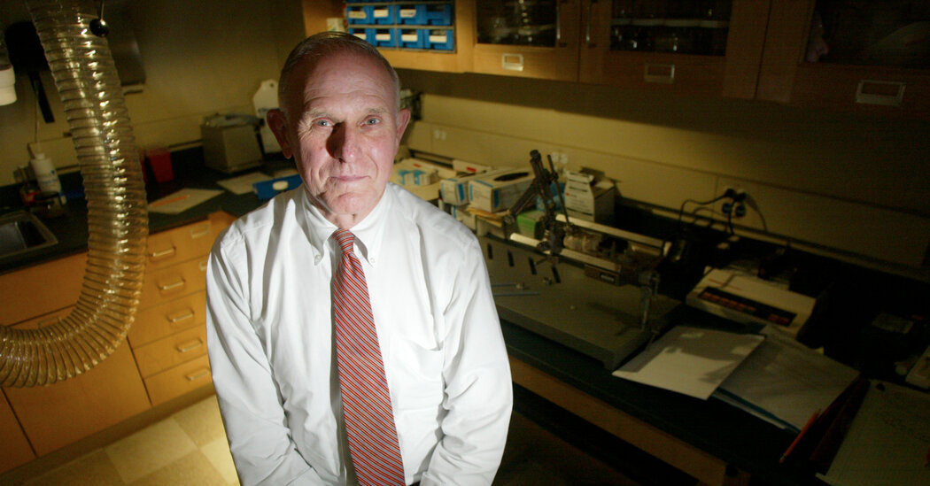
Twice a week, Stephanie, a 37-year-old artist based in Boston, meets with her therapist to work on improving symptoms associated with post-traumatic stress disorder, which include flashbacks, bad dreams and suicidal thoughts.“I was having nightmares where I was yelling out so loudly that my partner could actually hear and transcribe what I was saying on another floor of the house,” said Stephanie, who asked to withhold her last name because of the stigma surrounding mental health.Her sessions, however, are not your average armchair therapy. At some point during each meeting, her therapist, Ainat Rogel, places Stephanie in front of a computer, attaches several electrodes to her skull and instructs her to control a video that appears onscreen — using just her thoughts.The treatment is known as neurofeedback, a therapy some say can improve our attention, moods, sleeping habits and even our athletic ability by measuring brain activity and showing it to us — either on a screen or through headphones — in real time. A practitioner places electrodes on a patient’s head that detects (but does not stimulate) brain activity. This is then analyzed by a computer that sends it back to the patient as images or sounds. As the patient completes tasks, the computer encourages healthy patterns of brain activity.People suffering from anxiety, for instance, may lessen their symptoms by repeatedly being guided to generate brain patterns correlated with a calmer state of mind. Just as we learn to ride a bike by doing it repeatedly, the theory goes, our brains can learn to be less depressed, more focused and better primed for a good night’s sleep.In Stephanie’s case, Dr. Rogel sets a video to play at full screen when sensors detect brain activity corresponding to a reduction in her PTSD symptoms. The video shrinks and becomes more difficult to view when the electrodes detect unhealthy patterns.Stephanie said she isn’t entirely sure what she does to make the video play at full screen. “Sometimes I’ll put a thought into my mind or focus on something I need to work on,” she said. “Sometimes I’ll focus on a memory.”The important part, said Dr. Rogel, who founded the neurofeedback clinic Boston Neurodynamics, is repeated exposure to beneficial brain activity. If done often enough, she said, exercises like these can help patients retrain their brains into healthier patterns.“This is the only thing I’ve tried that’s given me any relief,” Stephanie said, adding that her symptoms have steadily improved since her first week of treatment this past May.Ainat Rogel, a neurofeedback practitioner, said the therapy is still proving itself. She said acupuncture took thousands of years to be taken seriously. “I really hope it doesn’t take us that long,” she added.Tony Luong for The New York TimesOver the last decade, neurofeedback has entered mainstream culture. Tobias Harris of the Philadelphia 76ers uses it to improve his focus. Motivational speaker Tony Robbins credits neurofeedback for increasing his ability to multitask. Well-heeled investors, including the former secretary of education, Betsy DeVos, continue to pour millions into neurofeedback companies that promise dramatic improvements to the ways our brains function.However, neurofeedback is still not accepted as a mainstream treatment within mental health circles — and the most robust research into the intervention so far suggests it is no more effective than a placebo.So … Does it Work?Neurofeedback is a type of biofeedback, which uses sensors to record and provide feedback about a range of biological processes like breathing, heart rate or muscle tension. Those suffering from panic attacks, for instance, might use a device that tracks the respiratory system and promotes measured breathing.Practitioners across the country use neurofeedback to treat conditions like attention deficit hyperactivity disorder, major depressive disorder, anxiety disorder, epilepsy and traumatic brain injuries. The Food and Drug Administrations has cleared a wide range of neurofeedback devices to treat these and other conditions, and the Centers for Disease Control and Prevention list it as an option in cases of ADHD in children, though they stop short of endorsing it.But a 2020 meta-analysis of 17 neurofeedback studies examining its impact on disorders like depression and anxiety was less inspiring. It suggested it is possible to coax our brains into producing certain patterns through repetitive feedback, but the impact on mood disorders was relatively small.“It’s one thing to be able to say, ‘Hey, we can change the brain,’ and another to show that inducing these changes is actually going to impact symptoms,” said David Dodell-Feder, an assistant psychology and neuroscience professor at the University of Rochester and an author of the study.Robert Thibault, a postdoctoral scholar at the Meta-Research Innovation Center at Stanford University, is also skeptical. He said neurofeedback advocates point to peer-reviewed research that have “impressive results,” but most are not rigorous double-blind, placebo-controlled trials. Of the dozen or so such trials, he said, “all but one concluded that fake neurofeedback works just as well as real neurofeedback.”Dr. Thibault said neurofeedback therapy success stories are likely caused by the placebo effect and not the treatment. He suggested that the therapy’s success may have something to do with the “healing environment” that practitioners create in their clinics or the allure of using sophisticated brain-monitoring technology.Though Dr. Rogel said she has personally witnessed the positive impact of neurofeedback on her patients, she would still “welcome the chance” to demonstrate neurofeedback’s effectiveness through double-blind, placebo-controlled trials. “I can say, well, this one client benefited from neurofeedback, and this is great,” she said. But she also wants to see more exhaustive studies that would back up the success she sees in her clinic each day.An Ill-Defined IndustryNeurofeedback practitioners vary widely in their training, background and the services they provide — contributing to confusion within the industry as to who should be administering the therapy and what it entails.“There are so many practitioners out there that, quite frankly, have no business doing neurofeedback,” said Rhonda Potter, an associate executive director of the Biofeedback Certification International Alliance, which offers a neurofeedback certificate program. To date, B.C.I.A. has certified 1,300 neurofeedback practitioners, mostly in North America. Ms. Potter guessed there are “many thousands” more operating across the country, however, that have not sought out certification.She said a formal background in mental health is not a prerequisite to becoming a neurofeedback provider. In many instances, an online course is all that is needed to earn the certificate required to operate one of the dozens of neurofeedback devices on the market.Dr. Rogel working with a patient at the offices of NeuroDynamics, a clinic in Boston she help found. Tony Luong for The New York TimesDuring a neurofeedback treatment, sensors relay signals detected in the brain to a computer that translates them into videos or songs that patient can interact with. Tony Luong for The New York TimesSome companies skip the practitioner entirely by selling pricey neurofeedback devices directly to consumers. Though unlikely to do much harm, Ms. Potter said she would be skeptical of any person or product “claiming you can simply take a piece of equipment out of a box, apply some sensors and say we’re doing neurofeedback.”Even among seasoned practitioners, the therapy varies widely. Some insist on creating a baseline “brain map” before a patient’s first neurofeedback session — applying sensors to the skull for two hours to observe and document brain activity. Others say such maps are optional or unnecessary.Through its accreditation program, B.C.I.A. is attempting to provide stricter standards for the industry, Ms. Potter said. To earn the group’s credentials, practitioners must have a degree in a relevant health care field, take coursework in neuroscience, complete a training program and pass an exam. However, the certificate program, the only one of its kind, is voluntary.Before You Try ItWhile its effectiveness is still debated, neurofeedback is generally thought to be safe. Even critics admit there are few side effects or downsides for those that have the time and money. So if you have read the studies, understand the criticisms, and still want to try your hand (or head) at neurofeedback — here are some things to keep in mind.Know the costs — and find out if your insurance will cover it.The costs of neurofeedback therapy can be prohibitive, and few insurance plans will cover it. A single session costs $100 to $300, but most practitioners say patients need at least 10 exposures, and often many more, to benefit. A brain map and analysis, which some practitioners use to set a baseline for future sessions, can cost $1,000 or more.Neurofeedback practitioners said costs are similar to what you might pay for traditional talk therapy — and potentially less, depending on how many sessions you undergo. Some insurers do cover it, so call your provider to learn about their policies. Many neurofeedback clinics offer work sheets with talking points and questions to help you advocate for coverage.Choose your practitioner carefully.Ms. Potter of B.C.I.A. pointed to a searchable database on the organization’s website as a good place to find a practitioner, though she added there are also plenty of practitioners who lack these credentials and still do good work. She suggested interviewing several practitioners before making a decision — and to prepare questions in advance.“Find out exactly what training they’ve had, and what type of machinery they are trained to use,” she said. “If there’s a specific disorder you’re looking to have treated, make sure they have experience treating that disorder.”Explore all recommended treatment options.Though there are no known side effects to neurofeedback, it could still pose a danger to those who pursue the therapy to the exclusion of other treatment options recommended by health professionals.“One of the biggest issues is delaying proper treatment that’s evidenced to actually work,” said Dr. Thibault. “If you have a learning disorder, and you’re pursuing neurofeedback in place of accepted therapies that would help you correct your learning disorder, then you’re doing yourself harm.”David Dodge is a freelance writer focusing on health, wellness, parenting, travel and the L.G.B.T.Q. community.

Read more →

Alan Scott, Doctor Behind the Medical Use of Botox, Dies at 89

An ophthalmologist and researcher, he discovered a drug that treated serious eye conditions. It also smoothed wrinkles — and an alternative industry was born.It is a neurotoxin 100 times more deadly than cyanide and the cause of the food-borne illness known as botulism. During World War II and for some years after, the Department of Defense hoped to develop it as a chemical weapon. But it wasn’t until the 1970s that Alan Scott, an ophthalmologist, turned this toxin, Clostridium botulinum, into a pharmaceutical, when he began to investigate it as a medical treatment for serious eye impairments.Little did he know at the time that the therapeutic drug he developed would become the basis of a billion-dollar industry famous for its cosmetic use as a temporary wrinkle eraser.Dr. Scott, who came to be called the “Father of Botox,” died on Dec. 16 at a hospital in Greenbrae, Calif. He was 89. The cause was complications of sepsis, his daughter Alison Ferguson said.When, in 1978, Dr. Scott first injected the powerful paralytic Clostridium botulinum into the eye muscles of a patient who had undergone retinal detachment surgery that had left his eye pulled to one side, he didn’t know who was more nervous, himself or the patient, he told Scientific American magazine in 2016.But the procedure succeeded, and Dr. Scott would go on to refine one of the world’s deadliest poisons into a life-altering treatment — he called it Oculinum — for those who suffered from conditions like strabismus, a misalignment of the eyes.Doctors also began using it to treat migraines and jaw-clenching, among other ailments, and as they did so many of their delighted patients noticed a curious byproduct: The toxin’s ability to paralyze targeted facial muscles smoothed the lines around them, though its effects wore off after a few months.Dr. Scott was amused by the drug’s off-label trajectory under a new name, Botox. His focus on it was always solely therapeutic.“I think that’s a charming, slightly frivolous use,” he told The San Francisco Chronicle in 2002, the year the FDA approved Botox for cosmetic purposes.Dr. Scott and his colleagues had spent decades researching and producing what they called Oculinim. But because they had no patent, no pharmaceutical company would manufacture it, and Dr. Scott resorted to taking out a mortgage on his house and asking for small donations from doctors, who then used it in clinical trials.He and his team had already developed Teflon-coated needles to accurately target muscles with various substances before settling on and then refining the toxin to treat strabismus and blepharospasm, a condition that causes the eyes to involuntarily shut tight. In 1989, the Food and Drug Administration approved it for those uses.Dr. Scott had no wish to continue to be a pharmaceutical manufacturer, and in 1991 he sold the rights to make Oculinum to its distributor, Allergan, for an undisclosed amount. The following year, the company changed the drug’s name to Botox.In the decades that followed, the public’s appetite for it as a facial enhancement exploded. Movie directors began complaining that actors were losing their ability to frown or smile properly — “frozen face” became a trope of the tabloids. It was derided as a pernicious enabler of a youth-obsessed society, a practice best left to the stars of reality television.But practitioners grew more skilled at deploying it, and the age of its adherents kept dropping as more and more women maintained that it was a necessary tool for job security in an ageist culture. Now, Botox is a household name, its use seemingly as common as a facial.A pharmaceutical to treat serious eye impairments, developed by Dr. Scott, was found to have a curious byproduct: an ability to paralyze targeted facial muscles and smooth the lines around them. Thus a billion-dollar cosmetic industry was born. Shannon Stapleton/ReutersAlan Brown Scott was born in Berkeley, Calif., on July 13, 1932. His father, Marion Irving Scott, was a dentist; his mother, Helen Elizabeth (Brown) Scott, worked in a laboratory at the University of California, Berkeley.Dr. Scott earned an undergraduate degree in medical sciences from UC Berkeley in 1953, and a medical degree from the University of California, San Francisco. He had a surgical internship and residency in neurosurgery at the University of Minnesota, followed by a residency in ophthalmology at Stanford University. He was a founding member of the Smith-Kettlewell Eye Research Institute in San Francisco and the institute’s senior scientist and co-director for over two decades.He married Ruth White, a teacher and homemaker, in 1956. She died in 2009. In addition to his daughter Alison, Dr. Scott is survived by his wife, Jacquelyn Lehmer; three other daughters, Jennifer, Heidi and Ann Scott; a son, Nathaniel; four stepdaughters, Suzanne, Mary, Sally and Phillis Lehmer; 20 grandchildren; and two great-grandchildren.Dr. Scott was not the first scientist to have explored the therapeutic potential of Clostridium botulinum. “Sausage poison” is what Justinus Kerner, a German poet and doctor, called the pathogen in the 1820s; he had observed the paralytic effects of food poisoning in his town after a single giant sausage sickened 13 people, six of whom died. After injecting it in snails, locusts and rabbits, Dr. Kerner finally injected into himself, noted its inhibiting effect on the autonomic and motor nervous systems, and hypothesized its use as a medical treatment for certain neural conditions. (Decades later, a microbiologist named it Bacillus botulinum, after botulus, the Latin word for sausage.)In 2013, Dr. Scott founded the Strabismus Research Foundation in Mill Valley, Calif., where he developed the use of bipuvicaine, a local anesthetic. At his death he was also working on a treatment procedure involving the electrical stimulation of the eye muscles by means of a tiny implanted pacemaker-like device.Meanwhile, sales of Botox for medical and cosmetic treatments have continued to soar. For the first nine months of 2021, it generated global revenues of more than $3.3 billion, with cosmetic sales accounting for slightly less than half of that figure, according to the earnings report for that period from AbbVie, the company that acquired Allergan in 2020.But Dr. Scott never regretted selling the drug.“I had my house paid for, my kids were educated,” he told The San Francisco Chronicle. “And I had the satisfaction of seeing absolutely wonderful medical results. So I was satisfied.” He added, “I’m not terribly good at giving away and spending money anyway.”

Read more →

Face masks cut distance airborne pathogens could travel in half, new study finds

The effectiveness of face masks has been a hotly debated topic since the emergence of COVID-19. However, a new study by researchers at the University of Central Florida offers more evidence that they work.
In a study appearing today in the Journal of Infectious Diseases, the researchers found that face masks reduce the distance airborne pathogens could travel, when speaking or coughing, by more than half compared to not wearing a mask.
The findings are important as airborne viral pathogens, such as SARS-CoV-2, can be encapsulated and transmitted through liquid droplets and aerosols formed during human respiratory functions such as speaking and coughing.
Knowing ways to reduce this transmission distance can help keep people safe and aid in managing responses to pandemics, such as COVD-19, which has resulted in global-scale infection, health care system overloads, and economic damage.
These responses could include relaxing some social distancing guidelines when masks are worn.
“The research provides clear evidence and guidelines that 3 feet of distancing with face coverings is better than 6 feet of distancing without face coverings,” says study co-author Kareem Ahmed, an associate professor in UCF’s Department of Mechanical and Aerospace Engineering.

Read more →

Skin-related side effects indicate better prognosis for patients taking certain cancer drugs

Immune checkpoint inhibitors, which strengthen the immune response against tumor cells, have become standard of care for many patients with advanced cancers; however, the medications can often cause side effects, most commonly affecting the skin. A new study led by researchers at Massachusetts General Hospital (MGH) and published in JAMA Dermatology indicates that these side effects may actually be an indicator that the medications are working.
For the study, investigators accessed the TriNetX Diamond network, a database of health records and claims data from more than 200 million U.S. and European patients. The team compared information for 14,016 patients with advanced cancer who were treated with immune checkpoint inhibitors: 7,008 who developed skin-related side effects and 7,008 who did not.
The median study follow-up was 3.2 years and 3,233 (26.1%) of the patients had died during that time. Patients who experienced at least one skin-related adverse event had a 22% decrease in mortality. Interestingly, this protective effect was not the same for all skin-related adverse events and was strongest among patients who developed vitiligo (loss of skin color in blotches), lichen planus (an inflammatory skin condition), itchiness, dryness, and non-specific rashes, ranging from a 30%-50% protection from mortality.
“These data provide oncologists and dermatologists with important prognostic information when counseling immunotherapy recipients on the clinical implications of the skin toxicities,” says senior author Yevgeniy R. Semenov, MD, an investigator in the Department of Dermatology at MGH. “Also, skin toxicities tend to occur early in the course of immunotherapy and present an opportunity to evaluate efficacy soon after initiating treatment. As such, our findings may help identify patients who are more likely to benefit from their current immunotherapy regimen versus those who may need to be considered for a stronger or alternative treatment regimen.”
Additional research is needed to understand the mechanisms behind the relationships between these skin reactions and a patient’s prognosis, and whether interventions used to treat or prevent them may affect survival.
Story Source:
Materials provided by Massachusetts General Hospital. Note: Content may be edited for style and length.

Read more →

Assessing Progression Risk in Cancer

A molecular feature in prostate cancer, called endogenous retroviral (ERV) RNA, has been found to have prognostic value and also distinguish differences between men of African and European or Middle Eastern ancestry, according to a study led by researchers at the University of California, Irvine. The team also identified ERV expression signatures that may be useful for identifying prostate cancer patients at greatest risk of progression regardless of ancestry, which may also extend to progression in other cancers.
Findings from the study, “Expression of Endogenous Retroviral RNA in Prostate Tumors has Prognostic Value and Shows Differences among Americans of African Versus European/Middle Eastern Ancestry,” were recently published in the online journal Cancers.
Prostate cancer is the most common cancer diagnosed in men in the U.S. and affects millions of men worldwide, but there are disparities in its aggressiveness between different ancestries. There is a higher burden among Black American men compared to White American men. Black American patients are diagnosed at an earlier age and at a more advanced stage than White American patients and being Black is an independent predictor of disease relapse in those undergoing radical prostatectomy.
“Measuring ERV expression may have the potential to help physicians predict which patients would most benefit from active surveillance or radical therapy, and they also have the potential to be useful in clinically relevant prognostic models for other cancers,” said Farahnaz Rahmatpanah, PhD, assistant professor in residency in the Department of Pathology & Laboratory Medicine at the UCI School of Medicine. “We also believe that in the future, experiments to knock out or overexpress ERVs in cells and tissue culture may further advance our understanding of the consequences of differential regulation of ERVs among people of different geographical ancestry.”
To better understand the biological basis for disparities, the team investigated two potential roles for ERVs in prostate cancer. They discovered differences in ERV expression among prostate tumors which may be associated with variations in the mechanism of progression between patients of primarily African versus primarily European or Middle Eastern ancestry and determined the pathways where these genes have important functions.
A biochemical recurrence risk-prediction model was developed using clinical data and ERV transcripts, which outperformed prediction models based on clinical data alone. The ERV expression signatures that correlated with biochemical relapse among prostate cancer patients of all ancestries were revealed, indicating that ERVs may be useful for identifying patients at greatest risk of progression and that the utility of ERV expression for studying prostate cancer progression may extend to other cancers.
This work was supported by the National Institutes of Health, American Cancer Society and the National Cancer Institute.
Story Source:
Materials provided by University of California – Irvine. Note: Content may be edited for style and length.

Read more →

'Kick and kill’ strategy aimed at eliminating HIV-infected cells

In a study using mice, a UCLA-led team of researchers have improved upon a method they developed in 2017 that was designed to kill HIV-infected cells. The advance could move scientists a step closer to being able to reduce the amount of virus, or even eliminate it, from infected people who are dependent on lifesaving medications to keep the virus from multiplying and illness at bay.
The strategy, described in the peer-reviewed journal Nature Communications, uses cells that are naturally produced by the immune system to kill infected cells that hide in the body, potentially eradicating them, said Dr. Jocelyn Kim, an assistant professor of medicine in the division of infectious diseases at the David Geffen School of Medicine at UCLA.
“These findings show proof-of-concept for a therapeutic strategy to potentially eliminate HIV from the body, a task that had been nearly insurmountable for many years,” said Kim, the study’s lead author. “The study opens a new paradigm for a possible HIV cure in the future.”
Worldwide, there are currently 38 million people living with HIV, and an estimated 36 million have died of HIV-related diseases in the decades since HIV began circulating, according to UNAIDS.
People with HIV take antiretroviral medication to keep the virus at bay. But HIV has the ability to elude antiretrovirals by lying dormant in cells called CD4+ T cells, which signal another type of T cell, the CD8, to destroy HIV-infected cells. When a person with HIV stops treatment, the virus emerges from those reservoirs and replicates in the body, weakening the immune system and raising the likelihood of opportunistic infections or cancers that can lead to illness or death.
The UCLA-led study continues research on a strategy called “kick and kill,” which many of the same scientists first described in a 2017 paper. The approach coaxes the dormant virus to reveal itself in infected cells, so it can then be targeted and killed. In the earlier study, the researchers gave antiretroviral drugs to mice whose immune systems had been altered to mimic those of humans, and then infected with HIV. They then administered a synthetic compound called SUW133, which was developed at Stanford University, to activate the mice’s dormant HIV. Up to 25% of the previously dormant cells that began expressing HIV died within 24 hours.
But a more effective way to kill those cells was needed.
In the new study, while the mice were receiving antiretrovirals, the researchers used SUW133 to flush HIV infected cells out of hiding. They then injected healthy natural killer cells into the mice’s blood to kill the infected cells. The combination of SUW133 and injections of healthy natural killer immune cells completely cleared the HIV in 40% of the HIV-infected mice.
The researchers also analyzed the mice’s spleens — because the spleen harbors immune cells, it’s a good place to look for latent HIV-infected cells — and did not detect the virus there, suggesting that cells harboring HIV were eliminated. In addition, the combination approach performed better than either the administration of the latency reversing agent alone or the natural killer cells alone.
Kim said the researchers’ next objective is to further refine the approach to eliminate HIV in 100% of the mice they test in future experiments. “We will also be moving this research toward preclinical studies in nonhuman primates with the ultimate goal of testing the same approach in humans,” she said.
The study was funded by the National Institutes of Health, the American Foundation for AIDS Research, the National Science Foundation, a National Center for Advancing Translational Sciences UCLA CTSI Grant and the McCarthy Family Foundation.
The study’s co-authors are Tian-Hao Zhang, Camille Carmona, Bryanna Lee, Dr. Christopher Seet, Matthew Kostelny, Nisarg Shah, Hongying Chen, Kylie Farrell, Dr. Mohamed Soliman, Melanie Dimapasoc, Michelle Sinani, Dr. Kenia Yazmin, Reyna Blanco, David Bojorquez, Hong Jiang, Yuan Shi, Yushen Du, Ren Sun and Jerome Zack of UCLA; Natalia Komarova, Dominik Wodarz and Matthew Marsden of UC Irvine; and Paul Wender of Stanford University. Sun is also a member of the faculty of the University of Hong Kong.

Read more →

Epigenetic mechanisms for parent-specific genetic activation decoded

Hereditary diseases as well as cancers and cardiovascular diseases may be associated with a phenomenon known as genomic imprinting, in which only the maternally or paternally inherited gene is active. An international research team involving scientists at the Technical University of Munich (TUM), the Max Planck Institute for Molecular Genetics (MPIMG) in Berlin and Harvard University in Cambridge (USA) has now investigated the mechanisms responsible for the deactivation of the genes.
Our cells contain the entire genetic information from our mother and our father. From each of them we inherit 23 chromosomes that contain our DNA. Two copies of each gene are therefore present in our genome and, as a general rule, both are active. This has the advantage that defective mutations inherited from the mother or father are generally cancelled out by the other copy of the gene.
However, for around one percent of our genes, only the gene inherited from the father or mother is active, while the other is deactivated, a phenomenon known as genomic imprinting.
Approach for treating diseases
“Many genetic and epigenetic diseases are associated with genomic imprinting, such as Beckwith-Wiedemann syndrome, Angelman syndrome and Prader-Willi syndrome,” explains Dr. Daniel Andergassen, the head of the Independent Junior Research Group at the Institute of Pharmacology and Toxicology at TUM. “If the healthy, deactivated gene could be reactivated, it would be theoretically possible to compensate for complications caused by the active, defective gene.”
“But before developing future treatments, we need to understand the fundamentals,” says Prof. Alexander Meissner, director at the MPIMG. “It has become clear in recent years that genomic imprinting is mediated by multiple molecular mechanisms.”
Read lock for the gene

Read more →

New treatment target ID’d for radiation-resistant cervical cancer

Understanding how cells die is key to developing new treatments for many diseases, whether the goal is to make cancer cells die or keep healthy cells alive in the face of other illnesses, such as massive infections or strokes. Two new studies from Washington University School of Medicine in St. Louis have identified a previously unrecognized pathway of cell death — named lysoptosis — and demonstrate how it could lead to new therapies for cervical cancer.
Both studies, which together analyzed data in roundworms, mice and human cells, appear Jan. 12 in the Nature journal Communications Biology.
The blood of patients with cervical cancer and other tumor types is dotted with a protein called SERPINB3. According to the new research, when the gene that manufactures SERPINB3 is absent in cervical cancer cells, the tumor cells die more easily when exposed to the stress of radiation. Similarly, microscopic roundworms called C. elegans that are missing the equivalent gene die more easily when exposed to stresses in their environments.
“It’s been known for a long time that high levels of this protein in the blood are a marker of cervical cancer and other squamous cell cancers — the higher the protein levels in the blood, the worse the prognosis,” said Stephanie Markovina, MD, PhD, an assistant professor of radiation oncology.
“We wondered if this protein may be doing something to protect the cancer. We thought it was possible that the gene was protecting the cancer cells from stress in the same way the equivalent roundworm gene was protecting C. elegans from stress.”
Markovina collaborated with Gary Silverman, MD, PhD, the Harriet B. Spoehrer Professor and head of the Department of Pediatrics; and Cliff J. Luke, PhD, an associate professor of pediatrics, who had been studying this pathway in C. elegans and mice.

Read more →

Mouse study identifies bacterial protein associated with colorectal cancers

Strains of bacteria that cause common food-poisoning symptoms often contain a toxin that can damage DNA in intestinal cells, potentially triggering colon cancer, according to a study from researchers at the Johns Hopkins Bloomberg School of Public Health.
The discovery raises the possibility that some of the roughly two million new cases of colorectal cancer every year around the world originate from brief and seemingly mild food-poisoning events. It also points to the possibility of future drugs that prevent colorectal cancers by neutralizing the newly identified toxin, UshA.
The findings were published January 12 in the January edition of Cancer Discovery.
Prior research has suggested that certain bacteria that reside in the gut can trigger colorectal cancer via persistent infections involving chronic gut inflammation. Short-term infections causing food poisoning, including traveler’s diarrhea, which normally resolve in a day or two, have traditionally been considered non-carcinogenic.
“We hope this study will motivate other researchers to do epidemiological studies to investigate this potential link between transient diarrheal infections and colon cancer development,” says study senior author Fengyi Wan, PhD, associate professor in the Department of Biochemistry and Molecular Biology at the Bloomberg School.
In the study, Wan’s team performed experiments with a mouse model of transient bacterial diarrheal disease using the bacterium Citrobacter rodentium, which has strong similarities to diarrhea-causing strains of the human-infecting microbe Escherichia coli. The researchers observed that Citrobacter infection quickly led to strong signs of DNA damage in gut-lining cells in the mice.

Read more →

Study challenges evolutionary theory that DNA mutations are random

A simple roadside weed may hold the key to understanding and predicting DNA mutation, according to new research from University of California, Davis, and the Max Planck Institute for Developmental Biology in Germany.
The findings, published January 12 in the journal Nature, radically change our understanding of evolution and could one day help researchers breed better crops or even help humans fight cancer.
Mutations occur when DNA is damaged and left unrepaired, creating a new variation. The scientists wanted to know if mutation was purely random or something deeper. What they found was unexpected.
“We always thought of mutation as basically random across the genome,” said Grey Monroe, an assistant professor in the UC Davis Department of Plant Sciences who is lead author on the paper. “It turns out that mutation is very non-random and it’s non-random in a way that benefits the plant. It’s a totally new way of thinking about mutation.”
Researchers spent three years sequencing the DNA of hundreds of Arabidopsis thaliana, or thale cress, a small, flowering weed considered the “lab rat among plants” because of its relatively small genome comprising around 120 million base pairs. Humans, by comparison, have roughly 3 billion base pairs.
“It’s a model organism for genetics,” Monroe said.

Read more →