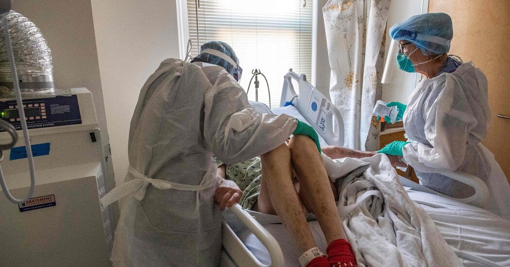
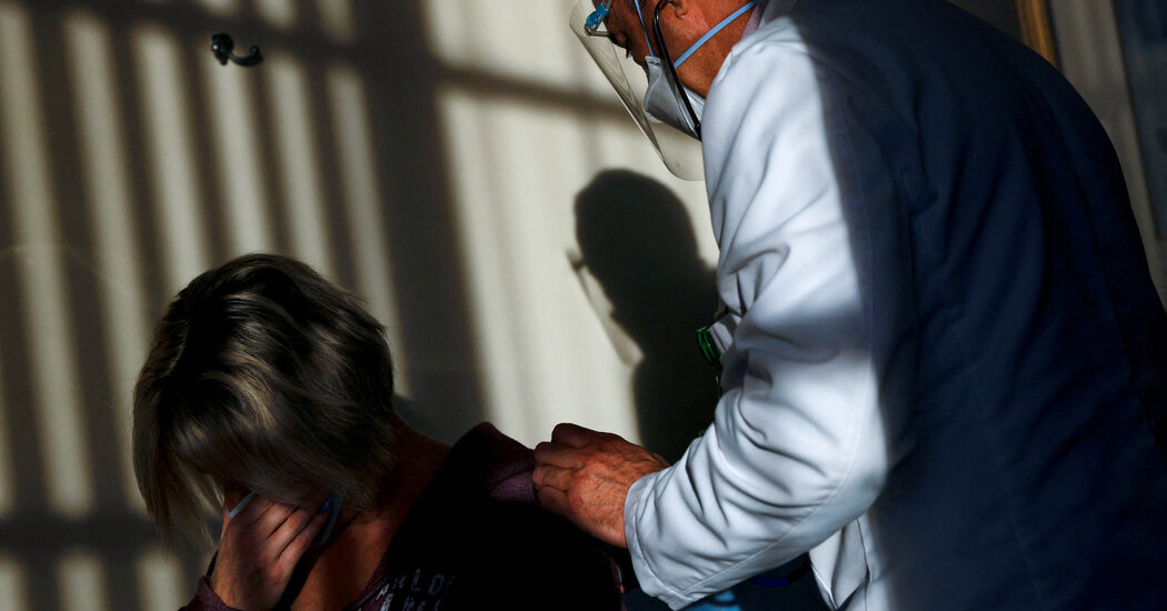
Covid patients may have increased risk of developing mental health problems.

Social isolation, economic stress, loss of loved ones and other struggles during the pandemic have contributed to rising mental health issues like anxiety and depression.But can having Covid itself increase the risk of developing mental health problems? A large new study suggests it can.The study, published Wednesday in the journal The BMJ, analyzed records of nearly 154,000 Covid patients in the Veterans Health Administration system and compared their experience in the year after they recovered from their initial infection with that of a similar group of people who did not contract the virus.The study included only patients who had no mental health diagnoses or treatment for at least two years before becoming infected with the coronavirus, allowing researchers to focus on psychiatric diagnoses and treatment that occurred after coronavirus infection.People who had Covid were 39 percent more likely to be diagnosed with depression and 35 percent more likely to be diagnosed with anxiety over the months following infection than people without Covid during the same period, the study found. Covid patients were 38 percent more likely to be diagnosed with stress and adjustment disorders and 41 percent more likely to be diagnosed with sleep disorders than uninfected people.“There appears to be a clear excess of mental health diagnoses in the months after Covid,” said Dr. Paul Harrison, a professor of psychiatry at the University of Oxford, who was not involved in the study.

Read more →

Blood samples from professional soccer players provides latest evidence that headers affect brain activity

Repetitive headers and accidental head impacts in soccer are leading to changes in blood patterns, linked to brain signalling pathways and potential alterations — the latest study to assess potential dangers of heading in soccer shows.
In this first-of-its-kind new research, experts analysed blood samples from 89 professional soccer players in a range of scenarios, including match-play and training — which involved heading. They also took samples generated by other high-intensity exercise aside from heading.
Results, published in the peer-reviewed journal Brain Injury, demonstrate “specific alterations” in the levels of the brain’s microRNAs — which are being recognized as blood biomarkers, associated with signalling pathways suggestive of brain alterations.
Looking at accidental head impacts specifically; the experts found the microRNAs (biomarkers) affected links to several signalling pathways involved in brain activity. When assessing heading, the experts detected deregulation of other microRNAs — linked to yet another molecular signalling pathway. Importantly, these microRNAs were found to be unaffected by other high-intensity exercise.
Reporting on the findings, Stian Bahr Sandmo, of the Oslo Sports Trauma Research Center at the Norwegian School of Sport Sciences, who led an international team of experts, says the research adds further knowledge to what we understand about head impacts in soccer.
“This is a relatively small sample-size exploratory study,” he says, “but, future findings expanding upon our research could ultimately lead to an improved understanding of the potential hazardous effects of repetitive head impacts. With millions of people playing soccer worldwide this might ultimately have a substantial influence on public health.”
MicroRNAs are small molecules found in our cells, that also circulate in different body fluids such as the blood (then referred to as “circulating microRNAs”). They help regulate gene expression — the process by which the instructions in our DNA are converted into a functional product, such as a protein. They are involved in many different physiological processes. Previous studies have suggested certain microRNAs in the blood are altered in response to mild traumatic brain injury (TBI). This has led to new research — such as this current study — to explore how, and if, microRNAs can be used as brain injury biomarkers. Until now, the effects of soccer-related head impacts on microRNAs have been largely unexplored.

Read more →

Covid: What's life like for unvaccinated people in the UK?

About five million adults in the UK still haven’t had a Covid vaccine, according to government figures and ONS population estimates.Covid vaccines offer very high levels of protection against infection and are safe. Leicester’s director of public health, who took the city through the UK’s first local lockdown, is warning unvaccinated people about the impact their behaviour could have on others. Some of those who’ve chosen not to be jabbed have been speaking to the BBC’s community affairs correspondent, Adina Campbell.Producer: Anthea Lee.Filmed and edited by Carl Ward, Steve Lammiman, John Boon, Raeph Ballantyne and Gemma Laister.

Read more →

Covid Patients May Have Increased Risk of Developing Mental Health Problems

A new, large study found that in the year after getting Covid, people were significantly more likely to be diagnosed with psychiatric disorders they hadn’t had than people who didn’t get infected.Social isolation, economic stress, loss of loved ones and other struggles during the pandemic have contributed to rising mental health issues like anxiety and depression.But can having Covid itself increase the risk of developing mental health problems? A large new study suggests it can.The study, published Wednesday in the journal The BMJ, analyzed records of nearly 154,000 Covid patients in the Veterans Health Administration system and compared their experience in the year after they recovered from their initial infection with that of a similar group of people who did not contract the virus.The study included only patients who had no mental health diagnoses or treatment for at least two years before becoming infected with the coronavirus, allowing researchers to focus on psychiatric diagnoses and treatment that occurred after coronavirus infection.People who had Covid were 39 percent more likely to be diagnosed with depression and 35 percent more likely to be diagnosed with anxiety over the months following infection than people without Covid during the same period, the study found. Covid patients were 38 percent more likely to be diagnosed with stress and adjustment disorders and 41 percent more likely to be diagnosed with sleep disorders than uninfected people.“There appears to be a clear excess of mental health diagnoses in the months after Covid,” said Dr. Paul Harrison, a professor of psychiatry at the University of Oxford, who was not involved in the study. He said the results echoed the emerging picture from other research, including a 2021 study on which he was an author, and “it strengthens the case that there is something about Covid that is leaving people at greater risk of common mental health conditions.”The data does not suggest that most Covid patients will develop mental health symptoms. Only between 4.4 percent and 5.6 percent of those in the study received diagnoses of depression, anxiety or stress and adjustment disorders.“It’s not an epidemic of anxiety and depression, fortunately,” Dr. Harrison said. “But it’s not trivial.”Researchers also found that Covid patients were 80 percent more likely to develop cognitive problems like brain fog, confusion and forgetfulness than those who didn’t have Covid. They were 34 percent more likely to develop opioid use disorders, possibly from drugs prescribed for pain, and 20 percent more likely to develop non-opioid substance use disorders including alcoholism, the study reported.After having Covid, people were 55 percent more likely to be taking prescribed antidepressants and 65 percent more likely to be taking prescribed anti-anxiety medications than contemporaries without Covid, the study found.Overall, more than 18 percent of the Covid patients received a diagnosis of or prescription for a neuropsychiatric issue in the following year, compared with less than 12 percent of the non-Covid group. Covid patients were 60 percent more likely to fall into those categories than people who didn’t have Covid, the study found.The study found that patients hospitalized for Covid were more likely to be diagnosed with mental health issues than those with less serious coronavirus infections. But people with mild initial infections were still at greater risk than people without Covid.“Some people always argue that ‘Oh, well, maybe people are depressed because they needed to go to the hospital and they spent like a week in the I.C.U.,’” said the senior author of the study, Dr. Ziyad Al-Aly, chief of research and development at the V.A. St. Louis Health Care System and a clinical epidemiologist at Washington University in St. Louis. “In people who weren’t hospitalized for Covid-19, the risk was lower but certainly significant. And most people don’t need to be hospitalized, so that is really the group that’s representative of most people with Covid-19.”The team also compared mental health diagnoses for people hospitalized for Covid with those hospitalized for any other reason. “Whether people were hospitalized for heart attacks or chemotherapy or whatever other conditions, the Covid-19 group exhibited a higher risk,” Dr. Al-Aly said.The study involved electronic medical records of 153,848 adults who tested positive for the coronavirus between March 1, 2020, and Jan. 15, 2021, and survived for at least 30 days. Because it was early in the pandemic, very few were vaccinated before infection. The patients were followed until Nov. 30, 2021. Dr. Al-Aly said his team was planning to analyze whether subsequent vaccination modified people’s mental health symptoms, as well as other post-Covid medical issues the group has studied.The Covid patients were compared with more than 5.6 million patients in the Veterans system who did not test positive for the coronavirus and more than 5.8 million patients from before the pandemic, in the period spanning March 2018 through January 2019. To try to gauge the mental health impact of Covid-19 against that of another virus, the patients were also compared with about 72,000 patients who had the flu during the two and a half years before the pandemic. (Dr. Al-Aly said there were too few flu cases during the pandemic to provide a contemporaneous comparison.)The researchers tried to minimize differences between groups by adjusting for many demographic characteristics, pre-Covid health conditions, residence in nursing homes and other variables.In the year after their infection, the Covid patients had higher rates of mental health diagnoses than the other groups.“It’s not really surprising to me because we’ve been seeing this,” said Dr. Maura Boldrini, an associate professor of psychiatry at NewYork-Presbyterian Columbia University Medical Center. “It’s striking to me how many times we’ve seen people with these new symptoms with no previous psychiatric history.”Most veterans in the study were men, three-quarters were white and their average age was 63, so the findings may not apply to all Americans. Still, the study included over 1.3 million women and 2.1 million Black patients, and Dr. Al-Aly said “we found evidence of increased risk regardless of age, race or gender.”There are several possible reasons for the increase in mental health diagnoses, Dr. Al-Aly and outside experts said. Dr. Boldrini said she believed the symptoms were most likely influenced by both biological factors and the psychological stresses associated with having an illness.“In psychiatry, it almost always is an interplay,” she said.Research, including brain autopsies of patients who died of Covid-19, has found evidence that Covid infection can generate inflammation or tiny blood clots in the brain, and can cause small and large strokes, said Dr. Boldrini, who has conducted some of these studies. In some people, the immune response that is activated to fight against a coronavirus infection may not shut down effectively once the infection is gone, which can fuel inflammation, she said.“Inflammatory markers can disrupt the ability of the brain to function in many ways, including the ability of the brain to make serotonin, which is fundamental for mood and sleep,” Dr. Boldrini said.By themselves, such brain changes may or may not cause psychological problems. But, if someone is experiencing stress from having felt physically ill or because having Covid disrupted their lives and routines, she said, “you may be more prone to not being able to cope because your brain is not functioning 100 percent.”Dr. Harrison, who has conducted other studies with large electronic medical databases, noted that such analyses can miss more granular information about patients. He also said that some people in the comparison groups might have had Covid and not been tested to confirm it, and that some Covid patients might have been more likely to receive diagnoses because they were more worried about their health after Covid or because doctors were quicker to identify psychological symptoms.“There’s no one analysis that tells you the whole story,” Dr. Al-Aly said. “Maybe all of us or most of us experienced some sort of an emotional distress or mental health stress or some sleep problem,” he added. “But people with Covid did worse.”

Read more →

A possible cure for sickle cell?

Sickle cell anemia is an inherited blood disorder where red blood cells become sickle/crescent shaped. It causes frequent infections, swelling in the hands and legs, pain, severe tiredness and delayed growth or puberty. Treatment typically focuses on controlling symptoms and may include pain medicines during crises; hydroxyurea to reduce the number of pain episodes; antibiotics and vaccines to prevent bacterial infections and blood transfusions.
While a remedy for this severe disease has remained elusive, a recent study in the New England Journal of Medicine (Biologic and Clinical Efficacy of LentiGlobin for Sickle Cell Disease/Kanter et. al.), if proven applicable, may be a possible cure.
In an editorial in this week’s New England Journal of Medicine, Martin Steinberg, MD, professor of medicine at Boston University School of Medicine, comments on the results of the study which was the first successful gene therapy for sickle cell disease that adds a gene to patient blood stem cells that prevents complications of sickle cell disease. “Gene therapy with autologous stem cells extends the possibility of a cure to all patients without the need for immunosuppression,” explains Steinberg who also is hematologist at Boston Medical Center.
While patients in the study no longer had symptoms of sickle cell disease, Steinberg points out that most continued to have a shorter than normal lifespan of their red blood cells which he thinks could be associated with some complications in the longer-term.
Steinberg believes that for patients to accept this difficult and very expensive treatment it must be curative or nearly curative and last a lifetime. “At this point we do not yet know the sustainability of the results but based on this study the prospects seem good,” he says.
Steinberg acknowledges that any highly effective gene therapy will not improve the health of most people with sickle cell disease throughout the world. “Most patients with this disease live in Africa and India where access to highly technological health care is limited. What is needed are more drugs that can be taken orally and increase fetal hemoglobin levels. This will have a higher likelihood of benefiting populations suffering the most from this disease.”
This editorial appears online in the New England Journal of Medicine.
Story Source:
Materials provided by Boston University School of Medicine. Note: Content may be edited for style and length.

Read more →

Researchers create personalized organoid models for rare spinal cancer

A new study adds to a growing body of evidence that organoids — lab-grown collections of cells that mimic a patient’s tumor — are a promising avenue for drug discovery to improve outcomes in patients with cancer, particularly rare cancers for which clinical data on drug effectiveness is often lacking.
Organoids are grown in a lab using a patient’s own tissue cells collected during surgery. These “mini tumors” are simpler, smaller versions of bodily organs or tumors that replicate the full-function structures. Scientists in the laboratory of Dr. Alice Soragni at UCLA have pioneered their growth and use to study diseases and possible treatments.
In the latest study, published online Wednesday in Science Advances, investigators led by Dr. Soragni obtained seven tumor samples from five patients with a rare bone cancer called a chordoma, which has few therapeutic options. Chordomas are tumors that arise in the sacrum or skull base. Their primary treatment is surgery, but because of their location, complete removal is not always feasible and recurrence rates are high. Chordomas do not respond to conventional chemotherapy, and their rarity (approximately one case for each million people a year in the U.S.) complicates running trials to identify effective therapies.
“There is a real need for clinically-relevant, personalized models to find therapeutics for chordoma and many other rare cancers,” said Soragni, who is an assistant professor in the Department of Orthopaedic Surgery and a member of the Jonsson Comprehensive Cancer Center. “Because this cancer is so rare and there are few models available, our ability to study how it responds to potential drugs is quite limited. What we have done is to optimize a platform to grow organoids using cells from the tumors to be able to investigate their biological characteristics and determine which pathways might be most promising for treatment by testing them against a wide range of therapeutics.”
As reported in the new study, investigators successfully created viable organoids from all samples. The patient-derived organoids showed morphologies and characteristics closely matching the actual chordoma tumors they were derived from, such as expressing a protein called Ki-67, associated with cell proliferation, and brachyury, a protein that is a well- established marker of chordoma.
The investigators then used the organoids to perform high-throughput drug screening, an automated drug discovery process that allows for large numbers of potential therapeutics to be tested at once, rather than one at a time, significantly accelerating the development and testing of potential targets and drugs. This type of large-scale screening has been key to drug development and testing but this is its first application to patient-derived chordoma organoids.
From the screening, the authors identified several drug targets and biological pathways that could be potentially pursued for therapy for chordoma. They also pinpoint the importance of a personalized approach, given the diverse responses seen among patients as well as between different tumors collected from the same patient.
“We’ve shown that the patient-derived tumor organoids we develop can be effectively screened against hundreds of drugs using our platform, which now includes machine learning approaches to study organoids growth patterns and pathway analysis to find targetable biological processes,” said Soragni. Over the past year, the laboratory of Dr. Soragni has extended its screening capabilities to be able to generate and test hundreds to thousands of drugs on tumor organoids each week thanks to funds from an NIH grant and the Department of Orthopaedic Surgery. “Our automation-compatible approach to grow and screen patient-derived tumor organoids works well for rare carcinomas (Phan et al, 2019) and now even slow growing bone tumors. Our work continues to be focused on generating individualized models for rare tumors, which often lack therapeutic options, with a long-term goal to leverage these results in clinic.”
The study’s first authors are Ahmad Al Shihabi, MD, Project scientist in the lab of Dr. Alice Soragni at UCLA and Ardalan Davarifar, MD, PhD, hematology/oncology clinical fellow in the Soragni Lab. The other authors are Huyen Thi Lam Nguyen, Nasrin Tavanaie, Scott D. Nelson, Jane Yanagawa, Noah Federman, Nicholas Bernthal, and Alice Soragni, all of UCLA, and Francis Hornicek, former UCLA now of University of Miami.
The research was supported by the NIH (R01CA244729 to A.S.) and a Seed Grant from UCLA David Geffen School of Medicine (to A.S., N.F. and J.Y.).

Read more →

Does 'bridging' therapy improve outcome for people with stroke?

There has been debate over the best treatment for a certain type of stroke caused by a blockage of a large artery in the brain. A new meta-analysis finds that people who have this kind of stroke who can be treated within four-and-a-half hours after their symptoms start may do better after their stroke when treated with both a clot-busting drug and physical clot removal, compared to physical removal only. The research is published in the February 16, 2022, online issue of Neurology®, the medical journal of the American Academy of Neurology. Combining the two therapies, called bridging therapy, was linked to better chances of a person surviving and living independently after stroke.
The most common type of stroke is an ischemic stroke, occurring when a vessel supplying blood to the brain is blocked. When the blockage is in a major artery, it is called a large vessel occlusion stroke. Large vessel occlusions of the anterior circulation, which were examined in this study, occur in the front of the brain and are a leading cause of adult disability.
Two-step bridging therapy involves the following: intravenous thrombolysis, injecting clot-busting drugs; and mechanical thrombectomy, a minimally invasive procedure in which the blood clot is removed through a small incision.
“For people with this kind of stroke, our analysis suggests that using clot-busting drug therapy combined with physical removal may be associated with better outcomes compared to treating people with physical removal of the clot only,” said meta-analysis author Gabriela Trifan, MD, of the University of Illinois Chicago and a member of the American Academy of Neurology. “We found that bridging therapy was also linked to better chances for more robust blood flow returning to the brain after stroke, and in turn, better functional independence for people after stroke.”
For the meta-analysis, researchers looked at 41 studies involving 14,885 people with large vessel occlusion strokes with an average age of 70. Of those, 8,238 people were treated with bridging therapy and 6,647 were treated with clot removal alone. The drug alteplase was used for the clot-busting therapy.
Researchers found that people who had bridging therapy had 29% higher odds of being able to live independently after three months. Trifan said that would translate into an anticipated additional 62 people out of every 1,000 people who would be able to live independently with bridging therapy versus clot removal alone. The people receiving bridging therapy also had 24% higher odds of blood flow returning to the parts of their brain affected by stroke compared to people who had clot removal alone, which translated into an anticipated 34 additional people out of every 1,000.
People who had bridging therapy also had 31% lower odds of dying 90 days after their stroke compared to people who had clot removal alone. Trifan said this would translate to an anticipated 59 fewer deaths per 1,000 people.
When the researchers restricted the analyses to the latest six high-quality randomized clinical trials, they found that functional independence and safety outcomes were similar between bringing therapy and clot removal alone. “While these results are not strong enough to change practice at this time, they constitute a step forward into the concept of individualized medicine, where for selected patients in the appropriate clinical settings, clot removal may be as efficient and safe as bridging therapy,” said Trifan.
The meta-analysis does not prove that people with this type of stroke will have better outcomes when treated with both therapies; it only shows an association.
“This meta-analysis demonstrates that bridging therapy is safe and does not increase the risk of hemorrhage or delay the start of clot removal,” Trifan said.
A limitation of the meta-analysis is that alteplase was the only drug allowed in this analysis. In addition, the results are only applicable to people who go to medical centers that provide thrombectomy.
Story Source:
Materials provided by American Academy of Neurology. Note: Content may be edited for style and length.

Read more →

Huntington's, ALS: Where the body fails

Huntington’s, Alzheimer’s, ALS, and multiple other neurodegenerative diseases share a commonality: they are all characterized by proteins (different ones for each disease) aggregating in neurons within the brain and nervous system. Now, Technion scientists have found that the cells have the mechanisms to clear those aggregates — they just fail to activate them. Their study was recently published in Nature Communications.
Proteins are the building blocks and the functioning units of our body. Any time the body needs something done, specific proteins are generated to accomplish it. To do this, the code for the particular protein is read from the DNA, and the protein is built from sub-units called amino acids. It is then folded into the 3D shape it needs to assume. Other proteins, called “chaperones,” assist in this folding process.
Aggregates form when certain proteins form incorrectly. Instead of performing the function they were supposed to perform, they attach to each other, creating sizeable clusters that not only are useless, but also disrupt the cells’ normal functionality. Ph.D. student Kinneret Rozales and M.D./Ph.D. student Amal Younis, working as part of the research group of Professor Reut Shalgi, examined how the cells respond to the aggregates building up inside them.
How can we know how a cell feels? We cannot ask it whether it is happy or in pain. But we can examine which genes the cell expresses. We know the cell would activate certain genes when it feels stress. On the other hand, if everything is fine, those genes would not be activated.
Some of what the cell does in response to stress is activating specific chaperones, in an attempt to correct or remove misfolded proteins. But which chaperones are activated? And which ones are needed to solve the problem? A great many different chaperones are encoded in the human DNA. Rozales and Younis examined 66 of them in cells with Huntington or ALS-associated protein aggregates. Some chaperones, they found, only make things worse. But quite surprisingly, they also found chaperones that could eliminate the aggregates, curing the cell. The tools to cure the disease are already within us, encoded by our own DNA!
Why then, if the necessary chaperones exist, do they not cure patients’ cells before neurons degenerate? “It is not enough that the tools exist in the cell’s toolbox,” said Prof. Shalgi. “The cell needs to realize there is a problem, and then it needs to know which out of the many tools available to it, it should use to solve the problem.”
Unfortunately, the group found, this is where the bottleneck lies. In cells with Huntington-associated protein aggregates, the cells sensed there was a problem, and activated some stress-response chaperones, but not the correct ones. The cells did not know what was causing the stress, or what they should do to correct the situation. With ALS-associated aggregates, things were even worse; the cells did not realize that they need to activate chaperones at all and displayed no signs of stress.
“The cell is a complicated system,” said Prof. Shalgi in explaining the surprising findings. “Think of your computer: when something is wrong, sometimes you do not realize it at first. It just responds a bit slower than it used to, perhaps, or it throws an error message that you ignore and forget. When you do realize something wrong — in the way of a blue screen or a refusal to start, you, or a technician on your behalf, attempt to diagnose and solve the problem. Sometimes the solution is found straight away, but other times it is something that you never encountered before, and you don’t know which driver needs to be installed, or piece of hardware needs to be replaced. It is the same with our cells: they do not always realize there is a problem, or know how to solve it, even when they do in fact have the tools to do so. The good news is that since the ability is there, we hope future treatments can be developed to activate it and employ the body’s own tools to cure these debilitating neurodegenerative diseases.”
The study was done in collaboration with the Berlin Lab at the Rappaport Faculty of Medicine and supported by the Israeli Science Foundation (ISF), the ERC, the Prince Center for Neurodegenerative Disorders, and the Rappaport Institute.

Read more →

Unexpected findings detailed in new portrait of HIV

Using powerful tools and techniques developed in the field of structural biology, researchers at the University of Washington and The Scripps Research Institute have discovered new details about the human immunodeficiency virus, HIV. The findings bring into focus the basic architecture of the virus just above and below its surface and may help in the design and development of a vaccine that can protect against AIDS.
These detailed findings include 3D views of the structure and position of the virus’ envelope “spike” proteins (the Env protein, used when the virus binds with cells) in the context of the full virus. Normally, researchers view the protein particles separated from the virus or expressed as engineered or purified proteins. In another key development, the scientists shed new light on the glycan shields — the sugars on the proteins that can hide it from the body’s immune system.
“We’re looking at the whole virus particle and how this protein on the surface relates to the rest of the virus,” said lead author Kelly Lee, associate professor of medicinal chemistry in the UW School of Pharmacy. “And by looking at the intact virus structure, we can see how the different facets of this ‘face of the virus’ are being displayed and how they would be recognized by or hidden from the immune system.”
This intact view of the virus also allowed the scientists to gain new insights into positioning of the envelope spike protein on the surface relative to the internal protein structure, called the Gag lattice.
“This finding overturns previous models of how the parts of the viruses are assembled and helps to focus our attention on where the docking interaction of these two proteins is likely to be,” Lee said. “This interaction needs to be resolved in more detail, but at least the current work gives us the correct architectural model for the virus assembly.”
It was this particular finding that led to the title of the paper — “Cryo-ET of Env on intact HIV virions reveals structural variations and positioning on the Gag lattice” — published Feb. 4 in the journal Cell. Another finding that had not been previously observed, the scientists pointed out, is that the “stalk” supporting the envelope proteins is flexible and can tilt, presenting both opportunity and challenges to the immune system’s neutralizing antibodies that protect cells from infection.
“Structural biology has driven HIV vaccine design, so as we get a better and better picture of what it is we’re targeting, that inspires innovation and may lead to improved vaccines,” said co-corresponding author Michael Zwick, associate professor of immunology and microbiology at the Scripps Research Institute.
HIV’s envelope presents a particularly difficult target for vaccine development, Zwick added, because the virus displays so few spikes and camouflages them with sugar molecules so as to evade our immune system.
“All these features increase the dynamic variability that the HIV spike protein presents to the immune system,” said Lee, who also directs a UW lab exploring virus structure and dynamics. “This is something that people in HIV vaccine development have grappled with from the very beginning — this virus mutates and changes itself astronomically and rapidly. Each time it infects an individual, you end up with literally thousands of different variants within that one individual, and if you look across populations, it’s multiplied even more.”
In fact, in February, an even more deadly strain of HIV was found to have been circulating in the Netherlands. Luckily, while the strain is a “highly virulent variant,” it still responds to treatment.
“This is just another reminder that these viruses are always changing, so we need scientists to continue studying them,” Zwick said.
Co-authors are Vidya Mangala Prasad, former acting instructor at UW School of Pharmacy, currently at the Indian Institute of Science; Daniel Leaman, Department of Immunology and Microbiology, The Scripps Research Institute; Klaus Lovendahl, Jacob Croft and Edgar Hodge, Department of Medicinal Chemistry, UW School of Pharmacy; and Mark Benhaim, who worked on the project as a graduate student at UW.
Story Source:
Materials provided by University of Washington. Original written by Jake Ellison. Note: Content may be edited for style and length.

Read more →