Study shows differences between brains of girls, boys with autism

Brain organization differs between boys and girls with autism, according to a new study from the Stanford University School of Medicine.
The differences, identified by analyzing hundreds of brain scans with artificial intelligence techniques, were unique to autism and not found in typically developing boys and girls. The research helps explain why autism symptoms differ between the sexes and may pave the way for better diagnostics for girls, according to the scientists.
Autism is a developmental disorder with a spectrum of severity. Affected children have social and communication deficits, show restricted interests and display repetitive behaviors. The original description of autism, published in 1943 by Leo Kanner, MD, was biased toward male patients. The disorder is diagnosed in four times as many boys as girls, and most autism research has focused on males.
“When a condition is described in a biased way, the diagnostic methods are biased,” said the study’s lead author, Kaustubh Supekar, PhD, a clinical assistant professor of psychiatry and behavioral sciences. “This study suggests we need to think differently.”
The study was published online Feb. 15 in The British Journal of Psychiatry.
“We detected significant differences between the brains of boys and girls with autism, and obtained individualized predictions of clinical symptoms in girls,” said the study’s senior author, Vinod Menon, PhD, a professor of psychiatry and behavioral sciences and the Rachael L. and Walter F. Nichols, MD, Professor. “We know that camouflaging of symptoms is a major challenge in the diagnosis of autism in girls, resulting in diagnostic and treatment delays.”
Girls with autism generally have fewer overt repetitive behaviors than boys, which may contribute to diagnostic delays, the researchers said.

Read more →

How superbugs uses mirror images to create antibiotic resistance

Methicillin-resistant Staphylococcus aureus (MRSA) is a bacterial infection that has become resistant to most of the antibiotics used to treat regular staph infections. Duke computer scientist Bruce Donald and collaborators at the University of Connecticut are working to develop new enzyme inhibitors to fight MRSA. In research published in PLOS Computational Biology, the team discovered how a single small mutation makes a big difference in drug efficacy.
They examined dihydrofolate reductase (DHFR), an enzyme that antibiotics target to fight MRSA. Drugs that inhibit DHFR work a bit like locks and keys; they bind to enzymes in MRSA, which have a specific three-dimensional structure that only allows molecules that fit precisely to attach to them.
A mutation can change the structure of a bacterial enzyme and cause drugs to lose effectiveness. The F98Y mutation is a well-known resistance mutation. A slight change in the 98th amino acid in the DHFR enzyme changes a phenylalanine to a tyrosine. “Those two amino acids are structurally similar,” said Graham Holt, grad student in the Donald lab, “but the mutation has a huge effect on the efficacy of the inhibitors.” In essence, it changes the lock.
Pablo Gainza, PhD, former graduate student in the Donald lab, thought he should be able to predict this mutation using OSPREY, a suite of programs for computational structure-based protein design developed in the Donald lab. But he couldn’t. After knocking down hypothesis after hypothesis to figure out why he was unable to predict this mutation, he went back to examine the starting structure.
“We looked at the electron density data from the crystallographer and found something strange,” Donald said. In trying to determine the structure of the F98Y mutant, crystallographers used a computer program that — unbeknownst to them — flipped the chirality, or made a mirror image, of the NADPH cofactor to get a better fit. The “flipped” chemical species they discovered through their analysis exists in experimental conditions in the laboratory and plausibly in vivo.
“Using OSPREY, we discovered this flipped chirality,” Donald said, “which we believe happened because of the F98Y mutation.” As in 2-factor authentication, the single enzyme mutation and the flipped cofactor appear to conspire together to evade the inhibitor.
This “chiral evasion” changes the structural basis for resistance. But now Donald and colleagues know not only how a single small mutation changed the lock, but also the structure they need to make a better key — a better drug inhibitor.
“This is the first example of an enzyme that exploits the chirality of its cofactor in order to evade its inhibitors,” Holt said. “Now that we see this happening, that will help inform computational strategies to develop better inhibitors.”
The Donald lab showed that, by taking flipped chirality into account, OSPREY’s predictions closely match experimental measurements of inhibitor potency. They worked with collaborators at the University of Connecticut who conducted biochemical experiments to test the theory and provide structural evidence.
“This is only the beginning of the story,” Donald said. “Our discovery of chiral evasion should lead to more resilient inhibitors: better drug designs.” Right now, most drug design is reactive, waiting for resistance to arise, which it always does. “We hope to make drug design proactive, by using our algorithms to anticipate resistance,” Donald said.
Story Source:
Materials provided by Duke University. Original written by Alissa Kocer. Note: Content may be edited for style and length.

Read more →

How motor neurons develop into subtypes that activate different muscles

Motor neurons play a vital role in movement, linking the central nervous system with different muscles in the body.
As such, scientists are very interested in understanding the biological mechanisms that control how these neurons form.
On Feb. 17 in Nature Communications, researchers report an exciting advance in this field. They have uncovered new details about the process through which motor neurons develop into subtypes that connect the spinal cord with different target muscles and help to control different body parts.
Led by biologists at the University at Buffalo, the research concludes that a gene called Kdm6b helps control motor neurons’ fate. The study, which was completed in mice, finds that Kdm6b: Encourages motor neurons to develop into subtypes found in the medial motor column. These neurons target dorsal axial muscles. Encourages motor neurons to develop into subtypes found in the hypaxial motor column. These neurons target intercostal and abdominal muscles. Discourages motor neurons from developing into subtypes found in the lateral motor column. These neurons target ventral and dorsal limb muscles. Discourages motor neurons from developing into subtypes found in the preganglionic motor column identities. These neurons target the sympathetic ganglia, which control internal organs, such as the heart.The study also reports that Kdm6b works cooperatively with a complex of proteins called Isl1-Lhx3 to influence the way motor neurons diversify.
“During early development, humans generate nerve cells that connect with muscles and control muscle activity,” says UB biologist Soo-Kyung Lee, the study’s senior author. “The formation of these nerve cells at the right time and place is critical for humans’ survival and movement controls. Our study in mice revealed how these nerve cells acquire their specialized identity. Our study could inform strategies to generate specialized nerve cells and treat motor system disorders and spinal cord injuries.”
Lee, PhD, is Empire Innovation Professor and Om P. Bahl Endowed Professor in the Department of Biological Sciences in the UB College of Arts and Sciences.
Jae W. Lee, PhD, UB professor of biological sciences, is also a co-author of the new paper in Nature Communications, and the study’s first authors are two former Lee lab postdoctoral researchers: Wenxian Wang, PhD, who worked with the Lees at UB, and Hyeyoung Cho, PhD, who worked with the Lees at Oregon Health and Science University.
“One of the most fundamental questions, but a poorly understood topic in neuroscience, is how a single neuronal population diversifies into subtypes with distinct synaptic targets,” says Jae W. Lee. “Our paper provides a crucial insight into this important topic, making a major contribution to understanding how motor neurons further develop to different columnar clusters.”
The study was funded by the U.S. National Institutes of Health.
Story Source:
Materials provided by University at Buffalo. Original written by Charlotte Hsu. Note: Content may be edited for style and length.

Read more →

Will Adults Need a Fourth Dose of Covid Vaccine? It’s Too Soon to Know.

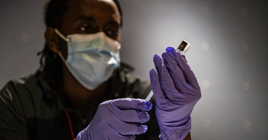
A Food and Drug Administration official said the best time for an additional shot may be fall, when the spread of the coronavirus is expected to pick up again WASHINGTON — Although new federal data suggests that the effectiveness of booster shots wanes after about four months, the Biden administration is not planning to recommend fourth doses of the coronavirus vaccine anytime soon.“We simply don’t have enough data to know that it’s a good thing to do,” Dr. Peter Marks, who heads the division of the Food and Drug Administration that regulates vaccines, said in an interview this week. In a separate interview, Dr. Anthony S. Fauci, the chief medical adviser to the White House, said the vaccines are still a firm bulwark against severe illness, despite data from the Centers for Disease Control and Prevention showing that booster shots lose some of their potency after four to five months.The C.D.C.’s research, released last Friday, analyzed hospitalizations and visits to emergency rooms and urgent care clinics in 10 states by people who had had booster shots of either Moderna’s or Pfizer-BioNTech’s vaccine. The study showed the level of protection against hospitalization fell from 91 percent in the two months after a third shot to 78 percent after four to five months. Effectiveness against visits to emergency rooms or urgent care clinics declined from 87 percent to 66 percent. The data came with major caveats: Researchers did not examine variations by age group, underlying medical conditions or the presence of immune deficiencies. Still, they said, the findings underscored the possible importance of a fourth shot.“‘Should I get a fourth shot?’ That’s what a lot of people are asking me,” Dr. Fauci said. “The answer is if you look at where we are now, it looks like it’s good protection. Seventy-eight percent is good.” The administration’s vaccine strategy has been under constant review since President Biden took office. What comes next, Dr. Fauci said, will depend on whether protection from boosters holds steady or continues to drop after four to five months — and if it keeps dropping, how steeply.“It’s not only the number, it’s the inflection of the curve,” he said.That means more uncertainty for Americans exhausted by frequent changes in vaccine recommendations — pivots largely forced by the onset of new variants. Dr. Sterling Ransone, president of the American Academy of Family Physicians, said his patients keep asking about whether a fourth shot will be necessary and if so, when.“It’s frustrating, right?” said Dr. Ransone, who practices in the small town of Deltaville, Va. “We humans want some certainty and control of the situation. And this is a case where we don’t know what’s going to happen in the future. We don’t know the exact recommendation.”In Bangor, Maine, Dr. James W. Jarvis, who leads Covid response for Northern Light Health, a local health care system, said that he stresses to his patients how well the vaccines are working, even if boosters are needed. Although they don’t offer complete protection, he said, “the most recent data really suggests that these vaccines are still doing a good job.”Data from Britain is similar to that from the C.D.C., indicating that boosters are about 75 percent to 85 percent effective against hospitalization four to six months after they are given. Israel has also noted waning of the Pfizer-BioNTech vaccine’s effectiveness in the months after a booster shot, according to the C.D.C.Israel began offering a fourth shot in late December, but only to health care workers. The C.D.C. has recommended that those with immune deficiencies get three shots as part of their initial series, followed by a fourth shot as a booster.Biden administration officials say two-thirds of eligible adults have gotten a booster shot since the additional injections were authorized in November. Uptake has been slower among children over 12, who only became eligible in early January.The Coronavirus Pandemic: Key Things to KnowCard 1 of 3The state of the virus in the U.S.

Read more →

An easier way to grow model organs

During the past ten years, scientists who study how the human body develops and functions on the most basic level have enjoyed a renaissance of sorts, thanks to structures called organoids-tiny 3D models of organs developed from pluripotent stem cells that grow in petri dishes.
Organoids are derived from human pluripotent stem cells, which can be coaxed into any cell type in the human body and have become an important research tool for understanding human development and disease. They have allowed scientists to move away from simple two-dimensional growth of cells in culture and have provided important insights into the complex three-dimensional form and function of various organs, such as the lungs, brain and heart. However, growing tiny organs in a dish is a tricky process.
A University of Michigan Medical School laboratory, headed by Jason Spence, Ph.D., of the Department of Internal Medicine, has developed a new, significantly simpler way of cultivating a 3D model of the intestine that leads to increased complexity and organization. The advance, published in Cell Reports, details how intestinal organoids now include cells that make up the serosal mesothelium, the protective, outermost layer of the intestine. This layer is also found lining many other organ systems and is critical for the production of a non-adhesive surface allowing relatively frictionless movement of organs within the abdominal cavity.
Past research growing various types of mini organs relied on a supportive gel called Matrigel, which forms a 3D scaffolding that allowed different cell types to develop into an organoid.
“Matrigel is the gold-standard for organoid cultures, but has limitations,” explained Meghan Capeling, a Ph.D. candidate in Spence’s lab and lead of the new research. For one, Matrigel is very expensive, at around $200 for 5mL of product. Second, it’s derived from mouse tumor cells, “so if you’re considering downstream clinical applications, it wouldn’t work well because it contains unknown biological components,” said Capeling.
In an earlier paper published in 2018, Capeling and her colleagues determined that intestinal organoids could be grown in a simpler, biologically inert alginate gel, as they form their own supportive mesenchymal cells — cells that in the developing fetus that turn into connective tissue and smooth muscle.
This finding led the team to wonder if the cells needed a 3D growth environment at all. The answer, they determined, is no. In the new paper, they describe their successful generation of the human intestinal organoid in a simple suspension culture.
“It actually looks somewhat like bubble tea,” said Capeling, “It is just a normal tissue culture plate filled with growth media.” (Growth media is a liquid with life-sustaining chemicals and nutrients for the growth of cells.) They compared the suspension organoids to actual human tissue, as well as to organoids formed using Matrigel and alginate and found that they looked similar at the molecular level. In fact, the suspension organoids more closely resembled the actual human tissue.
The team’s next step was to see whether these floating mini-intestines could actually function like a developing human intestine, and they sought to use the organoids to understand how the serosal layer forms, noting that little-to-nothing is known about this process in the context of human development. The team interrogated the chemical cues that cause the serosa to form in suspension culture, says Capeling.
“This is one of the first studies to gain an idea of the specific regulators that might play a role in the proper development of the intestinal serosa.”
Given that abnormal development of the serosa can lead to congenital defects, the team hoped to leverage the organoids to uncover how this tissue layer forms normally. Using drugs that block the activity of specific proteins, Capeling and the team identified two pathways, called Wnt and Hedgehog, that were essential for the normal formation of the serosa. Though the suspension method resulted in fewer organoids overall, for researchers using human pluripotent stem cells, the method could be a game-changer. The team hopes that suspension culture will open up the possibility of larger-scale organoid experiments and will be an improved system to study human development and disease.
Additional authors include Sha Huang, Charlie Childs, Joshua H. Wu, Yu-Hwai Tsai, Angeline Wu, Neil Garg, Emily M. Holloway, Nambirajan Sundaram, Carine Bouffi, Michael Helmrath and Jason R. Spence.

Read more →

Dr. Herbert Benson, Who Saw the Mind as Medicinal, Dies at 86

A cardiologist and best-selling author, he was initially a skeptic before finding that a person can influence bodily health through Eastern-style meditation.Herbert Benson, a Harvard-trained cardiologist whose research showing the power of mind over body helped move meditation into the mainstream, died on Feb. 3 at a hospital in Boston. He was 86.His wife, Marilyn Benson, said the cause was heart disease and kidney failure.Dr. Benson did not set out to champion meditation; in fact, even after his first pioneering studies, he remained a skeptic, picking up the practice himself only decades later.He was, however, open to the possibility that state of mind could affect a person’s health — common sense today, but a radical, even heretical idea when he began researching it in the mid-1960s.During a stint working for the U.S. Public Health Service in Puerto Rico, he noticed that island residents often had significantly lower blood pressure than their mainland counterparts, all else being equal. He began to wonder if part of the cause lay outside the usual explanations of diet and exercise, a question he took up when he returned to Harvard as a researcher in 1965.Working in a lab at Boston City Hospital (now Boston Medical Center), he and his colleagues devised a way to train monkeys to raise and lower their blood pressure, based on a reward system. The work was low-key; many medical researchers took it as fact that while a stressful situation could raise heart rates thanks to the fight-or-flight response — discovered, coincidentally, in the same lab where Dr. Benson worked — the mind itself had no control over it.Word got out, though, and one day he was approached by several followers of the founder of transcendental meditation, a technique that claims to allow practitioners to enter a higher state of consciousness through the repetition of a mantra. Why teach monkeys, they told him, when we have already perfected the practice?“At first I didn’t want to get involved with them,” Dr. Benson told The New York Times in 1975, referring to the meditation practitioners. “The whole thing seemed a bit far out and somewhat peripheral to the traditional study of medicine. But they were persistent, and so finally I did agree to study them.”To avoid attention, he insisted they come after hours, and through a side door. He attached sensors to their chests and masks to their faces, to measure their breathing, and then had them switch between periods of normal thinking and focused meditation.The meditators were right: Across a variety of metrics — heart rate, oxygen intake — they showed an immediate and significant drop during their contemplative moments, akin, Dr. Benson said, to entering a sleep state while still awake.“I wasn’t so shocked as I was wary because I knew what was ahead of me because the negative mind-body bias was so strong,” he told Brainworld magazine in 2019. “I remained a cardiologist and also being head of cardiovascular teaching at Harvard Medical School, but I sustained two professional lives. I kept respectability within cardiology while I also did work in the mind-body field.”Working with Robert Keith Wallace, a young physiologist at the University of California, Los Angeles, he published his first findings in the early 1970s. Press reports called him a renegade and a maverick, and many in his profession shunned him.But others were impressed by the strength of his research, and by his objectivity. Unlike some researchers at the time, including Dr. Wallace, Dr. Benson was not an advocate of transcendental meditation; in fact, he split with Dr. Wallace when he insisted that there was nothing special about the practice or the use of mantras — any word or phrase, repeated over and over, will do, he said.Dr. Benson called his approach the relaxation response — the opposite of the fight-or-flight response. But whereas a stressful situation will cause the body to automatically raise its heart rate and release adrenalin, the relaxation response has to be asserted consciously.He demonstrated just how to do that in his 1975 book, “The Relaxation Response.” It hit at the right time: That same year the transcendental meditation movement claimed more than 400,000 adherents, studying at more than 300 centers around the United States alone.Millions more Americans, if skeptical about alternative medicine and Eastern spirituality, were still meditation-curious, and Dr. Benson, with his Ivy League pedigree and clinical approach to research, gave them license to indulge. The book sold more than four million copies and was a New York Times best seller.Over time, Dr. Benson’s insistence on the connection between the mind and the body became accepted, even standard fare among establishment researchers. In 1992 he founded the Mind-Body Institute, which in 2006 moved to Massachusetts General Hospital and, with an infusion of money from the investor John W. Henry, changed its name to the Benson-Henry Institute for Mind Body Medicine, with Dr. Benson as its director emeritus.Dr. Benson was the first Western doctor allowed to interview Tibetan monks about their practices, and he became friends with the Dalai Lama, right, when that Buddhist spiritual leader visited Boston in 1979. via Benson-Henry Institute for Mind Body MedicineHerbert Benson was born on April 24, 1935 in Yonkers, N.Y. His father, Charles, ran a series of wholesale produce businesses, and his mother, Hannah (Schiller) Benson, was a homemaker.He graduated from Wesleyan University in 1957 with a degree in biology and received his medical degree from Harvard in 1961.Along with his wife, he is survived by a son, Gregory; a daughter, Jennifer Benson; and four grandchildren.Dr. Benson wrote 11 books after “The Relaxation Response,” several of which delved further into the physiological effects of spirituality and faith. He was the first Western doctor allowed to interview Tibetan monks about their practices, and he became friends with the Dalai Lama during that Buddhist spiritual leader’s visit to Boston in 1979.Dr. Benson found, among other things, that Buddhist monks could, during meditation, raise their body temperature enough to completely dry damp sheets that had been draped over their bodies.Such findings were later disputed, and Dr. Benson was rarely without his critics. But he was undeterred, comparing himself to William James, a Harvard predecessor and another pioneer at the intersection of the mind and the body.Dr. Benson was not a praying man himself, but by the 1990s he was convinced that prayer, and faith in general, had a physiological impact. For him, the explanation lay in a version of the placebo effect: If we believe something is helping us, our bodies will work harder to heal.With a $2.4 million grant from the John Templeton Foundation, in 1996 he undertook a decade-long study on the healing power of prayer — specifically, whether one person’s prayers could help another.The conclusions, released in 2006, were definitive, and disappointing (at least to believers): Intercessional prayer not only had no impact, but in some cases where people believed they were being prayed for, they got worse — a result, Dr. Benson said, of their conviction that if someone was praying for them, they must be very ill, with their body trying to match that impression by getting sicker.Still, Dr. Benson believed that prayer could help at least a sick person doing the praying. And he always took care to say that even if his research was 100 percent accurate, meditation and prayer could never replace drugs and surgery completely.Both medical treatment and spiritual care, he said, were necessary — a fact that Western medicine had long tried to ignore, and one that he spent his career trying to correct.

Read more →

New medicines for treating heart patients

New University of Cincinnati research discovered a unique class of medications that act as blood thinners by inhibiting an enzyme in the genes of tick saliva.
The research focused on novel direct thrombin inhibitors (DTI) from tick salivary transcriptomes, or messenger RNA molecules expressed by an organism. The result is the development of new anticoagulant medications that can be used to treat patients with a variety of coronary issues, including heart attacks. The study was published in Nature Communications.
“Interest in ticks as a model for developing drugs that prevent blood clotting — [often] the cause of heart attacks and strokes — is firmly rooted in evolutionary biology,” says Richard Becker, MD, professor and director of the UC Heart, Lung and Vascular Institute and UC Division of Cardiovascular Health and Disease at the UC College of Medicine.
“Analysis of backbone structures suggest a novel evolutionary pathway by which different blood clot inhibiting properties evolved through a series of gene duplication events. Comparison of naturally occurring blood clot inhibitors of differing tick species suggests an evolutionary divergence approximately 100 million years ago.”
Becker, a co-author on the study, collaborated with researchers from the National University of Singapore, Duke University and the University of North Carolina on the study, which discovered DTIs from tick salivary transcriptomes and optimized their use as a pharmaceutical. The most potent is a key regulating enzyme in blood clot formation with very high specificity and binding capacity that is almost 500 times that of bivalirudin, a drug used during a typical nonsurgical procedure used to treat narrowing of the coronary arteries. Those minimally invasive procedures are performed in roughly 1 million persons yearly in the United States.
“Despite their greater ability to reduce the incidence of the formation of blood clots, the drugs demonstrated less bleeding, achieving a wider therapeutic index in nonhuman models,” Becker says. “The higher potency of the drug means it’s not necessary to use a lot of it in treating patients, which holds the cost of goods and manufacturing down.”
Becker says tick saliva, as in others that feed on blood like mosquitoes, sand flies, tsetse and black flies, contains pharmacological and immunological active compounds, which modulate immune responses and induce antibody production. This research leveraged an understanding of tick-host interactions and antibody formation.
“The holy grail of anticoagulant therapy has always been specificity, selectivity, efficacy and safety,” says Becker. “Clinician-scientists must have the training and an environment that embraces asking questions and finding solutions, including those potential found deep within nature. An ability to both measure and adjust the drug dose and rapidly reverse its effects is particularly important for safety purposes. The next step is to complete pharmacology, toxicology, drug stability and other important regulatory steps before conducting clinical trials in humans.”
Story Source:
Materials provided by University of Cincinnati. Original written by Bill Bangert. Note: Content may be edited for style and length.

Read more →

Well-functioning fat may be the key to fewer old-age ailments

Fat tissue plays an important role in human health. However, our fat tissue loses function as we age, which can lead to type 2 diabetes, obesity, cancer and other ailments. High levels of lifelong exercise seem to counteract this deterioration. This, according to research at the University of Copenhagen, where biologists studied the link between aging, exercise and fat tissue function in Danish men.
How well does your fat function? It isn’t a question that one gets asked very often. Nonetheless, research in recent years suggests that the function of our fat tissue, or adipose tissue, is central to why our bodies decay with age, and strongly linked to human diseases like diabetes 2, cancer as obesity often develop and fat cells undergo functional changes as we get older. Thus, overall health is not just influenced by the amount of fat we bear, but about how well our fat tissue functions.
A new University of Copenhagen study demonstrates that even though our fatty tissue loses important function with age, a high volume of exercise can have a significant impact for the better.
“Overall health is closely linked with how well our fat tissue functions. In the past, we regarded fat as an energy depot. In fact, fat is an organ that interacts with other organs and can optimize metabolic function. Among other things, fat tissue releases substances that affect muscle and brain metabolism when we feel hungry and much more. So, it’s important that fat tissue works the way it should,” explains Assistant Professor Anders Gudiksen of the University of Copenhagen’s Department of Biology.
Fat cell function worsens with age
Gudiksen and a group of colleagues looked at the role of age and physical training in maintaining fat tissue function. Specifically, they studied mitochondria, the tiny power plants within fat cells. Mitochondria convert calories from food to supply cells with energy. To maintain the life processes within cells, they need to function optimally.

Read more →

More chemicals, fewer words: Exposure to chemical mixtures during pregnancy alters brain development

By linking human population studies with experiments in cell and animal models, researchers have provided evidence that complex mixtures of endocrine disrupting chemicals impact children’s brain development and language acquisition. With their novel approach, the scientists show that up to 54 per cent of pregnant women were exposed to experimentally defined levels of concern. While current risk assessment tackles chemicals one at a time, these findings show the need to take mixtures into account for future risk assessment approaches.
There is increasing evidence that environmental chemicals to which we are continuously exposed can have endocrine disrupting properties and can thus be dangerous to human and animal health and development. Every year sees the release of a huge number of new compounds as part of the market authorisation and production processes of a vast range of goods, chiefly but not only plastic derivatives, that enter the human body from several sources, including water, food and air. While exposure levels for individual chemicals are often below existing limit values, exposure to the same chemicals in complex mixtures can still impact human health. Yet all existing risk assessments, and thus established limit values, are based on chemicals being examined one at a time. There was thus a strong need to test whether an alternative strategy would be possible, in which the actual mixtures measured in real life exposures could be tested as such in both the epidemiological and experimental setting. The EU-funded EDC-MixRisk project set out to tackle this unmet need.
“The uniqueness of this comprehensive project is that we have linked population data with experimental studies, and then used this information to develop new methods for risk assessment of chemical mixtures,” says Carl-Gustaf Bornehag, Professor at Karlstad University, Project Manager of the SELMA study and responsible for the epidemiological part of EDC-MixRisk.
The study was conducted in three steps:
• First, a mixture of chemicals in the blood and urine of pregnant women was identified in the Swedish pregnancy cohort SELMA, associated with delayed language development in children at 30 months. This critical mixture included a number of phthalates, bisphenol A, and perfluorinated chemicals.
• Second, experimental studies uncovered the molecular targets through which human-relevant levels of this mixture disrupted the regulation of endocrine circuits and of genes involved in autism and intellectual disability.
• Third, the findings from the experimental studies were used to develop new principles for risk assessment of this mixture.

Read more →

Scientists think a peptide could stop, reverse damage to nerve cells

Researchers at the University of Illinois Chicago found promising results in their search for a treatment to stop nerve cell degeneration that happens in some types of disorders, such as hereditary spastic paraplegia and Parkinson’s disease, which can cause significant disability.
The UIC research, published in the clinical neurology and translational neuroscience journal Brain, was led by Xue-Jun Li, the Michael A. Werckle Professor of biomedical sciences at the College of Medicine Rockford.
The study looked at how the long axons that carry messages between nerve cells in the brain can break down, which causes increasingly worse tightening of the leg muscles, leading to imbalance and eventually paralysis, in addition to other symptoms.
Previous research that used animal models to study the causes of the nerve cell degradation showed it may be a problem with the mitochondria, the powerhouse that drives the cells, that leads to the axons breaking down or not growing long enough.
Studying human nerve cells is difficult, but Li’s team was able to use human cells that they transformed into stem cells and then modified to become nerve cells with the genetic disorder for a particular type of hereditary spastic paraplegia.
“What we found was that the mitochondria in these cells were breaking apart, what we call mitochondrial fission, and that caused the axons to be shorter and less effective at carrying messages to the brain,” Li said. “We then looked at whether a particular agent would change the way the nerve cells function — and it did. It inhibited the mitochondrial fission and let the nerve cells grow normally and also stopped further damage.”
What this means for the thousands of people affected by this type of genetic disorder is that this agent, a particular chain of amino acids called a peptide, could prove to be useful for a drug or other therapy to stop the nerve cells from becoming damaged or reverse the course of the damage. The researchers also suggest that using gene therapy could prevent mitochondrial damage, providing another strategy to reverse the nerve damage.
The research was supported by the National Institute of Neurological Disorders and Stroke of the National Institutes of Health (R21NS109837).
Story Source:
Materials provided by University of Illinois Chicago. Original written by Carrie Foust. Note: Content may be edited for style and length.

Read more →