Cellular rejuvenation therapy safely reverses signs of aging in mice

Age may be just a number, but it’s a number that often carries unwanted side effects, from brittle bones and weaker muscles to increased risks of cardiovascular disease and cancer. Now, scientists at the Salk Institute, in collaboration with Genentech, a member of the Roche group, have shown that they can safely and effectively reverse the aging process in middle-aged and elderly mice by partially resetting their cells to more youthful states.
“We are elated that we can use this approach across the life span to slow down aging in normal animals. The technique is both safe and effective in mice,” says Juan Carlos Izpisua Belmonte, co-corresponding author and a professor in Salk’s Gene Expression Laboratory. “In addition to tackling age-related diseases, this approach may provide the biomedical community with a new tool to restore tissue and organismal health by improving cell function and resilience in different disease situations, such as neurodegenerative diseases.”
As organisms age, it is not just their outward appearances and health that change; every cell in their bodies carries a molecular clock that records the passage of time. Cells isolated from older people or animals have different patterns of chemicals along their DNA — called epigenetic markers — compared to younger people or animals. Scientists know that adding a mixture of four reprogramming molecules — Oct4, Sox2, Klf4 and cMyc, also known as “Yamanaka factors” — to cells can reset these epigenetic marks to their original patterns. This approach is how researchers can dial back adult cells, developmentally speaking, into stem cells.
In 2016, Izpisua Belmonte’s lab reported for the first time that they could use the Yamanaka factors to counter the signs of aging and increase life span in mice with a premature aging disease. More recently, the team found that, even in young mice, the Yamanaka factors can accelerate muscle regeneration. Following these initial observations, other scientists have used the same approach to improve the function of other tissues like the heart, brain and optic nerve, which is involved in vision.
In the new study, Izpisua Belmonte and his colleagues tested variations of the cellular rejuvenation approach in healthy animals as they aged. One group of mice received regular doses of the Yamanaka factors from the time they were 15 months old until 22 months, approximately equivalent to age 50 through 70 in humans. Another group was treated from 12 through 22 months, approximately age 35 to 70 in humans. And a third group was treated for just one month at age 25 months, similar to age 80 in humans.
“What we really wanted to establish was that using this approach for a longer time span is safe,” says Pradeep Reddy, a Salk staff scientist and co-first author of the new paper. “Indeed, we did not see any negative effects on the health, behavior or body weight of these animals.”
Compared to control animals, there were no blood cell alterations or neurological changes in the mice that had received the Yamanaka factors. Moreover, the team found no cancers in any of the groups of animals.

Read more →

New early signs of Parkinson’s uncovered in diverse study

Hearing loss and epilepsy are early features of Parkinson’s, according to pioneering new research from Queen Mary University of London — the first UK study of the condition in such a diverse population, published today (7 March 2022) in JAMA Neurology.
Queen Mary researchers funded by Bart’s Charity used electronic primary healthcare records from over a million people living in East London between 1990 and 2018 to explore early symptoms and risk factors for Parkinson’s.
The researchers found that known symptoms associated with Parkinson’s, including tremor and memory problems, can appear up to ten and five years before diagnosis respectively. They also uncovered two new early features of Parkinson’s, epilepsy and hearing loss, and were able to replicate these findings using additional data from the UK Biobank.
Whilst early signs of Parkinson’s have been described previously, these studies have largely focused on affluent white populations, with patients from minority ethnic groups and those living in areas of high social deprivation largely under-represented in Parkinson’s research to date. The new study provides further evidence of risk factors and early signs of Parkinson’s, using data from such a diverse and deprived urban population for the first time.
In East London, conditions like hypertension and Type 2 diabetes were associated with increased odds of developing Parkinson’s. The researchers also observed a stronger association between memory complaints within this population than previously described.
East London has one of the highest proportions of Black, South Asian and mixed/other ethnic groups, which comprise around 45% of residents in the area, in comparison to 14% in the rest of the UK. It also has some of the highest levels of deprivation in the UK, and 80% of patients included in the study were from low-income households.

Read more →

Field-based patient trial for cell-free Zika testing delivers highly accurate results

An international team of researchers, headed by experts from the University of Toronto’s Leslie Dan Faculty of Pharmacy, has led one of the first field trials for a synthetic biology-based diagnostic using patient samples. This work, conducted on-site in Latin America, reveals the potential for cell-free synthetic biology tools and companion hardware for providing rapid, de-centralized, and low-cost patient testing for infectious diseases like the Zika virus.
Study results, published today in Nature Biomedical Engineering, show that the novel diagnostic platform has analytical specificity and sensitivity equivalent to a US Centre for Disease Control PCR test for Zika and a diagnostic accuracy of 98.5 per cent with 268 patient samples collected in Recife, Brazil. The platform is also programmable and can be similarly applied to detect any pathogen sequence. In addition to validating highly accurate diagnostic results for Zika, the team also achieved similar diagnostic performance for chikungunya virus, another mosquito-borne arbovirus.
“We see emerging diagnostics, like the paper-based tests we’ve developed, as having tremendous near-term potential to augment existing PCR capacity, improve equity in access to health care, and aid in the responses to public health crises,” said Keith Pardee, assistant professor in the department of pharmaceutical sciences, Leslie Dan Faculty of Pharmacy, University of Toronto.
Prior to the current global COVID-19 pandemic, the 2015/2016 outbreak of Zika virus in Latin America emphasized the urgent need for rapid and low-cost testing that can be deployed beyond the reach of centralized diagnostic labs, explains Pardee who has a Canada Research Chair in Synthetic Biology and Human Health. “We were investigating and developing this technology well before the COVID-19 pandemic brought these issues to light at the global level. We’ve now been able to apply it and validate it in a region of endemic disease, which is really promising because these tools are meant to enable health systems to better respond to future outbreaks of infectious disease, particularly in low-resource settings,” he said.
The portable diagnostic platform is a combination of a cell-free, paper-based test and a field-ready companion device that allows data to be collected using image-based color analysis — purple for positive and yellow for negative. Called “PLUM” (Portable, Low-cost, User-friendly, Multimode), the toaster-size reader presents results from up to 384 samples and displays them in a single image capture. The hardware and software that make up PLUM were originally developed by co-authors Livia Guo and Seray Çiçek as part of their graduate work in the Pardee lab. To keep production costs low, Guo and Çiçek, co-founders of LSK Technologies, used customizable software programs and off-the shelf electronics, enabling PLUM to be built for approximately $500 USD per unit.
On the molecular side, the cell-free tests can be freeze-dried, allowing for distribution without refrigeration and, significantly, all of the molecular components of the test are independent of the PCR-supply chain. “Here we have demonstrated that these two technologies combined create a low-cost, highly accurate diagnostic tool,” said study lead author Margot Karlikow, a postdoctoral fellow in the Pardee lab from 2016 to 2021 and now co-founder of En Carta Diagnostics. “We also demonstrated that it is feasible to transport the platform across a significant distance and implement it effectively in another country. In many low- and middle-income countries, there is no PCR testing available outside of main cities, so the ultimate goal is that this platform be used as a high-quality alternative to PCR in more regional settings,” she said.
Dr. Lindomar Pena, department of virology, Oswaldo Cruz Foundation (Fiocruz), led the Brazilian team that collaborated on the project. “This robust diagnostic platform displayed desirable features to be used in developing countries such as Brazil and in laboratories with basic infrastructure. We hope it can be further developed and deployed in the Brazilian network of public health laboratories to diagnose Zika patients, trace contacts and identify hot-spot areas with active community transmission,” he said.
Showing that the platform could be transported and accurately detect Zika virus in patient samples is a significant step forward in creating more accessible and de-centralized testing, says Pardee. However, the extraction of RNA from patient samples still requires liquid handling by skilled technicians at this stage. “With performance on patient samples now validated, we are tackling these next challenges, like sample preparation, so that the platform and PCR-like diagnostic capacity can be distributed more broadly into the communities where they are needed.”

Read more →

Genetic study gives extensive insights into severe COVID-19

The world’s largest study of the genetics of critical Covid-19, involving more than 57,000 people, has revealed fresh details about some of the biological mechanisms behind the severe form of the disease.
Some 16 new genetic variants associated with severe Covid-19, including some related to blood clotting, immune response and intensity of inflammation, have been identified.
These findings will act as a roadmap for future efforts, opening new fields of research focused on potential new therapies and diagnostics with pinpoint accuracy, experts say.
Researchers from the GenOMICC consortium — a global collaboration to study genetics in critical illness — led by University of Edinburgh in partnership with Genomics England, made these discoveries by sequencing the genomes of 7,491 patients from 224 intensive care units in the UK.
Their DNA was compared with 48,400 other people who had not had Covid-19, participants in Genomics England’s 100,000 Genomes Project and that of a further 1,630 people who had experienced mild Covid.
Determining the whole genome sequence for all participants in the study allowed the team to create a precise map and identify genetic variation linked to severity of Covid-19. The team found key differences in 16 genes in the ICU patients when compared with the DNA of the other groups.

Read more →

Lensless camera captures cellular-level, 3D details in living tissue

First, catch the tiger.
Then attach Bio-FlatScope, the latest iteration of lensless microscopy being developed at Rice University.
That particular use is fanciful but not far-fetched, according to Jacob Robinson, an electrical and computer engineer at Rice’s George R. Brown School of Engineering who led the recent effort to test Bio-FlatScope in living creatures.
The research team’s FlatCam, a lensless device that channels light through a mask and directly onto a camera sensor, aimed primarily outward at the world at large. The raw images looked like static, but a custom algorithm used the data they contained to reconstruct what the camera saw.
The new device looks inward to image micron-scale targets like cells and blood vessels inside the body, even through the skin. Bio-FlatScope captures images that no lensed camera can see — showing, for example, dynamic changes in the fluorescent-tagged neurons in running mice.

Read more →

Scans reveal how Covid may change the brain

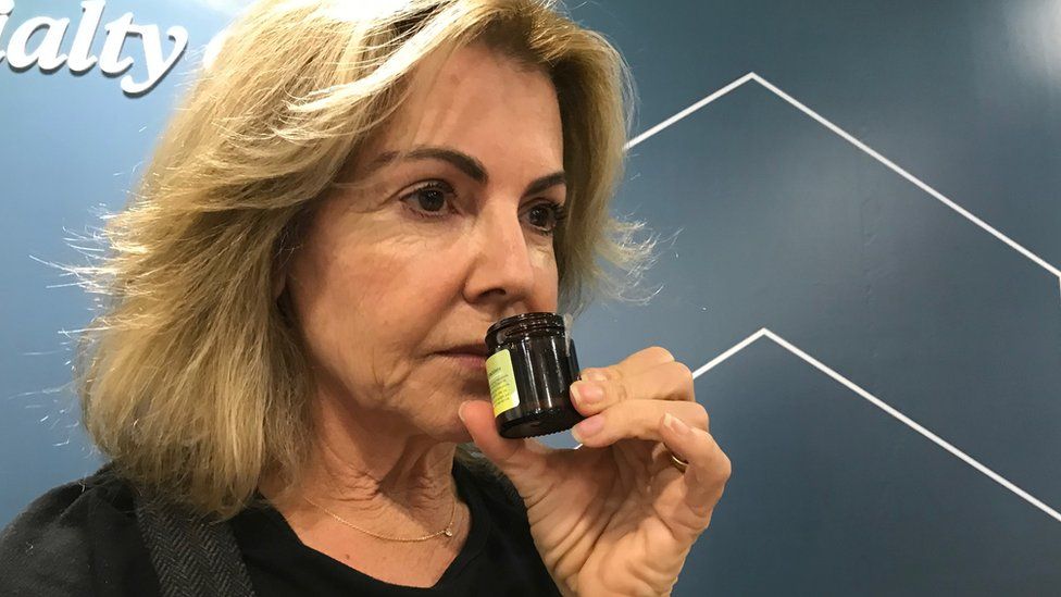
SharecloseShare pageCopy linkAbout sharingCatching Covid may cause changes to the brain, a study suggests.Scientists found significant differences in MRI (magnetic resonance imaging) scans before and after infection.Even after a mild infection, the overall size of the brain had shrunk slightly, with less grey matter in the parts related to smell and memory.The researchers do not know whether the changes are permanent but stressed the brain could heal.The study is published in the journal Nature.Lead author Prof Gwenaelle Douaud, from the Wellcome Centre for Integrative Neuroimaging, at the University of Oxford, said: “We were looking at essentially mild infection, so to see that we could really see some differences in their brain and how much their brain had changed compared with those who had not been infected was quite a surprise.”The UK Biobank project has followed the health of 500,000 people for about 15 years and has a database of scans recorded before the pandemic so provided a unique opportunity to study the long-term health impacts of the virus. The scientists rescanned:401 participants 4.5 months, on average, after their infection, 96% of whom had had mild Covid 384 participants who had not had CovidThey found:The overall brain size in infected participants had shrunk between 0.2 and 2%There were losses in grey matter in the olfactory areas, linked to smell, and regions linked to memoryThose who had recently recovered from Covid found it a bit harder to perform complex mental tasksBut the researchers do not know whether the changes are reversible or truly matter for health and wellbeing.”We need to bear in mind that the brain is really plastic – by that we mean it can heal itself – so there is a really good chance that, over time, the harmful effects of infection will ease,” Prof Douaud said.The most significant loss of grey matter was in the olfactory areas – but it is unclear whether the virus directly attacks this region or cells simply die off through lack of use after people with Covid lose their sense of smell.It is also unclear whether all variants of the virus cause this damage. The scans were performed when the original virus and alpha variant were prevalent and loss of smell and taste a primary symptom. But the number of people infected with the more recent Omicron variant reporting this symptom has fallen dramatically.’Your mind is what is being exercised’Paula Totaro lost her sense of smell when she caught Covid, in March 2020.”When it was gone, it was like living in a bubble or a vacuum – I found it really isolating,” she told BBC News.But after contacting the charity AbScent, which supports people who have lost their ability to smell and taste, she began smell training.”What smell training does – particularly if you do it twice a day, regularly, religiously – is it forces you to take the smell, allow it to go back into your nose and then to think about what it is that you’re smelling,” she said.”And that connection between what’s in the external world and what goes into your brain and your mind is what is being exercised.”Ms Totaro has now recovered most of her sense of smell – although she still has trouble identifying what different smells are.”It’s a mix of joy that the sense has come back but still a little bit of anxiety that I’m not quite there yet,” she said.UK Biobank chief scientist Prof Naomi Allen said: “It opens up all sorts of questions that other researchers can follow up about the effect of coronavirus infection on cognitive function, on brain fog and on other areas of the brain – and to really focus research on how best to mitigate that.”Prof David Werring, from the University College London Institute of Neurology, said other health-related behaviour could have contributed to the changes seen. “The changes in cognitive function were also subtle and of unclear relevance to day-to-day function,” he said.”And these changes are not necessarily seen in every infected individual and may not be relevant for more recent strains.”Follow @BBCMorelle on Twitter.More on this storyScans aim to reveal Covid health legacyUK BiobankWellcome Centre for Integrative NeuroimagingAbScentNature: SARS-CoV-2 is associated with changes in brain structure in UK BiobankThe BBC is not responsible for the content of external sites.

Read more →

Millions of girls fall out of love with sports by teens

SharecloseShare pageCopy linkAbout sharingImage source, Getty ImagesMany girls who used to be very sporty have “fallen out of love” with physical activity as teenagers, a study reveals. The reasons include body image, puberty and fear of judgement, Women in Sport, which surveyed 4,000 teenagers, says. It is a myth girls drop sport simply because their priorities change, the charity says, urging the sport, leisure and education sectors to work harder.Half said they disliked being watched if they exercised and some seven in 10 avoided sport on their period. Other reasons included:lack of confidence (61%), schoolwork pressures (47%) feeling unsafe outdoors (43%) Feeling self-conscious in gym or sports gear was another worry. One girl shared: “My school has a lot of unisex clothes, by unisex they’re made for boys, when you are in puberty and have wider hips it doesn’t fit right. “Skort – you feel vulnerable in them, even if no immediate threat and you’re not around the boys, you still don’t feel comfortable.”Some said they did not “have the right body shape”, while others dislike becoming hot and sweaty. Sport became too competitive as they grew older, some said, and they no longer felt able to join in just for fun. Asked what would motivate them, many said making exercise more fun, with more opportunities outside school with friends.Periods and low confidence put some girls off sportExercise addicts urged to build in rest daysWomen in Sport chief executive Stephanie Hilborne said: “It’s an absolute travesty that teenage girls are being pushed out of sport at such a scale. “Teenage girls are not voluntarily leaving sport, they are being pushed out as a consequence of deep-rooted gender stereotypes. “We must all do more to reverse this trend and not continue to accept this as inevitable.”Mental rewardsKate Dale, from Sport England’s This Girl Can campaign, said: “Sadly, this research is not surprising – the gender activity gap starts young. “Over two-thirds of teenage girls have quit sport altogether by the time they are 16 and 17. “This means that many girls grow into adults who miss out on the physical, social and mental rewards of an active lifestyle. “Positive experiences with physical activity at a young age are vital for building healthy habits for life. “It’s also vital that girls see women and girls who look like them playing sport, to challenge the stereotypes of what women getting active should look like.”This Girl Can has made Studio You, a video library of workouts for teenage girls, free to all schools.More on this storyExercise addicts urged to build in rest daysPeriods and low confidence put some girls off sportCovid’s ‘impact’ on children’s exerciseSport EnglandThis Girl CanWomen In SportThe BBC is not responsible for the content of external sites.

Read more →

Survivors of heart attack plus cardiac arrest at increased risk of early death

Patients who survive a heart attack together with sudden cardiac arrest are at increased risk of dying within six years following hospital discharge.
This is the conclusion of new research published today in the Journal of the American Heart Association.
A heart attack is when blood flow to the heart is blocked, and sudden cardiac arrest is when the heart malfunctions and suddenly stops beating unexpectedly.  Cardiac arrest can be fatal if not treated immediately. 
Most heart attacks do not lead to sudden cardiac arrest. But when sudden cardiac arrest occurs, heart attack is a common cause.
The study, by researchers from the National Institute of Health Research Health Informatics Collaborative (NIHR-HIC) led by Imperial College Healthcare NHS Trust and Imperial College London, draws on data from 13,444 patients collected between 2010 and 2017.  The study was funded by the National Institute for Health Research (NIHR) Imperial Biomedical Research Centre (BRC). 
It shows for the first time that patients who had heart attacks and then a sudden cardiac arrest were at a greater risk of developing further complications. Those who had a cardiac arrest at the time of their heart attack were twice as likely as those suffering heart attack alone to go on to develop abnormal heart rhythms known as ventricular arrhythmia (VA).  Those with a cardiac arrest were also 36 per cent more likely to die on average within three years following discharge from hospital.

Read more →

Is it working? Scientists say gene variant indicates effectiveness of immunotherapy for allergies

Seasonal allergies are very widespread in certain parts of the world. In Japan, it is estimated that about one third of the population is allergic to the pollen of the Japanese cedar, a native tree species, making Japanese cedar pollinosis one of the most common allergic diseases in the country. Luckily, allergen immunotherapy has progressed much over the past few decades, becoming the closest thing we have to a cure for both seasonal and year-round allergies. In sublingual immunotherapy (SLIT), patients can gradually develop immunity to a given allergen by regularly placing small, concentrated doses of it under their tongue. After several months, a good percentage of patients find themselves reacting much less severely when exposed to the same allergens in their daily lives.
While SLIT is beneficial for most people with allergic rhinitis — that is, those who suffer from inflammation of the inside of the nose after allergen exposure — the treatment is ineffective for about 20-30% of them. Unfortunately, there is currently no way of telling if SLIT will be effective for a patient short of trying it out and observing their response over a period of as long as two years, meaning that 20-30% of patients would have to tolerate all the side effects of the treatment for no benefit, for that long a period.
Against this backdrop, a team of researchers from Japan set out to find a biomarker that could be used to predict the responsiveness of a person to SLIT for Japanese cedar pollinosis. In their most recent paper, published online on 12th February 2022 in Allergy, they report a newly discovered association between a specific variant of the HLA-DPB1 gene and poor response to SLIT. This work was the result of a collaborative effort led by Prof. Shigeharu Fujieda and Dr. Masanori Kidoguchi of the University of Fukui, and Prof. Emiko Noguchi of the University of Tsukuba.
So, what is the HLA-DPB1 gene and why would it be related to one’s responsiveness to SLIT? This gene provides instructions for making a protein that plays a critical role in the immune system: helping it distinguish the body’s own proteins from proteins made by foreign invaders, such as bacteria and viruses. The protein encoded by the HLA-DPB1 gene forms a functional protein complex with the protein encoded by the HLA-DPA1 gene. However, these genes are highly polymorphic, meaning that a large number of genetic variants (alleles) exist. In previous studies, this research team had discovered that certain structural differences in the antigen-binding pockets between HLA-DPB1 alleles could make an individual more susceptible to Japanese cedar pollinosis and sensitization.
This led them to think that there might also be a connection between the alleles of HLA-DPB1 and an individual’s responsiveness to SLIT. To test their hypothesis, they enrolled over 200 patients with Japanese cedar pollinosis who underwent SLIT. The researchers determined the HLA-DPB1 alleles in these patients and conducted statistical analyses to see if they were related to their responsiveness to SLIT. “Our results suggest that patients carrying at least one HLA-DPB1*05:01 allele have an increased risk of being non-responders to SLIT in their second season of the immunotherapy,” says Prof. Fujieda. “This implies that differences in the antigen-binding pocket on the HLA-DPB1 protein may influence the effect of allergen immunotherapy,” he adds.
It is worth noting that this may be the first study ever to find an association between a genetic biomarker and an individual’s response to allergen immunotherapy. Genotyping the HLA-DPB1 gene could serve as a cost-effective biomarker to predict the responsiveness of a given patient to SLIT for Japanese cedar pollinosis, saving valuable time. Moreover, these findings may help researchers worldwide rethink how genetic biomarkers can be used both in research and clinical practice, as Prof. Fujieda remarks: “Our study could prompt updates in current international guidelines and consensus documents on the potential of genetic biomarkers.”
Let us hope immunotherapy keeps advancing until no one has to suffer the consequences of severe allergies.
Story Source:
Materials provided by University of Fukui. Note: Content may be edited for style and length.

Read more →

People with heart defects may be at greater risk for severe COVID-19 illness

People with a congenital heart defect who were hospitalized with COVID-19 infection were at higher risk for severe illness or death than those without a heart defect, according to new research published today in the American Heart Association’s flagship, peer-reviewed journal Circulation. Researchers found people with a congenital heart defect who contracted COVID-19 were also more likely to require treatment in the intensive care unit (ICU) or need a ventilator.
Among those at the highest risk for the most severe COVID-19 illness were patients who had a heart defect and another health condition, were ages 50 and older, or were men, according to the study.
There are more than a dozen types of congenital heart defects, which result when the heart, or blood vessels near the heart don’t develop normally before birth. According to the American Heart Association’s Heart Disease and Stroke Statistics — 2022 Update, congenital heart defects are the most common birth defect worldwide, with a global prevalence of 157 per 100,000 in 2017.
“Data comparing COVID-19 outcomes among individuals with and without congenital heart defects has been limited,” said lead author Karrie Downing, M.P.H., an epidemiologist at the National Center on Birth Defects and Developmental Disabilities and the COVID-19 Response Team at the U.S. Centers for Disease Control and Prevention in Atlanta.
Researchers examined data on hospitalized COVID-19 patients from March 2020 to January 2021, collected in the Premier Healthcare Database Special COVID-19 Release, a database representing approximately 20% of all U.S. hospitalizations. The COVID-19 patients with and without heart defects in this study received care in the same hospitals. Differences in age, gender, race/ethnicity, health insurance types and other high-risk conditions (specifically heart failure, pulmonary hypertension, Down syndrome, diabetes and obesity) were accounted for across those populations.
During this period, the database had more than 235,000 patients, ages 1 to 64 years old, who were hospitalized for COVID-19. Patients were divided into two groups: those who had a congenital heart defect and those who did not. Across these two categories, researchers then determined how many required an admission to the ICU, needed a ventilator to help with breathing or died. Researchers also reviewed other characteristics including other health conditions.
Of the 235,638 hospitalized COVID-19 patients evaluated for this study, 421 or 0.2% had a congenital heart defect. The analysis found: among the patients with a heart defect, most were over the age of 30 (73%), and 61% were male; 55% were non-Hispanic white people, 19% were Hispanic people and 16% were non-Hispanic Black people; overall, 68% of the patients with a heart defect also had at least one other health condition noted, compared to 59% among those without a congenital heart defect; 54% of patients with a congenital heart defect were admitted to the ICU compared to 43% of those without a congenital heart defect; 24% of patients with a congenital heart defect required a ventilator to breathe compared to 15% of those without a congenital heart defect; and 11% of patients with a congenital heart defect died during hospitalization compared to 7% of those without a congenital heart defect.In addition, people with congenital heart defects consistently remained at high-risk for severe COVID-19 illness, even when divided into categories by age or other health conditions noted in the study, according to the researchers.
Downing believes these findings have immediate, practical relevance for health care professionals as the COVID-19 pandemic continues to evolve: “People with heart defects should be encouraged to receive the COVID-19 vaccines and boosters and to continue to practice additional preventive measures for COVID-19, such as mask-wearing and physical distancing. People with heart defects should also consult with their health care teams about additional steps to manage personal risks related to COVID-19, given the significantly increased risk of severe infection and serious complications.”
Downing noted that not all patients with heart defects who were hospitalized with COVID-19 had poor outcomes. “More work is needed to identify why the clinical course of COVID-19 disease results in significantly worse outcomes for some hospitalized patients with risk factors for critical COVID-19 illness, like heart defects, and not for others,” she said.
There are several limitations to this study. Only people already hospitalized with COVID-19 were included, the clinical details about the underlying heart defect were not available, and lab testing to identify and/or confirm COVID-19 diagnoses may vary by hospital. Lastly, COVID-19 vaccination status was not considered, since the vaccines became available in the U.S. starting in December 2020.
Co-authors are Regina Simeone, Ph.D.; Matthew Oster, M.D., M.P.H.; and Sherry Farr, Ph.D. Authors’ disclosures are listed in the manuscript.

Read more →