Sleep apnea accelerates aging, but treatment may reverse it

Obstructive sleep apnea (OSA) affects 22 million people in the U.S. and is linked to a higher risk of hypertension, heart attacks, stroke, diabetes and many other chronic conditions. But now researchers from the University of Missouri School of Medicine have found that untreated OSA also accelerates the biological aging process and that appropriate treatment can slow or possibly reverse the trend.
Age acceleration testing involves a blood test that analyzes DNA and uses an algorithm to measure a person’s biological age. The phenomenon of a person’s biological age surpassing their chronological age is called “epigenetic age acceleration,” and is linked to overall mortality and to chronic diseases.
“Age acceleration isn’t unique to OSA — it can be caused by a variety of environmental factors like smoking, poor diet or pollution,” said Rene Cortese, PhD, assistant professor in the Department of Child Health and the Department of Obstetrics, Gynecology and Women’s Health. “In Western culture, it’s not uncommon for people to experience epigenetic age acceleration, but we wanted to know how OSA affects systemic age acceleration compared to those who don’t suffer from this condition.”
Cortese’s team studied 16 adult nonsmokers who were diagnosed with OSA and compared them to eight control subjects without the condition to assess the impact of OSA on epigenetic age acceleration over a one-year period. After a baseline blood test, the OSA group received continuous positive airway pressure (CPAP) treatment for one year before being tested again.
“Our results found that OSA-induced sleep disruptions and lower oxygen levels during sleep promoted faster biological age acceleration compared to the control group,” Cortese said. “However, the OSA patients who adhered to CPAP showed a deceleration of the epigenetic age, while the age acceleration trends did not change for the control group. Our results suggest that biological age acceleration is at least partially reversible when effective treatment of OSA is implemented.”
Cortese said the key to CPAP’s success in slowing age acceleration is strong adherence to using the device for at least four hours per night. It’s not clear how age acceleration will affect clinical outcomes and how it applies to other risk groups or children with OSA.
“Since children with OSA are treated differently from adults, this research raises a lot of questions,” Cortese said. “We need to learn more about the mechanisms and the biology behind these findings. It’s very exciting and thought-provoking research.”
In addition to Cortese, the study authors include MU colleagues Leila Kheirandish-Gozal, MD, director of the Child Health Research Institute; and David Gozal, MD, the Marie M. and Harry L. Smith Endowed Chair of Child Health.
This work was partially supported by grants from the National Institutes of Health, Tier 2 and TRIUMPH grants from the University of Missouri, and a Leda J. Sears Charitable Trust grant.
Story Source:
Materials provided by University of Missouri-Columbia. Note: Content may be edited for style and length.
Read more →

New technology enables unprecedented glimpse inside single brain cells

Salk Institute researchers have developed a new genomic technology to simultaneously analyze the DNA, RNA and chromatin — a combination of DNA and protein — from a single cell. The method, which took five years to develop, is an important step forward for large collaborations where multiple teams are working simultaneously to classify thousands of new cell types. The new technology, published in Cell Genomics on March 9, 2022, will help streamline analyses.
“This multimodal platform is going to be useful by providing a comprehensive database that can be used by the groups trying to integrate their single-modality data,” says Joseph Ecker, director of the Genomic Analysis Laboratory at Salk, the Salk International Council Chair in Genetics and Howard Hughes Medical Institute Investigator. “This new information can also inform and guide future cell-type classification.”
Ecker believes this technology will be vital for large-scale efforts, such as the National Institutes of Health’s BRAIN Initiative Cell Census Network, which he co-chairs. A major effort of the BRAIN Initiative is to develop catalogues of mouse and human brain cell types. This information can then be used to better understand how the brain grows and develops, as well as the role different cell types play in neurodegenerative diseases, such as Alzheimer’s.
Current single-cell technology works by extracting either DNA, RNA or chromatin from a cell’s nucleus, and then analyzing its molecular structure for patterns. However, this method destroys the cell in the process, requiring researchers to rely on computational algorithms to analyze more than one of these components per cell or to compare the results.
For the new method, called snmCAT-seq, scientists used biomarkers to tag DNA, RNA and chromatin without removing them from the cell. This allowed the researchers to measure all three types of molecular information in the same cell. The scientists then used this method to identify 63 cell types in the frontal cortex region of the human brain and benchmarked the efficacy of computational methods for integrating multiple single-cell technologies. The team found the computational methods have high accuracy in characterizing broadly defined brain-cell populations but show significant ambiguity in analyzing finely defined cell types, suggesting the necessity to define cell types by diverse measurements for more accurate classification.
The technology could also be used to better understand how genes and cells interact to cause neurodegenerative diseases.
“These diseases can broadly affect many cell types. But there could be certain cell populations that are particularly vulnerable,” says co-first author Chongyuan Luo, assistant professor of human genetics at the David Geffen School of Medicine at UCLA. “Genetic research has pinpointed the regions of the genome that are relevant for diseases like Alzheimer’s. We’re providing another data dimension and identifying the cell types affected by these genomic regions.”
As a next step, the team plans to use the new platform to survey other areas of the brain, and to compare cells from healthy human brains with those from brains affected by Alzheimer’s and other neurodegenerative diseases.
Other authors included Hanqing Liu, Bang-An Wang, Zhuzhu Zhang, Dong-Sung Lee, Jingtian Zhou, Sheng-Yong Niu, Rosa Castanon, Anna Bartlett, Angeline Rivkin, Jacinta Lucero, Joseph R. Nery, Jesse R. Dixon and M. Margarita Behrens of Salk; Fangming Xie, Ethan J. Armand, Wayne I. Doyle, Sebastian Preissl and Eran A. Mukamel of the University of California San Diego; Kimberly Siletti, Lijuan Hu and Sten Linnarsson of the Karolinska Institutet in Sweden; Trygve E. Bakken, Rebecca D. Hodge and Ed Lein of the Allen Institute for Brain Science in Seattle; Rongxin Fang, Xinxin Wang, and Bing Ren of the Ludwig Institute for Cancer Research in La Jolla, California; Tim Stuart and Rahul Satija of the New York Genome Center; and David A. Davis and Deborah C. Mash of the University of Miami.
The research was supported by the National Institutes of Health (5R21HG009274, 5R21MH112161, 5U19MH11483, R01MH125252, U01HG012079, 5T32MH020002, R01HG010634 and U01MH114812), the Howard Hughes Medical Institute and UC San Diego School of Medicine.
Story Source:
Materials provided by Salk Institute. Note: Content may be edited for style and length.

Read more →

An ‘oracle’ for predicting the evolution of gene regulation

Computational biologists have created a neural network model capable of predicting how changes to non-coding DNA sequences in yeast affect gene expression. They also devised a unique way of representing this data in two dimensions, making it easy to understand the past and future evolution of non-coding sequences in organisms beyond yeast — and even design custom gene expression patterns for gene therapies and industrial applications.
Despite the sheer number of genes that each human cell contains, these so-called “coding” DNA sequences comprise just 1% of our entire genome. The remaining 99% is made up of “non-coding” DNA — which, unlike coding DNA, does not carry the instructions to build proteins.
One vital function of this non-coding DNA, also called “regulatory” DNA, is to help turn genes on and off, controlling how much (if any) of a protein is made. Over time, as cells replicate their DNA to grow and divide, mutations often crop up in these non-coding regions — sometimes tweaking their function and changing the way they control gene expression. Many of these mutations are trivial, and some are even beneficial. Occasionally, though, they can be associated with increased risk of common diseases, such as type 2 diabetes, or more life-threatening ones, including cancer.
To better understand the repercussions of such mutations, researchers have been hard at work on mathematical maps that allow them to look at an organism’s genome, predict which genes will be expressed, and determine how that expression will affect the organism’s observable traits. These maps, called fitness landscapes, were conceptualized roughly a century ago to understand how genetic makeup influences one common measure of organismal fitness in particular: reproductive success. Early fitness landscapes were very simple, often focusing on a limited number of mutations. Much richer data sets are now available, but researchers still require additional tools to characterize and visualize such complex data. This ability would not only facilitate a better understanding of how individual genes have evolved over time, but would also help to predict what sequence and expression changes might occur in the future.
In a new study published on March 9 in Nature, a team of scientists has developed a framework for studying the fitness landscapes of regulatory DNA. They created a neural network model that, when trained on hundreds of millions of experimental measurements, was capable of predicting how changes to these non-coding sequences in yeast affected gene expression. They also devised a unique way of representing the landscapes in two dimensions, making it easy to understand the past and forecast the future evolution of non-coding sequences in organisms beyond yeast — and even design custom gene expression patterns for gene therapies and industrial applications.
“We now have an ‘oracle’ that can be queried to ask: What if we tried all possible mutations of this sequence? Or, what new sequence should we design to give us a desired expression?” says Aviv Regev, a professor of biology at MIT (on leave), core member of the Broad Institute of Harvard and MIT (on leave), head of Genentech Research and Early Development, and the study’s senior author. “Scientists can now use the model for their own evolutionary question or scenario, and for other problems like making sequences that control gene expression in desired ways. I am also excited about the possibilities for machine learning researchers interested in interpretability; they can ask their questions in reverse, to better understand the underlying biology.”
Prior to this study, many researchers had simply trained their models on known mutations (or slight variations thereof) that exist in nature. However, Regev’s team wanted to go a step further by creating their own unbiased models capable of predicting an organism’s fitness and gene expression based on any possible DNA sequence — even sequences they’d never seen before. This would also enable researchers to use such models to engineer cells for pharmaceutical purposes, including new treatments for cancer and autoimmune disorders.

Read more →

How the transition to agriculture affects populations in the present day

The transition of human societies from hunter-gatherers to farmers and pastoralists is a more nuanced process than generally thought, according to a new study of peoples living in the highlands of southwest Ethiopia. The work was published March 9 in Current Biology.
Much of the study of how people transitioned away from a lifestyle based mostly on food collected from the wild to one based on cultivated crops has focused on Europe, where the shift to agriculture, or “Neolithic transition,” concluded thousands of years ago. Based largely on genetic studies, the prevailing view is that the transition occurred mainly by population replacement rather than cultural change, said first author Shyamalika Gopalan, a graduate student at the time of the work advised by Brenna Henn, associate professor of anthropology at the University of California, Davis.
“The prevailing view has been that in Europe it was a wave of people that came through and replaced everyone,” Gopalan said.
The transition to agriculture is still underway in the highlands of southwest Ethiopia. Farmers and pastoralists started moving into the area 1,500 to 2,000 years ago, encroaching on the resident hunter-gatherers, and the groups have since been living alongside each other. That presents an opportunity to study this transition and the degree to which it represents replacement versus cultural change in the present day and a different global context.
The team, led by Henn and Barry Hewlett at Washington State University, Vancouver, collected DNA samples from five groups of people in the southwest highlands: the hunter-gatherer Chabu; the Majang, who practice small-scale cultivation of crops; and the Shekkacho, Bench and Sheko, who practice more intensive farming. The goals were to assess both the genetic ancestry of the different groups and demographic trends in the recent past.
“Based on genetics we can estimate the effective population size over the past 60 generations, or about 2,000 years,” Gopalan said.

Read more →

Scientists identify possible new treatment for COVID-19

Investigators at Cedars-Sinai have identified a potential new therapy for COVID-19: a biologic substance created by reengineered human skin cells.
Scientists found the substance stopped SARS-CoV-2, the virus that causes COVID-19, from reproducing itself and also protected infected cells when tested in human lung cells. Although still in the early stages, the findings open the possibility of having a new therapy for COVID-19 patients. The details of the potential therapy are published in the journal Biomaterials and Biosystems.
“We were surprised to find this potential therapy shuts down a novel pathway for viral replication and also protects infected cells,” saidAhmed G. Ibrahim, PhD, MPH, assistant professor in the Smidt Heart Institute at Cedars-Sinai and first author of the study.
Few treatments currently exist for COVID-19 and the ones that do primarily focus solely on preventing the virus from replicating. This new potential treatment inhibits replication but also protects or repairs tissue, which is important because COVID-19 can cause symptoms that affect patients long after the viral infection has been cleared.
The potential therapy investigated in this study was created by scientists using skin cells called dermal fibroblasts. The investigators engineered the cells to produce therapeutic extracellular vesicles (EVs), which are nanoparticles that serve as a communication system between cells and tissue. Engineering these fibroblasts allowed them to secrete EVs — which the investigators dubbed “ASTEX” — with the ability to repair tissue.
In previous experiments, the investigators demonstrated that ASTEX can repair heart tissue, lung tissue and muscle damage in laboratory mice. When the COVID-19 pandemic hit in 2020, the investigators turned to studying whether ASTEX could be used as treatment against SARS-CoV-2.

Read more →

Man given genetically modified pig heart dies

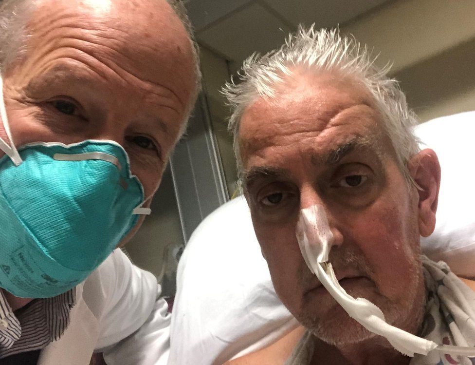
SharecloseShare pageCopy linkAbout sharingImage source, University of Maryland School of MedicineThe first person in the world to get a heart transplant from a genetically-modified pig has died.David Bennett, who had terminal heart disease, survived for two months following the surgery in the US.But his condition began to deteriorate several days ago, his doctors in Baltimore said, and the 57-year-old died on 8 March.Mr Bennett knew the risks attached to the surgery, acknowledging before the procedure it was “a shot in the dark”.Doctors at the University of Maryland Medical Center were granted a special dispensation by the US medical regulator to carry out the procedure, on the basis that Mr Bennett – who was ineligible for a human transplant – would otherwise have died.He had already been bedridden for six weeks leading up to the surgery, attached to a machine which was keeping him alive.Pig kidney transplanted into brain-dead personMr Bennett underwent the surgery on 7 January, and doctors say in the weeks afterwards he spent time with his family, watched the Super Bowl and spoke about wanting to get home to his dog, Lucky.But his condition deteriorated, leaving doctors “devastated”.”He proved to be a brave and noble patient who fought all the way to the end,” surgeon Bartley Griffith, who performed the transplant, said in a statement released by the hospital.But Mr Bennett’s son, David Jr, said he hoped his father’s transplant would “be the beginning of hope and not the end”, according to news agency AP. “We are grateful for every innovative moment, every crazy dream, every sleepless night that went into this historic effort,” he added.Dr Griffith said previously the surgery would bring the world “one step closer to solving the organ shortage crisis”. Currently 17 people die every day in the US waiting for a transplant, with more than 100,000 reportedly on the waiting list.The possibility of using animal organs for so-called xenotransplantation to meet the demand has long been considered, and using pig heart valves is already common.In October 2021, surgeons in New York announced that they had successfully transplanted a pig’s kidney into a person. At the time, the operation was the most advanced experiment in the field so far. However, the recipient on that occasion was brain dead with no hope of recovery.More on this storyUS man gets pig heart in world-first transplantBid to grow transplant organs in pigsThree ethical issues around pig heart transplantsPig kidney transplanted into brain-dead personGM pigs take step to being organ donors

Read more →

Treatment of long COVID could be hampered by lack of consensus in identifying and diagnosing the condition, researchers say

In a new report, researchers say the challenges of treating long COVID are amplified by a critical issue: we do not know what constitutes long COVID or how to formally diagnose it, an issue that is further exacerbated by limited research data of varying quality and consistency.
Early reports foretell a difficult challenge with long COVID, which researchers call Post-Acute Sequelae of SARS-CoV-2 infection (PASC). Some patients with prior acute COVID-19 cases have continued to report new or persistent health issues affecting nearly every organ system.
Writing in the March 8 Annals of Internal Medicine, researchers from UCLA Health and the David Geffen School of Medicine at UCLA, with a colleague at the University of Washington in Seattle, point out that while PASC has been approved for inclusion and protections within the Americans with Disabilities Act, which has strict medical and legal paperwork requirements, there is limited study data or medical consensus on what constitutes long COVID.
“The first challenge when studying any disease is knowing how to diagnose it, and although we have seen serious medical consequences stemming from COVID-19, we do not yet have definitive diagnostic criteria,” said Lauren E. Wisk, PhD, a researcher with the Division of Internal Medicine and Health Services Research in the David Geffen School of Medicine at UCLA and the UCLA Fielding School of Public Health, the article’s first author. “We believe that as more high-quality data emerges, the current list of symptoms will become better refined, and the timing and duration of symptoms will become clearer. So far, however, these have remained elusive.”
“We need high-quality data and information that supports an accurate diagnosis before patients can receive appropriate supportive care and effective, disease-specific therapy,” said Joann G. Elmore, MD, MPH, professor at the David Geffen School of Medicine at UCLA and the UCLA Fielding School of Public Health, the article’s senior author. “The scientific research community will need to be able to provide data that helps the medical community to distinguish long COVID symptoms from those of other illnesses.”
Although multiple studies are in progress, the authors say making useful comparisons across studies are nearly impossible without uniformly applied criteria. They also point out that researchers must contend with confounding issues in study design that can skew results, such as biases that can result from patient’s own recollection and clinicians’ interpretation of symptoms.
“Due to the dynamic nature of the virus itself and the technology available to test, monitor, and treat infection, substantial variation may exist in apparent clinical presentation of PASC,” the authors write. “Now more than ever, we must implement robust, standardized, longitudinal assessments of health and well-being across systems and settings, including premorbid evaluation, to facilitate real-time monitoring of trends.”
In addition to recall and surveillance bias, study selection bias and health care access could produce misleading results, according to the article.
“People who were already vulnerable to socioeconomic and racial or ethnic disparities — people who often have limited access to health care — have disproportionately borne the burden of the COVID-19 pandemic. Now, inequities in the development, presentation and documentation of long COVID-19 may also be accentuated,” said Dr. Wisk.
The authors offer potential solutions to ensure equity in future study and treatment, first urging the medical community to come together on a case definition that can be consistently applied. They further recommend that researchers implement robust and standardized measures of potential risk factors and outcomes; consider risk of bias when designing studies; take steps to facilitate cross-study comparisons; and to “be judicious in application of this evolving evidence as we all strive to provide effective and efficient care that reduces prior inequities.”

Read more →

A nanoscale look at coronavirus infection

A human cell being infected by a coronavirus is a crowded place as the virus turns its host into a virus-replicating machine. Now, for the first time, Stanford scientists have used super-resolution light microscopy to sift through the crowd and determine where in the cell viral molecules lie.
W.E. Moerner, professor of chemistry, and Stanley Qi, assistant professor of bioengineering and Institute Scholar at Stanford ChEM-H, have used the method, which gives scientists a nanoscale view into the cell, to pinpoint exactly where in the cell certain pieces of the coronavirus — like the spike protein and the genetic material — are at different points post-infection. They found that, unlike what lower-resolution confocal microscopy has indicated, the virus-replicating machinery and the RNA product of that process are physically separated in the cell, which could indicate new details about the viral life cycle.
Moerner, the Harry S. Mosher Professor in the School of Humanities and Sciences and professor, by courtesy, of applied physics, and Qi studied a coronavirus called HCoV-229E that, like its cousin SARS-CoV-2, is made up of a spike protein-studded envelope surrounding a strand of RNA, the virus’ genetic material. That single strand of genomic RNA, or gRNA, contains the instructions for making all the proteins that the virus needs, including those that make copies of the gRNA and those that assemble into the packaging that wraps around the RNA to make a new, intact virus.
“When infected, the cell turns itself into a zombie, completely mind controlled into producing more virus,” said Qi, who is also an assistant professor of chemical and systems biology.
Scientists know a lot about which molecules are involved in which steps of viral life cycle. But precisely where in the cell all the virus’ molecules are during those steps has remained largely unanswered. Understanding these subtle details could give greater insight into precisely how the virus infects cells and help researchers find vulnerabilities or develop better treatments for infection.
In the study, which was published in Cell Reports Methods Feb. 28, the team zeroed in on two different forms of RNA: double-stranded RNA, or dsRNA, which is an intermediate along the way to making new copies of the virus, and gRNA, one strand of which gets injected into the cell, replicated and then packaged into new viruses. Knowing exactly where in the cell those pieces are could tell scientists not only where the virus-replicating steps (dsRNA) and virus-assembly steps (gRNA) are taking place, but how those steps are coordinated spatially.

Read more →

Patients with persistent postural-perceptual dizziness show warning signs early on

People who suffer from persistent postural-perceptual dizziness (PPPD) experience unsteadiness, non-spinning vertigo and dizziness. These symptoms are exacerbated by movement, upright posture, and visual stimuli. In a new study, scientists have tried to find out if these exacerbating factors are present in the period before PPPD is diagnosed. They found that patients developing PPPD are likely to have them early on after the onset of balance disorder symptoms.
The vestibular system, which is the link between the inner ear and the brain, helps the body maintain its balance. When people experience vestibular symptoms, i.e., symptoms of balance disorder, it can develop into persistent postural-perceptual dizziness (PPPD), a chronic disorder where patients experience dizziness and non-spinning vertigo, particularly during moving, maintaining an upright posture, and when exposed to complex visual stimuli. However, not all individuals suffering from vestibular symptoms go on to develop PPPD, and it is not clear if people showing exacerbating factors for PPPD tend to develop PPPD or not.
Recently, a research team comprising Assistant Professor Kayoko Kabaya, Dr. Masaki Kondo, Dr. Shinichi Iwasaki, and other researchers from Nagoya City University, Japan, analyzed medical records of patients who were tested for vestibular symptoms for the first time to identify predictive factors for developing PPPD later on, and explore the possibility that patients showing exacerbating factors early on are more likely to develop chronic PPPD following the onset of vestibular symptoms. “PPPD is often severe and resistant to treatment. We believe that it is important to provide preventive interventions before PPPD develops, and wanted to identify the characteristics of patients who are prone to PPPD,” explains Dr. Kabaya, the lead author of the study. This paper was published in the journal Laryngoscope Investigative Otolaryngology.
In their study, the severity of the symptoms experienced by the patients was evaluated using the Niigata PPPD Questionnaire (NPQ), which involved questions on the exacerbating factors (upright posture, movement, and visual stimulation). Additionally, the perception of handicap due to dizziness was evaluated using a self-assessment scale called “The Dizziness Handicap Inventory.” The patients were then followed up for more than 3 months, and the NPQ scores of patients developing PPPD during the follow-up were compared with that of patients who did not develop PPPD.
More than half of the patients reported experiencing exacerbating factors shortly after the vestibular symptoms, worsening their symptoms. About 10% of these patients developed PPPD during the follow-up period, and the exacerbating factors were found to have a more severe effect on the vestibular symptoms in these patients. Notably, the NPQ scores of those who developed PPPD were significantly higher than that of those who did not.
“Our results suggest that patients who develop PPPD are likely to have its exacerbating factors at the early stages of the disease following the onset of vestibular symptoms,” says Dr. Kabaya.
With these findings, the researchers are optimistic that their study could help establish preventive measures against the disease. “PPPD is a disease that causes long-term social loss and occurs following acute vestibular symptoms. Based on our finding that patients with exacerbating factors during acute vestibular symptom are more likely to develop PPPD, our study could encourage the development of intervention protocols for such patients before they develop PPPD,” says Dr. Kabaya.
Story Source:
Materials provided by Nagoya City University. Note: Content may be edited for style and length.

Read more →

Ongoing 'immune injuries' might cause persistent breathlessness after COVID-19

Long-lasting immune activity in the airways might be the cause of persistent breathlessness following COVID-19.
This is according to a new study of 38 people who were previously hospitalised with severe COVID-19.
The results, published in Immunity, suggest these patients have an altered landscape of immune cells in their airways and signs of ongoing lung damage. However, the preliminary results hint that this might improve over time.
The researchers say that their findings need to be confirmed by a larger study, but suggest that recovery from COVID-19 might be accelerated by treatments that dampen the immune system and reduce inflammation.
Joint lead author, Dr James Harker, from Imperial’s National Heart & Lung Institute, said: “Our study found that many months after SARS-CoV-2 infection, there were still abnormal immune cells in the airways of patients with persistent breathlessness. We also identified a protein ‘signature’ in the lungs indicating ongoing injury to the airways.”
Joint senior author, Professor Pallav Shah, also from Imperial’s National Heart & Lung Institute, said: “These findings suggest that persistent breathlessness in our group of COVID-19 patients is being caused by failure to turn off the immune response, which leads to airway inflammation and injury. The next steps of our research will be to see if there are treatments that can reduce the immune activity and whether they help to reduce the persistent breathlessness some patients experience.”
Previous studies have examined the causes of post-COVID-19 breathlessness by looking at markers in the blood, but the new study looks directly at which immune cells are active in the lungs too.

Read more →