Molecular imaging uncovers effects of COVID-19 on the brain

A significant number of COVID-19 neurological complications — such as fatigue, headache, and cognitive impairment — are ultimately reversible, according to new research summarized by The Journal of Nuclear Medicine. The comprehensive literature review of molecular imaging findings sheds light on how COVID-19 affects the brain and identifies important implications for other neurological conditions, like Parkinson’s disease or Alzheimer’s disease.
Neurological symptoms are present in roughly two-thirds of hospitalized COVID-19 patients. Symptoms include fatigue, loss of smell, attention problems and memory loss. Patients who have more severe COVID-19 cases, are older, or have pre-existing conditions are more likely to experience these neurological issues.
Molecular imaging with PET or SPECT has been used to determine how COVID-19 affects the brain; however, these scans often show conflicting results. To make sense of the data, researchers conducted a comprehensive, systematic and critical review of molecular imaging studies in neuropsychiatric COVID-19 cases.
Their report was structured according to neurological symptoms and how they developed over time. The five symptoms included in the report were encephalitis, Parkinsonism and other neurodegenerative diseases, focal symptoms/lesions, encephalopathy, and post-COVID-19 syndrome. This helped the researchers to understand potential underlying (and most likely diverse) causes of the symptoms and to unravel discrepancies in the PET and SPECT literature.
“The presented studies are of high importance for patients struggling with neurological or cognitive aftermaths of COVID-19,” said Philipp T. Meyer, MD, PhD, head of the Department of Nuclear Medicine of the Medical Center-University of Freiburg, in Freiburg, Germany. “To the best of our knowledge there are no convincing studies clearly demonstrating relevant and irreversible brain damage, except for disease complications like brain infarcts and bleedings. Thus, from our perspective, in the vast majority of cases there is no reason to assume that reported impairments will be permanent and not responsive to treatment.”
What are the implications of this research for the future of molecular imaging of COVID-19 neurological symptoms? First, there is a clear need for further well-designed studies. “These need to be prospective, recruit larger patient cohorts, follow accepted syndrome or stage definitions, and use proper methodology,” noted Jonas A. Hosp, MD, attending physician of the Department of Neurology and Clinical Neuroscience of the Medical Center-University of Freiburg, in Freiburg, Germany. “Carefully designed studies of COVID-19 populations will be of great interest moving forward.”
Second, there are several potential clinical applications of molecular imaging in COVID-19 patients with cognitive or neurological impairment. “It may be the case that COVID-19 unmasked or hastened a pre-existing neurodegenerative disease like Parkinson’s or Alzheimer’s,” said Meyer. “Molecular imaging could be used to identify these patients.”
This study was made available online in February 2022.
The authors of “Molecular imaging findings on acute and long-term effects of COVID-19 on the brain: A systematic review” include Philipp T. Meyer and Ganna Blazhenets, Department of Nuclear Medicine, Medical Center-University of Freiburg, Freiburg, Germany; Sabine Hellwig, Department of Psychiatry and Psychotherapy, Medical Center-University of Freiburg, Freiburg, Germany; and Jonas A. Hosp, Department of Neurology and Clinical Neuroscience, Medical Center-University of Freiburg, Freiburg, Germany.
Read more →

New acoustic fabric converts audible sounds into electrical signals

Having trouble hearing? Just turn up your shirt. That’s the idea behind a new “acoustic fabric” developed by engineers at MIT and collaborators at Rhode Island School of Design.
The team has designed a fabric that works like a microphone, converting sound first into mechanical vibrations, then into electrical signals, similarly to how our ears hear.
All fabrics vibrate in response to audible sounds, though these vibrations are on the scale of nanometers — far too small to ordinarily be sensed. To capture these imperceptible signals, the researchers created a flexible fiber that, when woven into a fabric, bends with the fabric like seaweed on the ocean’s surface.
The fiber is designed from a “piezoelectric” material that produces an electrical signal when bent or mechanically deformed, providing a means for the fabric to convert sound vibrations into electrical signals.
The fabric can capture sounds ranging in decibel from a quiet library to heavy road traffic, and determine the precise direction of sudden sounds like handclaps. When woven into a shirt’s lining, the fabric can detect a wearer’s subtle heartbeat features. The fibers can also be made to generate sound, such as a recording of spoken words, that another fabric can detect.
A study detailing the team’s design appears in Nature. Lead author Wei Yan, who helped develop the fiber as an MIT postdoc, sees many uses for fabrics that hear.

Read more →

For accuracy, brain studies of complex behavior require thousands of people

As brain scans have become more detailed and informative in recent decades, neuroimaging has seemed to promise a way for doctors and scientists to “see” what’s going wrong inside the brains of people with mental illnesses or neurological conditions. Such imaging has revealed correlations between brain anatomy or function and illness, suggesting potential new ways to diagnose and treat psychiatric, psychological and neurological conditions. But the promise has yet to turn into reality, and a new study explains why: The results of most studies are unreliable because they involved too few participants.
Scientists rely on brainwide association studies to measure brain structure and function — using MRI brain scans — and link them to complex characteristics such as personality, behavior, cognition, neurological conditions, and mental illness. But a study by researchers at Washington University School of Medicine in St. Louis and the University of Minnesota, published March 16 in Nature, shows that most published brainwide association studies are performed with too few participants to yield reliable findings.
Using publicly available data sets — involving a total of nearly 50,000 participants — the researchers analyzed a range of sample sizes and found that brainwide association studies need thousands of individuals to achieve higher reproducibility. Typical brainwide association studies enroll just a couple dozen people.
Such so-called underpowered studies are susceptible to uncovering strong but spurious associations by chance while missing real but weaker associations. Routinely underpowered brainwide association studies result in a glut of astonishingly strong yet irreproducible findings that slow progress toward understanding how the brain works, the researchers said.
“Our findings reflect a systemic, structural problem with studies that are designed to find correlations between two complex things, such as the brain and behavior,” said senior author Nico Dosenbach, MD, PhD, an associate professor of neurology at Washington University. “It’s not a problem with any individual researcher or study. It’s not even unique to neuroimaging. The field of genomics discovered a similar problem about a decade ago with genomic data and took steps to address it. The NIH (National Institutes of Health) began funding larger data-collection efforts and mandating that data must be shared publicly, which reduces bias and as a result, genome science has gotten much better. Sometimes you just have to change the research paradigm. Genomics has shown us the way.”
First author Scott Marek, PhD, an instructor in psychiatry at Washington University, and co-first author Brenden Tervo-Clemmens, PhD, a postdoctoral researcher at Massachusetts General Hospital/Harvard Medical School, realized something was wrong with how brainwide association studies typically are conducted when they could not replicate the results of their own study.

Read more →

Discovery of an immune escape mechanism promoting Listeria infection of the central nervous system

Some “hypervirulent” strains of Listeria monocytogenes have a greater capacity to infect the central nervous system. Scientists from the Institut Pasteur, Université Paris Cité, Inserm and the Paris Public Hospital Network (AP-HP) have discovered a mechanism that enables cells infected with Listeria monocytogenes to escape immune responses. This mechanism provides infected cells circulating in the blood with a higher probability of adhering to and infecting cells of cerebral vessels, thereby enabling bacteria to cross the blood-brain barrier and infect the brain. The study will be published in Nature on March 16, 2022.
The central nervous system is separated from the bloodstream by a physiological barrier known as the blood-brain barrier, which is very tight. But some pathogens manage to cross it and are therefore able to infect the central nervous system, using mechanisms that are not yet well understood.
Listeria monocytogenes is the bacterium responsible for human listeriosis, a severe foodborne illness that can lead to a central nervous system infection known as neurolisteriosis. This central nervous system infection is particularly serious, proving fatal in 30% of cases.
Scientists from the Biology of Infection Unit at the Institut Pasteur (Université Paris Cité, Inserm) and the Listeria National Reference Center and WHO Collaborating Center led by Marc Lecuit (Université Paris Cité and Necker-Enfants Malades Hospital (AP-HP)) recently discovered the mechanism by which Listeria monocytogenes infects the central nervous system. They developed a clinically relevant experimental model that reproduces the different stages of human listeriosis, and involves virulent strains of Listeria isolated from patients with neurolisteriosis.
The scientists first observed that inflammatory monocytes, a type of white blood cell, are infected by the bacteria. These infected monocytes circulate in the bloodstream and adhere to the cerebral vessels’ cells, allowing Listeria to infect the brain tissue.
The research team then demonstrated that InIB, a Listeria monocytogenes surface protein, enables the bacteria to evade the immune system and survive in the protective niche provided by the infected monocytes. The interaction between InlB and its cellular receptor c-Met blocks the cell death mediated by cytotoxic T lymphocytes, which specifically target Listeria-infected cells. InIB therefore enables infected cells to survive cytotoxic T lymphocytes.
This mechanism extends the life span of infected cells, raising the number of infected monocytes in the blood and facilitating bacterial spread to host tissues, including the brain. It also favors the persistence of Listeria in the gut tissue, its fecal excretion and transmission back to the environment.
“We discovered a specific, unexpected mechanism by which a pathogen increases the life span of the cells it infects by specifically blocking an immune system function that is crucial for controlling infection,” explains Marc Lecuit (Université Paris Cité and Necker-Enfants Malades Hospital (AP-HP)), head of the Biology of Infection Unit at the Institut Pasteur (Université Paris Cité, Inserm).
It is possible that other intracellular pathogens such as Toxoplasma gondii and Mycobacterium tuberculosis use similar mechanisms to infect the brain. Identifying and understanding the immune escape mechanisms of infected cells could give rise to new therapeutic strategies to prevent infection and also pave the way for new immunosuppressive approaches for organ transplantation.
This research was funded by the Institut Pasteur, Inserm and the European Research Council (ERC) and also received funding from the Le Roch-Les Mousquetaires Foundation.
Story Source:
Materials provided by Institut Pasteur. Note: Content may be edited for style and length.

Read more →

Toward a quantum computer that calculates molecular energy

Quantum computers are getting bigger, but there are still few practical ways to take advantage of their extra computing power. To get over this hurdle, researchers are designing algorithms to ease the transition from classical to quantum computers. In a new study in Nature, researchers unveil an algorithm that reduces the statistical errors, or noise, produced by quantum bits, or qubits, in crunching chemistry equations.
Developed by Columbia chemistry professor David Reichman and postdoc Joonho Lee with researchers at Google Quantum AI, the algorithm uses up to 16 qubits on Sycamore, Google’s 53-qubit computer, to calculate ground state energy, the lowest energy state of a molecule. “These are the largest quantum chemistry calculations that have ever been done on a real quantum device,” Reichman said.
The ability to accurately calculate ground state energy, will enable chemists to develop new materials, said Lee, who is also a visiting researcher at Google Quantum AI. The algorithm could be used to design materials to speed up nitrogen fixation for farming and hydrolysis for making clean energy, among other sustainability goals, he said.
The algorithm uses a quantum Monte Carlo, a system of methods for calculating probabilities when there are a large number of random, unknown variables at play, like in a game of roulette. Here, the researchers used their algorithm to determine the ground state energy of three molecules: heliocide (H4), using eight qubits for the calculation; molecular nitrogen (N2), using 12 qubits; and solid diamond, using 16 qubits.
Ground state energy is influenced by variables such as the number of electrons in a molecule, the direction in which they spin, and the paths they take as they orbit a nucleus. This electronic energy is encoded in the Schrodinger equation. Solving the equation on a classical computer becomes exponentially harder as molecules get bigger, although methods for estimating the solution have made the process easier. How quantum computers might circumvent the exponential scaling problem has been an open question in the field.
In principle, quantum computers should be able to handle exponentially larger and more complex calculations, like those needed to solve the Schrodinger equation, because the qubits that make them up take advantage of quantum states. Unlike binary digits, or bits, made up of ones and zeros, qubits can exist in two states simultaneously. Qubits, however, are fragile and error-prone: the more qubits used, the less accurate the final answer. Lee’s algorithm harnesses the combined power of classical and quantum computers to solve chemistry equations more efficiently while minimizing the quantum computer’s mistakes.
“It’s the best of both worlds,” Lee said. “We leveraged tools that we already had as well as tools that are considered state-of-the-art in quantum information science to refine quantum computational chemistry.”
A classical computer can handle most of Lee’s quantum Monte Carlo simulation. Sycamore jumps in for the last, most computationally complex step: the calculation of the overlap between a trial wave function — a guess at the mathematical description of the ground state energy that can be implemented by the quantum computer — and a sample wave function, which is part of the Monte Carlo’s statistical process. This overlap provides a set of constraints, known as the boundary condition, to the Monte Carlo sampling, which ensures the statistical efficiency of the calculation.
The prior record for solving ground state energy used 12 qubits and a method called the variational quantum eigensolver, or VQE. But VQE ignored the effects of interacting electrons, an important variable in calculating ground state energy that Lee’s quantum Monte Carlo algorithm now includes. Adding virtual correlation techniques from classic computers could help chemists tackle even larger molecules, Lee said.
The hybrid classical-quantum calculations in this new work were found to be as accurate as some of the best classical methods. This suggests that problems could be solved more accurately and/or quickly with a quantum computer than without — a key milestone for quantum computing. Lee and his colleagues will continue to tweak their algorithm to make it more efficient, while engineers work to build better quantum hardware.
“The feasibility of solving larger and more challenging chemical problems will only increase with time,” Lee said. “This gives us hope that quantum technologies that are being developed will be practically useful.”
Story Source:
Materials provided by Columbia University. Original written by Ellen Neff. Note: Content may be edited for style and length.

Read more →

How gut microbes work to tame intestinal inflammation

Bile acids made by the liver have long been known for their critical role in helping to absorb the food we ingest.
But, according to a series of new studies from Harvard Medical School, these fat- and vitamin-dissolving substances are also important players in gut immunity and inflammation because they regulate the activity of key immune cells linked to a range of inflammatory bowel conditions, such as ulcerative colitis and Crohn’s disease.
An initial report in 2020 mapped out the effects of bile acids on mouse gut immunity, but left some key questions unanswered: First, just how do bile acids get gut immune cells to perform their immune-regulatory work? Second, which bacteria and bacterial enzymes produce these bile acids? Third, do these bile acids play a role in human intestinal inflammation?
Now, two studies led by the same team of investigators — one published March 16 in Nature and one published in Cell Host & Microbe in 2021 — answer these questions and add further clarity to the initial observations. The research, conducted at the intersection of chemical biology, microbiology, and immunology, was co-led by Sloan Devlin, assistant professor of biological chemistry and molecular pharmacology, and Jun Huh, associate professor of immunology at HMS.
The studies identify three bile acid metabolites and corresponding bacterial genes that produce molecules that affect the activity of inflammation-regulating immune cells. The work also demonstrates that the presence and activity of these bacteria and the immune molecules they produce are notably reduced in patients with inflammatory bowel disease (IBD).
“We carry trillions of bacteria in and on our bodies, and a growing body of research indicates that gut bacteria can affect host immune responses,” Huh said. “Our findings provide a novel mechanistic insight into how these bacteria work to mediate immune regulation in the gut.”
The findings, based on experiments in mice and human stool samples, reveal the identity of three critical microbial players in this cascade and the bacterial genes that regulate bile acid modification. Furthermore, they show that intestinal samples from patients with conditions such as ulcerative colitis or Crohn’s disease have markedly lower levels of both the anti-inflammatory molecules and the bacterial genes responsible for their production.
The findings bring scientists a step closer to developing small-molecule treatments and live bacterial therapeutics that regulate intestinal inflammation.
“All three molecules and the bacterial genes that we discovered that produce these molecules are reduced in patients with IBD,” Devlin said. “Restoring the presence of either the compounds or the bacteria that make them offers a possible therapeutic avenue to treat a range of inflammatory diseases marked by these deficiencies and affecting millions of people worldwide.”
Story Source:
Materials provided by Harvard Medical School. Original written by Ekaterina Pesheva. Note: Content may be edited for style and length.

Read more →

Brain-Imaging Studies Hampered by Small Data Sets, Study Finds

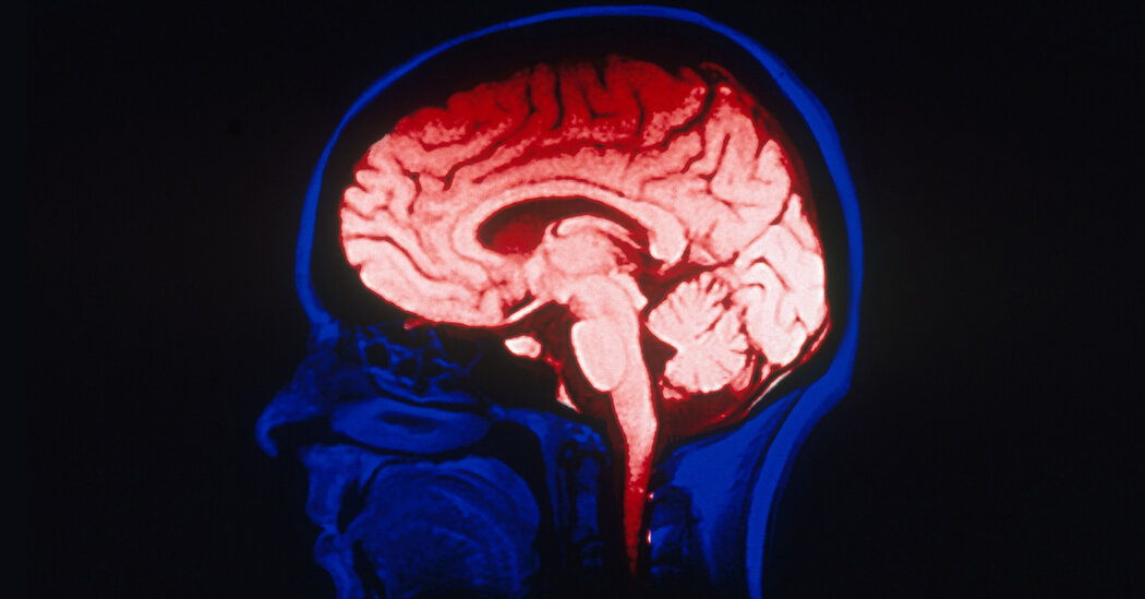
Researchers have long used imaging technology to try to understand mental-health ailments. But with relatively few participants, such studies may not be producing valid findings.For two decades, researchers have used brain-imaging technology to try to identify how the structure and function of a person’s brain connects to a range of mental-health ailments, from anxiety and depression to suicidal tendencies.But a new paper, published Wednesday in Nature, calls into question whether much of this research is actually yielding valid findings. Many such studies, the paper’s authors found, tend to include fewer than two dozen participants, far shy of the number needed to generate reliable results.“You need thousands of individuals,” said Scott Marek, a psychiatric researcher at the Washington University School of Medicine in St. Louis and an author of the paper. He described the finding as a “gut punch” for the typical studies that use imaging to try to better understand mental health.Studies that use magnetic-resonance imaging technology commonly temper their conclusions with a cautionary statement noting the small sample size. But enlisting participants can be time-consuming and expensive, ranging from $600 to $2,000 an hour, said Dr. Nico Dosenbach, a neurologist at Washington University School of Medicine and another author on the paper. The median number of subjects in mental-health-related studies that use brain imaging is around 23, he added.But the Nature paper demonstrates that the data drawn from just two dozen subjects is generally insufficient to be reliable and can in fact yield “massively inflated” findings,” Dr. Dosenbach said.For their analysis, the researchers examined three of the largest studies using brain-imaging technology to reach conclusions about brain structure and mental health. All three studies are ongoing: the Human Connectome Project, which has 1,200 participants; the Adolescent Brain Cognitive Development, or A.B.C.D., study, with 12,000 participants; and the U.K. Biobank study, with 35,700 participants.The authors of the Nature paper looked at subsets of data within those three studies to determine whether smaller slices were misleading or “reproducible,” meaning that the findings could be considered scientifically valid.For instance, the A.B.C.D. study looks, among other things, at whether thickness of the brain’s gray matter can be correlated to mental health and problem-solving ability. The authors of the Nature paper looked at small subsets within the big study and found that the subsets produced results that were unreliable when compared with the results yielded by the full data set.On the other hand, the authors found, when results were generated from sample sizes involving several thousand subjects, the findings were similar to those from the full data set.The authors ran millions of calculations by using different sample sizes and the hundreds of brain regions explored in the various major studies. Time and again, the researchers found that subsets of data from fewer than several thousand people did not produce results consistent with those of the full data set.Dr. Marek said that the paper’s findings “absolutely” applied beyond mental health. Other fields, like genomics and cancer research, have had their own reckonings with the limits of small sample sizes and have tried to correct course, he noted.“My hunch this is much more about population science than it is about any one of those fields,” he said.

Read more →

Longer, more intense allergy seasons could result from climate change

Allergy seasons are likely to become longer and grow more intense as a result of increasing temperatures caused by humanmade climate change, according to new research from the University of Michigan.
By the end of this century, pollen emissions could begin 40 days earlier in the spring than we saw between 1995 and 2014. Allergy sufferers could see that season last an additional 19 days before high pollen counts may subside.
In addition, thanks to rising temperatures and increasing CO2 levels, the annual amount of pollen emitted each year could increase up to 200%.
“Pollen-induced respiratory allergies are getting worse with climate change,” said Yingxiao Zhang, a U-M graduate student research assistant in climate and space sciences and engineering and first author of the paper in Nature Communications. “Our findings can be a starting point for further investigations into the consequence of climate change on pollen and corresponding health effects.”
U-M researchers developed a predictive model that examines 15 of the most common pollen types and how their production will be impacted by projected changes in temperatures and precipitation. They combined climate data along with socioeconomic scenarios, correlating their modeling with the data from 1995 through 2014. They then used their model to predict pollen emissions for the last two decades of the 21st century.
Allergies symptoms run the gamut from the mildly irritating, such as watery eyes, sneezing or rashes, to more serious conditions, such as difficulty breathing or anaphylaxis. According to the Asthma and Allergy Foundation of America, 30% of adults and 40% of children suffer from allergies in the U.S.
The grasses, weeds and trees that produce pollen are affected by climate change. Increased temperatures cause them to activate earlier than their historical norms. Hotter temperatures can also increase the amount of pollen produced.
Allison Steiner, U-M professor of climate and space sciences and engineering, said the modeling developed by her team could eventually allow for allergy season predictions targeted to different geographical regions.
“We’re hoping to include our pollen emissions model within a national air quality forecasting system to provide improved and climate-sensitive forecasts to the public,” she said.
The research was supported by the National Science Foundation.
Story Source:
Materials provided by University of Michigan. Note: Content may be edited for style and length.

Read more →

AI to predict antidepressant outcomes in youth

Mayo Clinic researchers have taken the first step in using artificial intelligence (AI) to predict early outcomes with antidepressants in children and adolescents with major depressive disorder, in a study published in The Journal of Child Psychology and Psychiatry. This work resulted from a collaborative effort between the departments of Molecular Pharmacology and Experimental Therapeutics, and Psychiatry and Psychology, at Mayo Clinic, with support from Mayo Clinic’s Center for Individualized Medicine.
“This preliminary work suggests that AI has promise for assisting clinical decisions by informing physicians on the selection, use and dosing of antidepressants for children and adolescents with major depressive disorder,” says Paul Croarkin, D.O., a Mayo Clinic psychiatrist and senior author of the study. “We saw improved predictions of treatment outcomes in samples of children and adolescents across two classes of antidepressants.”
In the study, researchers identified variation in six depressive symptoms: difficulty having fun, social withdrawal, excessive fatigue, irritability, low self-esteem and depressed feelings.
They assessed these symptoms with the Children’s Depression Rating Scale-Revised to predict outcomes to 10 to 12 weeks of antidepressant pharmacotherapy: The six symptoms predicted 10- to 12-week outcomes at four to six weeks in fluoxetine testing datasets, with an average accuracy of 73%. The same six symptoms predicted 10- to 12-week outcomes at four to six weeks in duloxetine testing datasets, with an average accuracy of 76%. In placebo-treated patients, predicting response and remission accuracy was significantly lower than for antidepressants at 67%.These outcomes show the potential of AI and patient data to ensure children and adolescents receive treatment that has the highest likelihood of delivering therapeutic benefits with minimized side effects, explains Arjun Athreya, Ph.D., a Mayo Clinic researcher and lead author of the study.
“We designed the algorithm to mimic a clinician’s logic of treatment management at an interim time point based on their estimated guess of whether a patient will likely or not benefit from pharmacotherapy at the current dose,” says Dr. Athreya. “Hence, it was essential for me as a computer engineer to embed and observe the practice closely to not only understand the needs of the patient, but also how AI can be consumed and useful to the clinician to benefit the patient.”
Next steps
The research findings are a foundation for future work incorporating physiological information, brain-based measures and pharmacogenomic data for precision medicine approaches in treating youth with depression. This will improve the care of young patients with depression, and help clinicians initiate and dose antidepressants in patients who benefit most.
“Technological advances are understudied tools that could enhance treatment approaches,” says Liewei Wang, M.D., Ph.D., the Bernard and Edith Waterman Director of the Pharmacogenomics Program and Director of the Center for Individualized Medicine at the Mayo Clinic. “Predicting outcomes in children and adolescents treated for depression is critical in managing what could become a lifelong disease burden.”
Acknowledgments
This work was supported by Mayo Clinic Foundation for Medical Education and Research; the National Science Foundation under award No. 2041339; and the National Institute of Mental Health under awards R01MH113700, R01MH124655 and R01AA027486. The content is solely the authors’ responsibility and does not necessarily represent the official views of the funding agencies. The authors have declared no competing or potential conflicts of interest.
Story Source:
Materials provided by Mayo Clinic. Original written by Colette Gallagher. Note: Content may be edited for style and length.

Read more →

Those with facial scars rate their own appearance more critically than surgeons and strangers

Patients who undergo facial surgery think their surgical scars look worse than surgeons and independent observers do, according to a new study from the Perelman School of Medicine at the University of Pennsylvania. Surgeons and those not tied personally to the particular scarring felt similarly about how significant a scar appeared, but those who looked at their own faces had more negative feelings about the condition and appearance of the scar. Researchers say that surgeons should explain to their patients in detail how their scar will likely look post-surgery and explicitly say to their patients that they themselves will likely perceive their scars to be more significant than others will. The study is published in the journal Facial Plastic Surgery & Aesthetic Medicine.
Eighty-one patients who had facial skin cancer and then received Mohs micrographic surgery (a type of precise skin surgery where layers of skin are removed a little at a time) rated their scars a week after surgery and then three months after. While their feelings about their scar improved by roughly 40% from week one to the three-month mark, they still judged their scars more critically than Mohs surgeons and independent observers after three months.
“Our research seems to support the saying ‘we are our own worst critics,'” said senior author Joseph F. Sobanko, MD, director of Dermatologic Surgery Education and an associate professor of Dermatology at Penn. “Patients are probably going to view scarring on their faces as more severe than their own surgeon will and even someone they walk by on the street.”
Armed with that knowledge, surgeons should speak to their patients not just about the process of surgery but also what to expect during the healing process and what their face will look like after the incision is completely healed, Sobanko said.
“Our goal as surgeons should be to remove cancer effectively while minimizing scarring,” Sobanko added. “Nevertheless, skin cancer surgery will produce highly visible changes early in the healing process and our job as surgeons is to prepare patients for how their skin will look during the healing process. We should also be direct with our patients and tell them that they are going to be the most critical of their appearance.”
The Penn researchers made very specific choices when designing the study. The team decided to use facial scarring because of the obviously personal relationship people have with their faces. Previous research from Sobanko and colleagues showed that people are the most sensitive about scars on their faces compared to scars on other parts of their body. The researchers also chose to have participants assess scars at the one-week mark and at three months.
“At one week, incisions from surgery are quite visible, and that can be very jarring for patients,” Sobanko said. “As weeks progress the incisions heal predictably and our prior research has shown that most patients return to their baseline quality of life approximately 3 months after surgery.”
While the advice for providers is to be honest and clear with their patients about scarring, Sobanko and his team are planning to study specific ways that surgeons can help patients feel better about their surgical mark.
“One method we have used in our practice is to connect people about to go through Mohs facial surgery with willing individuals who already have been through the surgery,” said Sobanko. “Anecdotally, our patients have appreciated the opportunity to ask questions to someone who has experienced what they are about to go through and also see first-hand how someone else’s face healed. We’re excited to study whether that and other interventions can ease patients’ minds and help them feel better about the entire surgery experience.”
This research supported by funding from the Dermatology Foundation Career Development Award.
Story Source:
Materials provided by University of Pennsylvania School of Medicine. Note: Content may be edited for style and length.

Read more →