COVID-19 Increases risk of type 2 diabetes, study finds

People who have had Covid-19 are at increased risk of developing type 2 diabetes. This is the result of a study by DDZ, DZD and IQVIA, which has now been published in Diabetologia.
Studies show that the human pancreas can also be a target of the SARS-CoV-2 (severe acute respiratory syndrome coronavirus type 2 virus). Following a Covid-19 infection, reduced numbers of insulin secretory granules in beta cells and impaired glucose-stimulated insulin secretion have been observed. In addition, after Covid-19 disease, some patients developed insulin resistance and had elevated blood glucose levels although they had no previous history of diabetes. SARS-CoV-2 infection may lead to a strong release of pro-inflammatory signaling substances (cytokines). Activation of the immune system may persist for months after a SARS-CoV-2 infection and impair insulin effectiveness (muscle, fat cells, liver).
To date, however, it is unclear whether these metabolic changes are transient or whether Covid-19 disease increases the risk of persisting diabetes. To investigate this question, researchers from the German Diabetes Center (DDZ), German Center for Diabetes Research (DZD) and IQVIA (Frankfurt) conducted a retrospective cohort study.
The cohort study included a representative panel of 1,171 physician practices across Germany (March 2020 to January 2021: 8.8 million patients). Follow-up continued until July 2021. “The aim of our study was to investigate the incidence of diabetes after infection with SARS-CoV-2,” said first author Wolfgang Rathmann, head of the Epidemiology Research Group at the DDZ. As a control group, the researchers selected people with acute upper respiratory tract infections (AURI), which are also frequently caused by viruses. The two cohorts were matched for sex, age, health insurance, month of Covid-19 or AURI diagnosis, and comorbidities (obesity, hypertension, high cholesterol, heart attack, stroke). Patients on corticosteroid therapy were excluded from the study.
During the study period, 35,865 people were diagnosed with Covid-19. “Our analyses showed that patients with Covid-19 developed type 2 diabetes more frequently than people with AURI. The incidence of diabetes with Covid-19 infection was 15.8 compared to 12.3 per 1000 people per year with AURI. Statistical analysis resulted in an incidence rate ratio (IRR) of 1.28. Put simply, this means that the relative risk of developing type 2 diabetes was 28% higher in the Covid-19 group than in the AURI group,” Rathmann said, summarizing the results.
Although type 2 diabetes is unlikely to be a problem for the vast majority of people with mild Covid-19 disease, the authors recommend that anyone who has recovered from Covid-19 be alert to the warning signs and symptoms, such as fatigue, frequent urination and increased thirst and seek immediate treatment.
Story Source:
Materials provided by Deutsches Zentrum fuer Diabetesforschung DZD. Note: Content may be edited for style and length.

Read more →

The secret to staying young: New research highlights power of life long exercise to keep muscles healthy

Lifelong physical activity could protect against age-related loss of muscle mass and function, according to research published in The Journal of Physiology. Individuals aged 68 and above who were physically active throughout their life have healthier ageing muscle that has superior function and is more resistant to fatigue compared to inactive individuals, both young and old.
This is the first study to investigate muscle, stem cell and nerve activity in humans. The researchers from University of Copenhagen, Denmark, found that elderly individuals who keep physically active throughout their adult life, whether by taking part in resistance exercise, ball games, racket sports, swimming, cycling, running and/or rowing had a greater number of muscle stem cells, otherwise known as satellite cells in their muscle. These cells are important for muscle regeneration and long-term growth and protect against nerve decay.
46 male participants took part in the study. They were divided into three groups: young sedentary (15), elderly lifelong exercise (16) and elderly sedentary (15). They performed a heavy resistance exercise, sitting in a mechanical chair performing a knee extension movement to evaluate muscle function. The amount of force produced was measured. Blood samples were taken, and muscle biopsies were analysed from both legs. The researchers found elderly lifelong exercisers outperformed both the elderly and young sedentary adults.
Lead author, Casper Soendenbroe, University of Copenhagen, Denmark said:
“This is the first study in humans to find that lifelong exercise at a recreational level could delay some detrimental effects of ageing. Using muscle tissue biopsies, we’ve found positive effects of exercise on the general ageing population. This has been missing from the literature as previous studies have mostly focused on master athletes, which is a minority group. Our study is more representative of the general population aged 60 and above, as the average person is more likely to take part in a mixture of activities at a moderate level. That’s why we wanted to explore the relation between satellite cell content and muscle health in recreationally active individuals. We can now use this as a biomarker to further investigate the link between exercise, ageing and muscle health.”
“The single most important message from this study, is that even a little exercise seems to go a long way, when it comes to protecting against the age-related decline in muscle function. This is an encouraging finding which can hopefully spur more people to engage in an activity that they enjoy. We still have much to learn about the mechanisms and interactions between nerves and muscles and how these change as we age. Our research takes us one step closer.”
It is worth noting that the study was only carried out in males and the average age was 73. As the effects of ageing on muscle health become more pronounced at 80+ years, follow up studies are needed to see if the benefits of lifelong exercise are maintained later in life. Further, investigation on recreational activity and muscle health need to be carried out in females.
Story Source:
Materials provided by The Physiological Society. Note: Content may be edited for style and length.

Read more →

Study shows that intranasal Rx halts memory decay in experimental Alzheimer’s model

A research collaboration between scientists at LSU Health New Orleans and the Karolinska Institutet in Sweden has found that applying specialized pro-resolving lipid mediators intranasally arrested memory loss and brain degeneration in an experimental model of Alzheimer’s Disease (AD). The results are published in the Nature journal, Communications Biology.
Neuroinflammation is a hallmark of neurodegenerative disorders, including Alzheimer’s Disease. Specialized pro-resolving lipid mediators are bioactive compounds composed of fatty acids like omega-3 or their derivatives that resolve inflammation. Neuroprotectin D1 (NPD1), discovered by Nicolas Bazan, MD, PhD, Boyd Professor and Director of the LSU Health New Orleans Neuroscience Center of Excellence, and colleagues is one. Previous studies by the Bazan lab demonstrated that NPD1 is protective in experimental stroke as well as retinal damage and that it is in short supply in the memory area of the brains from AD donors.
Resolving inflammation is a complex process involving mediators, cell subtypes and communication pathways. Response includes cell communications that order the activation of protective, pro-survival mechanisms and silence pro-inflammatory signaling pathways. Specialized pro-resolving lipid mediators such as NPD1 are the key signaling molecules in the process.
The paper discloses solid data on microglial activation, pro-inflammatory signaling, chronic inflammation and neuronal damage in Alzheimer’s disease. The study uses the AppNL-G-F/NL-G-F mouse model for AD. The paper extensively studied amyloid load, cognition, neuronal network oscillations, glial activation, receptors and inflammatory factors.
The authors conclude that the noninvasive administration route, intranasal delivery, of biologically active lipid messengers opens avenues for therapeutic exploration for AD and other neurodegenerative diseases.
“AD lacks prevention or cure and exerts a horrendous toll on patients and their families due to crippling progression and devastating adverse events,” notes Dr. Bazan, who led the research at LSU Health. “Millions of Americans currently suffer from AD, and the number is expected to escalate rapidly in the coming years.”
Bazan has been collaborating with Marianne Schultzberg, Senior Professor of Clinical Neuroscience at the Department of Neurobiology, Care Sciences and Society at the Karolinska Institutet (KI). Projects include looking at the novel signals that protect the brain in the cerebral spinal fluid (CSF) of the early stages of Alzheimer’s disease.
“This productive collaboration is uncovering important aspects of early stages of Alzheimer’s Disease, and the novel evolving mechanisms are promising paths for innovative therapies like the one disclosed in the current paper,” says Professor Schultzberg.
According to the National Institute on Aging, Alzheimer’s disease is currently ranked as the seventh leading cause of death in the United States and is the most common cause of dementia among older adults.
Other members of the LSU Health New Orleans research team include Drs. Khanh V. Do, Bokkyoo Jun and Megan L. Cothern. Ceren Emre, who recently completed her PhD at the Karolinska Institutet, was co-supervised by Dr. Bazan and worked at LSU Health New Orleans Neuroscience Center of Excellence for eight months just before the pandemic.

Read more →

Booster for immune protection after coronavirus infection

When our immune system comes into contact with the SARS-CoV-2 coronavirus, it fights back and produces antibodies. A similar immune response is triggered by Corona vaccines. However, there is still little data available on the strength and durability of immune protection. A team led by Prof. Carsten Watzl from the Leibniz Research Centre for Working Environment and Human Factors Institute for Occupational Research (IfADo), in cooperation with the Max Planck Institute of Molecular Physiology (MPI) and the Klinikum Dortmund, has now been able to detect high levels of neutralising antibodies in test persons even 300 days after a coronavirus infection with the original variant of the coronavirus. And what’s more: after complete vaccination, the recovered probands showed antibody levels about 5 times higher than those vaccinated without prior infection. This would provide much better protection against a severe course of the disease in the event of a new infection with other coronavirus variants.
Our immune protection is provided by two systems working hand in hand. When infected with a virus, the immune system reacts by producing antibodies that can prevent the virus from infecting further cells. At the same time, so-called T-killer cells can recognise the foreign virus components and thus kill already infected cells. During the immune reaction, the antibodies constantly improve and are finally tailor-made for the pathogen. The amount of these neutralising antibodies indicates how well a new infection can be fought off by the body.
Unique collaboration between biochemists, immunologists, clinicians and the Dortmund health department
“When the corona pandemic broke out, we as immunologists were of course interested in how our immune system defends itself against the corona virus. That’s why, together with our colleagues from the Max Planck Institute and the Dortmund Hospital, we developed a reliable test system to detect neutralising antibodies,” IfADo Director Carsten Watzl says. In order to be able to fish antibodies out of the blood in a targeted manner, you need an appropriate bait. One of the main targets of the immune system is the spike protein, which is used by the virus to bind to human cells and then infect them. “We have been able to produce a part of this protein, or more precisely the area that docks with the cell, in high purity in the test tube,” reports Jan-Erik Hoffmann, head of protein production at the MPI Dortmund. With this exact copy and blood samples from the “Klinikum Dortmund” the researchers at IfADo were able to develop a reliable and meaningful detection system for coronavirus antibodies. In close exchange with the Dortmund health department and the Dortmund hospital, the scientists used this system to perform a study with about 140 volunteers from a Dortmund health facility with several documented cases of SARS-CoV-2 infection at the beginning of the pandemic (March 2020)
5 times higher antibody levels after vaccination in recovered patients
Effective amounts of neutralising antibodies against the spike protein could be detected in almost all of the subjects tested positive for SARS-CoV-2. And even after 300 days, the antibody levels had hardly decreased in three out of four subjects. However, test persons were infected with the original variant of the coronavirus and neutralising antibodies against the original spike protein were measured. As we know, the virus has now evolved in such a way that immunity to the original virus currently offers significantly less protection. Therefore, the researchers also investigated the effect of vaccination with the vaccines from AstraZeneca and BioNTech on the immune system. The astonishing result: After complete vaccination, recovered test persons developed up to five times more neutralising antibodies than vaccinated persons without prior infection. This should also provide better protection against current variants.
“There are now several studies on the immune response after a COVID-19. Our study differs from this in that we had blood samples before and from the first weeks of the pandemic. So we knew exactly whether a test person was already infected or not. In addition to this unbiased data, the long period of the study of almost one year is also remarkable,” Watzl says. “The rules of the game have changed in the meantime, of course, because there are new variants like Omikron. However, it is important to know how long immunity actually lasts, because this can also protect against a severe course of the disease in the case of a new infection with other coronavirus variants. Currently, we are also using our jointly developed test systems to study the immune response to the COVID-19 vaccines and their protection against different coronavirus variants.”
“This study is a prime example of successful interdisciplinary cooperation that not only yields important scientific findings but is also highly relevant to society,” emphasises PD Dr. Bernhard Schaaf, Director of the Department of Pneumology and Infectious Diseases at Klinikum Dortmund. “This is transfer of knowledge into everyday life and at the same time transparent cooperation at eye level.”
Story Source:
Materials provided by Max Planck Institute of Molecular Physiology. Note: Content may be edited for style and length.

Read more →

COVID-19 pandemic fueled massive growth in green industry

Most people would say the COVID-19 pandemic hasn’t been a great couple of years. But for the green industry, like plant nurseries and greenhouses, it’s been a boon.
But will the uptick in gardening last once the last coronavirus restrictions are lifted?
Probably not to the same extreme levels, according to new research from the University of Georgia. But for some, the introduction to gardening may have been just what they needed to dive into a new hobby.
Of its more than 4,200 participants, the study found about one out of every three people began gardening in 2020 because they were home more. Many also put in new grass lawns and did outdoor renovations, such as installing new plant beds and other landscaping.
“You had low interest rates, so you had a lot of people refinancing, which gave them money to invest in their homes,” said Benjamin Campbell, lead author of the study and an associate professor in the College of Agricultural and Environmental Sciences. “You had people at home looking for something to do, whether by themselves or with their kids. That led to a huge demand for plants.”
Just under half of respondents said they didn’t plan to garden in the future, even if they had in 2020. But one out of 10 said they gardened in 2020 and planned to keep it up going forward, including 11% of Gen Xers and 13% of millennials and younger.

Read more →

A novel painless and reliable allergy test

Although allergies are widespread, their diagnosis is complex and, depending on the type of allergy, the prospects of success with therapy are not always clear. Skin tests so far have been unpleasant, time-consuming and associated with a certain risk of triggering an allergic overreaction. Researchers at the University of Bern and Inselspital, Bern University Hospital, have now developed a novel test that massively simplifies the diagnosis of allergies and can reliably predict the success of a therapy.
Approximately one third of the world’s population suffers from one or more allergies, with the trend increasing every year. By far the most widespread form of allergy is the so-called type I allergy, also known as immediate-type allergy. This includes, for example, allergic rhinitis (hay fever), allergic asthma, food allergies, or allergies against insect venoms, pollen, grasses or house dust mites. It is an overreaction of the immune system to actually harmless foreign components (allergens), which typically occurs within seconds or minutes of contact with the allergen. Allergic symptoms can range from redness and swelling of the skin, itching or shortness of breath to anaphylactic shock and death.
Diagnosing an allergy is complex: In addition to the medical history (anamnesis), test parameters of often unclear diagnostic value are taken into account and patients undergo skin tests. Such skin tests are unpleasant, at times painful, time-consuming and associated with a certain risk of triggering an allergic overreaction. Allergies are treated with symptom control, in severe cases also with immunotherapy. This involves injecting doses of an allergen in increasing concentrations under the patient’s skin over a period of up to five years, with the aim of desensitizing the patient to the allergen. Immunotherapy is not always successful: At present, there is no reliable method for predicting the chances of success before concluding such therapy.
A research group led by Alexander Eggel from the Department for BioMedical Research (DBMR) at the University of Bern and the Department of Rheumatology and Immunology, Inselspital, Bern University Hospital, together with Thomas Kaufmann from the Institute of Pharmacology at the University of Bern, has now developed an allergy test that, on the one hand, greatly simplifies diagnosis and, on the other hand, can reliably predict the success of immunotherapy. The test was recently presented in a publication of the Journal of Allergy and Clinical Immunology.
In vitro mast cells provide unprecedented reliability
Type I allergy occurs when the body produces immunoglobulin E (IgE) class antibodies in response to allergens. The IgE antibodies are bound by IgE receptors on the surface of specialized immune cells in the body called mast cells. Subsequent contact with the same allergens then leads to activation of the mast cells and therefore to the release of inflammatory mediators such as histamine or leukotrienes, which are responsible for the allergic symptoms.

Read more →

MRI innovation makes cancerous tissue light up and easier to see

A new form of magnetic resonance imaging (MRI) that makes cancerous tissue glow in medical images could help doctors more accurately detect and track the progression of cancer over time.
The innovation, developed by researchers at the University of Waterloo, creates images in which cancerous tissue appears to light up compared to healthy tissue, making it easier to see.
“Our studies show this new technology has promising potential to improve cancer screening, prognosis and treatment planning,” said Alexander Wong, Canada Research Chair in Artificial Intelligence and Medical Imaging and a professor of systems design engineering at Waterloo.
Irregular packing of cells leads to differences in the way water molecules move in cancerous tissue compared to healthy tissue. The new technology, called synthetic correlated diffusion imaging, highlights these differences by capturing, synthesizing and mixing MRI signals at different gradient pulse strengths and timings.
In the largest study of its kind, the researchers collaborated with medical experts at the Lunenfeld-Tanenbaum Research Institute, several Toronto hospitals and the Ontario Institute for Cancer Research to apply the technology to a cohort of 200 patients with prostate cancer.
Compared to standard MRI techniques, synthetic correlated diffusion imaging was better at delineating significant cancerous tissue, making it a potentially powerful tool for doctors and radiologists.
“Prostate cancer is the second most common cancer in men worldwide and the most frequently diagnosed cancer among men in more developed countries,” said Wong, also a director of the Vision and Image Processing (VIP) Lab at Waterloo. “That’s why we targeted it first in our research.
“We also have very promising results for breast cancer screening, detection, and treatment planning. This could be a game-changer for many kinds of cancer imaging and clinical decision support.”
The core research team also included Hayden Gunraj and Vignesh Sivan, engineering graduate students at Waterloo, and Dr. Masoom Haider of the Lunenfeld-Tanenbaum Research Institute.
Story Source:
Materials provided by University of Waterloo. Note: Content may be edited for style and length.

Read more →

‘He Goes Where the Fire Is’: A Virus Hunter in the Wuhan Market

As soon as Edward Holmes saw the dark-ringed eyes of the raccoon dogs staring at him through the bars of the iron cage, he knew he had to capture the moment.It was October 2014. Dr. Holmes, a biologist at the University of Sydney, had come to China to survey hundreds of species of animals, looking for new types of viruses.On a visit to Wuhan, a commercial center of 11 million people, scientists from the city’s Center for Disease Control and Prevention brought him to Huanan Seafood Wholesale Market. In stall after stall of the poorly ventilated space, he saw live wild animals — snakes, badgers, muskrats, birds — being sold for food. But it was the raccoon dogs that made him pull out his iPhone.As one of the world’s experts on virus evolution, Dr. Holmes had an intimate understanding of how viruses can jump from one species to another — sometimes with deadly consequences. The SARS outbreak of 2002 was caused by a bat coronavirus in China that infected some kind of wild mammal before infecting humans. Among the top suspects for that intermediate animal: the fluffy raccoon dog.“You could not get a better textbook example of disease emergence waiting to happen,” Dr. Holmes, 57, said in an interview.The tall, bald Englishman did his best not to draw attention to himself as he snapped a picture of the raccoon dogs, which look like long-legged raccoons but are more closely related to foxes. He then took a few more pictures of other animals in cages of their own. As a vendor began clubbing one of the creatures, Dr. Holmes pocketed his phone and slipped away.The photos faded from his mind until the last day of 2019. As Dr. Holmes was browsing Twitter from his Sydney home, he learned of an alarming outbreak in Wuhan — a SARS-like pneumonia with early cases linked to the Huanan market. The raccoon dogs, he thought.“It was a pandemic waiting to happen, and then it bloody well happened,” he said.The Huanan Market in Wuhan in 2014.Eddie HolmesDr. Holmes believes raccoon dogs at the market could have set off the coronavirus pandemic.Eddie HolmesFrom that day on, Dr. Holmes was swept into a vortex of discoveries and controversies related to the origins of the virus — making him feel like “the Forrest Gump of Covid,” he joked. He and a Chinese colleague were the first to share the genome of the new coronavirus with the world. He then discovered crucial clues about how the pathogen most likely evolved from bat coronaviruses.And in the contentious geopolitical debate over whether the virus may have leaked from a Wuhan laboratory, Dr. Holmes has become one of the strongest proponents of an opposing theory: that the virus spilled over from a wild animal. With colleagues in the United States, he recently published tantalizing clues that raccoon dogs kept in the very iron cage he photographed in 2014 could have set off the pandemic.Dr. Holmes’s Covid research has won him international acclaim, including Australia’s top science prize. But it has also garnered claims that his research had been overseen by the Chinese military, along with a flood of attacks on social media and even death threats.Through it all, Dr. Holmes has continued to publish a torrent of studies on Covid. Longtime colleagues attribute his steady output through unsteady times to an exceptional knack for building big scientific teams, and a willingness to dive into controversial debates if he thinks they are important.“He’s the right kind of person with the right kind of mind-set, because of the fact that he can be open-minded and engaged and thoughtful, and not become defensive,” said Pardis Sabeti, a geneticist at the Broad Institute of M.I.T. and Harvard who worked with Dr. Holmes on Ebola.Hunting for VirusesGrowing up in western England, a young Edward Holmes had a biology teacher who put a poster of an orangutan on the wall that read, “I’m not your cousin.”The Great ReadMore fascinating tales you can’t help but read all the way to the end.A family from India froze to death yards away from crossing into the U.S. at the northern border, where desperate migrants are trying their luck.Wolves have been steadily returning to California after being wiped out a century ago. But not everyone is rolling out the welcome mat.A waiter. A cab driver. An engineer. Ukrainians across the U.S., many of whom have never fired a weapon, have put their lives on hold to defend their homeland.The teacher told the class not to read the garbage in their textbook about evolution. That made the 14-year-old eager to dive in.He went on to study the evolution of apes and humans, and then turned to viruses. Over three decades — working in Edinburgh, Oxford, Pennsylvania and finally Sydney — Dr. Holmes has published more than 600 papers on the evolution of viruses including H.I.V., influenza and Ebola.When he was invited to come to the University of Sydney, in 2012, he seized the chance to move closer to Asia, where he feared that the wildlife trade could set off a new pandemic.“He goes where the fire is,” said Andrew Read, an evolutionary biologist at Penn State University, who worked with Dr. Holmes at the time.As he was preparing for the move, Dr. Holmes got an email out of the blue from a Chinese virologist named Yong-Zhen Zhang, asking if he’d like to study viruses with him in China. Their collaboration quickly expanded into a sweeping search for new viruses in hundreds of species of animals. They studied spiders plucked off the walls of huts and fish hauled up from the South China Sea.Westen Hubei province during one of Dr. Holmes’s survey trips in 2016.Eddie HolmesThey ultimately found more than 2,000 virus species new to science, with many surprises among them. Scientists used to think that influenza viruses infected primarily birds, for example, which could then pass them along to mammals like ourselves. But Dr. Holmes and Dr. Zhang found that fish and frogs get the flu, too.“That’s been quite eye opening,” said Andrew Rambaut, an evolutionary biologist at the University of Edinburgh who was not involved in the surveys. “The diversity of viruses that are out there is just enormous.”On one of their survey trips in 2014, Dr. Holmes and Dr. Zhang formed a partnership with scientists at the Wuhan Center for Disease Control and Prevention to survey animals in the surrounding Hubei Province. The C.D.C. scientists brought them to the Huanan market to see a worrying case of wildlife trade.After the visit, Dr. Holmes hoped he and his colleagues could use the genetic sequencing techniques they had developed for their animal surveys to look for viruses in the animals at the market. But his colleagues were more interested in searching for viruses in sick people.Dr. Zhang and Dr. Holmes began working with doctors at Wuhan Central Hospital, fishing for viral RNA in samples of lung fluid from people with pneumonia. Because of this collaboration, he was named a guest professor with the Chinese Center for Disease Control and Prevention from 2014 to 2020.Last month, Dr. Holmes and his colleagues published their first report on the project, based on samples from 408 patients collected in 2016 and 2017. Many were sick with more than one virus, it turned out, and some were also infected with bacteria or fungi. The researchers even saw evidence of a hidden outbreak: Six patients were infected with genetically identical enteroviruses.Dr. Holmes and Dr. Zhang also continued surveying the virosphere, examining soil, sediments and animal feces from across China. But in late December 2019, that work ground to a halt.Covid’s ArrivalA Covid-only hospital in Wuhan in March 2020.Agence France-Presse — Getty ImagesWhen Dr. Zhang got wind of a new pneumonia in Wuhan, he asked colleagues at the Wuhan Central Hospital to ship him lung fluid from a patient. It arrived on Jan. 3, and he used the techniques he and Dr. Holmes had perfected to search for viruses. Two days later, Dr. Zhang’s team had assembled the genome of a new coronavirus, SARS-CoV-2.Other scientific teams in China had also sequenced the virus. But none made it public, because the Chinese government had barred scientists from publishing information about it.Dr. Zhang and Dr. Holmes began writing a paper about the genome, which would later appear in the journal Nature. Dr. Zhang flouted the ban and uploaded the virus genome to a public database hosted by the U.S. National Institutes of Health. But the database requires a lengthy review of new genomes, and so days passed without the information going online.Dr. Holmes urged his collaborator to find another way to share the genome with the world. “It felt like it had to happen,” Dr. Holmes said.On Jan. 10, they agreed to share it on a forum for virologists, and Dr. Holmes put it online.That decision was a turning point, according to Jason McLellan, a structural biologist at the University of Texas at Austin who worked on the mRNA technology powering the Moderna vaccine. Only with that genetic sequence could researchers start working on tests, drugs and vaccines. Until then, Dr. McLellan said, scientists like himself were like runners in their starting blocks, waiting for a starter’s pistol.“It fired the moment Edward and Yong-Zhen posted the genome sequence,” he said. “Immediately, Twitter was abuzz, emails were being exchanged, and the race was on.”But according to Chinese media reports, Dr. Zhang paid a price for defying his country’s information ban. The day after the genome sequence went live, his laboratory at the Shanghai Public Health Clinical Center was reportedly ordered to close for “rectification.”Dr. Zhang later insisted to a reporter at Nature that the move was not a punishment, and that his lab later reopened. Email requests to Dr. Zhang to comment for this story went unanswered. Dr. Holmes declined to comment about Dr. Zhang’s current situation.Yong-Zhen Zhang, a Chinese virologist, and his team assembled the genome of SARS-CoV-2 and made it public, defying China’s ban on publishing information about it.Keith Brasher/The New York TimesAfter the coronavirus genome was sequenced, Dr. Holmes was puzzled to see some bits of genetic material that looked like they might have been put there through genetic engineering.On a Feb. 1, 2020, telephone conference, Dr. Holmes shared his worries with other virus experts, including Dr. Francis Collins, the director of the N.I.H., and Dr. Anthony S. Fauci, America’s top infectious disease expert. Other scientists explained on the call that those features of the genome could easily have been produced through the natural evolution of viruses.Soon afterward, Dr. Holmes helped researchers at the University of Hong Kong analyze a coronavirus, found in a pangolin, that was closely related to SARS-CoV-2. The virus looked especially similar in its surface protein, called spike, which the virus uses to enter cells.Finding such a distinct biological signature in a virus from a wild animal strengthened Dr. Holmes’s confidence that SARS-CoV-2 was not the product of genetic engineering. “Suddenly what looks odd is clearly natural,” Dr. Holmes said.Dr. Holmes and his colleagues laid out some of these findings in a letter published in March 2020. That same month, he published some of his photos of caged animals at the Huanan market in a commentary he wrote with Dr. Zhang, suggesting that it might have been the site of an animal spillover.But the idea that the virus had been engineered in a lab continued to gain traction, and Dr. Holmes came under attack for his work with Chinese scientists.In May 2020, The Daily Telegraph, an Australian newspaper, linked him to the Chinese military with an article titled, “How the Red Army Oversaw Coronavirus Research.”The newspaper based its claim on the fact that two scientists involved in the pangolin study had secondary affiliations with a Chinese military lab. Dr. Holmes, who said he never met the scientists, noted that they had helped with sequencing RNA from the pangolin tissue.The University of Sydney responded on Dr. Holmes’s behalf with a statement: “We strongly defend the right of our researchers to collaborate with scientists around the world in line with all relevant Australian laws and government guidelines.” The university noted that Dr. Holmes’s research was entirely supported by Australian grants.In late 2020, the World Health Organization organized a group of experts to travel to China to investigate the origin of the novel coronavirus. Dr. Holmes sent them his 2014 market photos, but they never made it into the W.H.O.’s report.“Some of the Chinese delegation suggested that I might have fabricated those pictures,” Dr. Holmes said. (Peter Daszak, the president of EcoHealth Alliance and one of the investigators of the W.H.O. report, corroborated this account: The Chinese investigators said the photos were “not verifiable, and could have been faked,” Dr. Daszak said.)Preventing Future SpilloversThe shuttered Huanan Market in October.Getty ImagesIn reports published last month, Dr. Holmes and over 30 collaborators analyzed early Covid cases, finding that they clustered around the market, and examined the mutations in early coronavirus samples.Chris Newman, a wildlife biologist at the University of Oxford and a co-author of one of the studies, said that his Chinese colleagues saw a number of wild mammals for sale at the Huanan market in late 2019. Any of them might have been responsible for the pandemic, Dr. Holmes said.“You can’t prove raccoon dogs yet, but they’re certainly a suspect,” he said.Some critics have questioned how sure Dr. Holmes and his colleagues can be that a Huanan animal was to blame. Although many of the earliest Covid cases were linked to the market, it’s possible that other cases of pneumonia have not yet been recognized as early Covid cases.“We still know far too little about the earliest cases — and there are likely additional cases we don’t know about — to draw final conclusions,” said Filippa Lentzos, an expert on biosecurity at King’s College London. “I remain open to both natural spillover and research-related origins.”Another problem: If infected animals indeed started the pandemic, they’ll never be found. In January 2020, when researchers from the Chinese C.D.C. arrived at the market to investigate, all the animals were gone.But Dr. Holmes argues that there’s more than enough evidence that animal markets could spark another pandemic. Last month, he and Chinese colleagues published a study of 18 animal species often sold at markets, obtaining them either in the wild or on breeding farms.“They were absolutely full of virus,” Dr. Holmes said.Over 100 vertebrate-infecting viruses came to light, including a number of potential human pathogens. And some of these viruses had recently jumped the species barrier — bird flu infecting badgers, dog coronaviruses infecting raccoon dogs. Some of the animals were sick with human viruses, too.The simplest way to reduce the odds of future pandemics, Dr. Holmes has argued, is to carry out studies like this one at the interface between humans and wildlife. His own experience discovering new viruses has convinced him that it doesn’t make sense to try to catalog every potential threat in wildlife.“You could never possibly sample every virus out there and then work out which one of those can infect humans,” Dr. Holmes said. “I don’t think that’s viable.”

Read more →

Hospitalizations of young children with the virus surged during the U.S. Omicron wave.

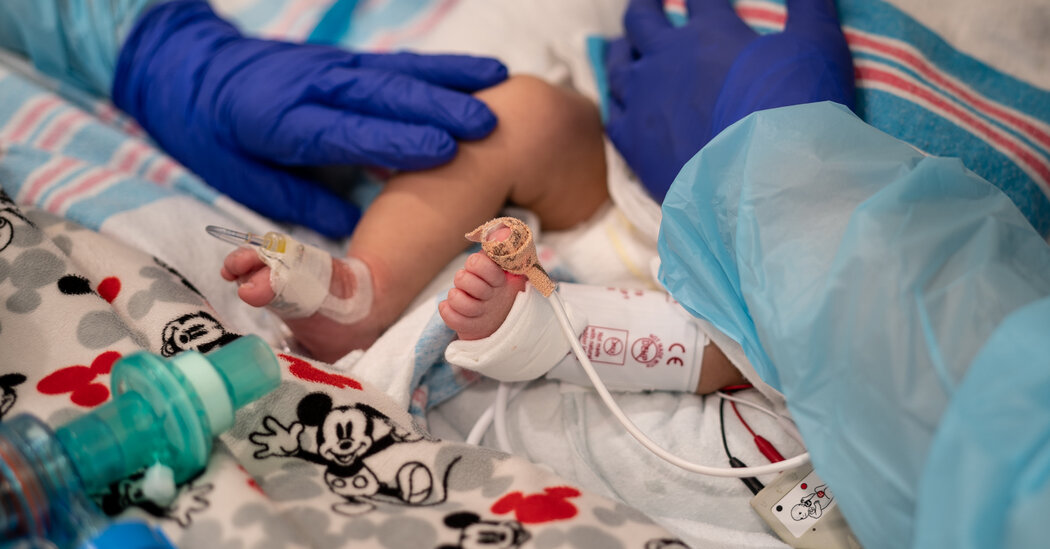
Babies and children younger than age 5 were hospitalized with coronavirus at much higher rates during the latest U.S. surge, when the highly transmissible Omicron variant was dominant, compared with earlier periods in the pandemic, according to the Centers for Disease Control and Prevention.Hospitalizations of these children were about five times higher during the Omicron surge, between Dec. 19 and Feb. 19, than during the period when the Delta variant was dominant, between June 27 and Dec. 18.Rates of admission to intensive care also rose dramatically among young children, reaching a peak on Jan. 8 of this year.Children of color younger than age 5 wound up in hospitals at disproportionate rates. Only one-third of the children were white, while 28 percent were Hispanic and 23 percent were Black. Hispanic people represent just 18 percent of the population, and Black Americans make up 13 percent.(Six percent of these hospitalizations were among Asian or other Pacific Islander children, about the same as their representation in the population.)Experts say children of color are infected at higher rates because they are more likely to have parents who work in public-facing jobs, and more likely to live in poverty and in multigenerational households.Though hospitalization rates for young children are still relatively low, compared to the rates among older Americans, the virus poses special risks to the youngest children and especially to babies.Infants six months old and younger were the most vulnerable, representing nearly half of the hospitalizations among young children during the Omicron period. They were hospitalized at rates about six times as high at the peak of the Omicron surge, compared with the peak of the Delta wave. Two infants died, the C.D.C. found.“People should know there are risks to children under 1 that are pretty serious, especially during surges, and they might want to take extra precautions to reduce exposure,” said Julia Raifman, an assistant professor of health law, policy and management at Boston University School of Public Health, who was not involved in the research.More than 1,000 children younger than age 18 have died of Covid since the pandemic started, including 350 children under 5. But experts also worry about the long-term effects, as well as multisystem inflammatory syndrome, a rare but serious condition.The C.D.C. study found that most of the children and babies who were hospitalized — about two-thirds — were healthy and did not have underlying medical conditions, as has been the case throughout the pandemic.No Covid vaccines are currently authorized in the United States for children younger than 5, and the regulatory process has been fraught with delays and setbacks. Public health experts strongly recommend that anyone who comes into regular contact with young children get vaccinated.“To help protect children too young to be vaccinated, everyone ages five and older, including pregnant women, family members and caregivers, should stay up to date with Covid-19 vaccines,” Dr. Kristin J. Marks, the study’s first author and an epidemic intelligence service officer with the CDC, said in an email.The study, published on March 15, examined hospitalizations of children in counties in 14 states whose catchment areas represent about 10 percent of the U.S. population.

Read more →

Covid trapped me at home for more than seven months

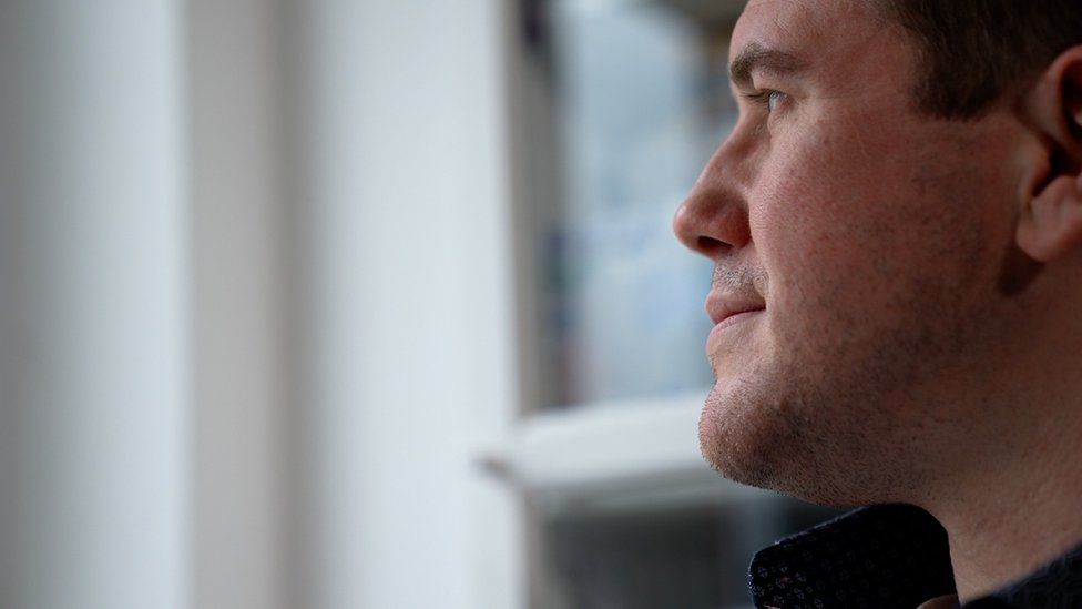
SharecloseShare pageCopy linkAbout sharingA vaccine has been used to free a man who was trapped at home by a Covid infection that lasted for more than seven months. It is the first time that a vaccine has been used to “treat” Covid rather than “prevent” it. Ian Lester, 37, has a weakened immune system that was unable to defeat the virus on its own. He says he became a prisoner in his home in Caerphilly, Wales, as he isolated for months on end. Ian was born with Wiskott-Aldrich syndrome, which makes it harder for him to fight off infections. Even a common cold can linger. He shielded during the first wave of Covid, but coronavirus eventually found him in December 2020. He had one of the classic symptoms – a slight loss of sense of taste and smell – which cleared up within a month. For most of us that would be the end of it, but Ian’s Covid journey was only just beginning. His doctors wanted him to keep on testing because his weakened immune system meant there was a risk he could be contagious for longer than normal.But month after month, test after test came back positive. Ian had to give up work at the opticians where he’d be in close contact with others and stay at home. Initially he tried to make the most of it, and the Christmas tree stayed up through January, but eventually the isolation took its toll.”People would feel like it’s going to be a long holiday, but after the three-month mark it wasn’t,” Ian told me.Slowly every day began to feel the same – a pattern of cooking, TV, reading, playing guitar and just waiting for his wife Katie to get home.Ian said: “It was like living in groundhog day, you just end up sitting there staring at the wall. “It slowly became my prison cell, especially when it got to the summertime and the restrictions were lifting for everyone else, you could see family and friends starting to get back to a real life, and I was still getting these positive results.” Ian started to become ill again after the three-month mark. Fatigue, headaches, insomnia, and a tight chest all set in, he was struggling to concentrate and every three to four weeks there would be a build-up of sticky mucous on his lungs. It was never enough to need hospital treatment, but it was clear his body wasn’t easily shrugging off Covid.”I was worried that I’d keep on getting worse and worse and worse and never get rid of it,” he said.Scientists and doctors were monitoring the battle between the virus and Ian’s immune system at Cardiff University and at the Immunodeficiency Centre for Wales in the University Hospital of Wales. The analysis showed Ian had a long-term infection, it was not just “dead virus” being detected, and his symptoms were not long-Covid. “Ian really didn’t have much of an immune response at all against the Covid virus,” Dr Mark Ponsford, a clinician scientist at Cardiff University, told the BBC.They could not find antibodies that would stick to and neutralise the coronavirus, and there were only limited signs of T-cells, another wing of the immune system, that could attack Covid. At the time, in the early summer of 2021, there were limited treatments, so the medical team decided to try something radical. Instead of giving a vaccine to prevent an infection, they decided to use the Pfizer vaccine to treat one. The difference in Ian’s body “was like night and day”, says Dr Ponsford. The first dose started to build his immunity, but it took a second dose to reach the point where his body could fight off the virus. By the end of August, Ian was testing negative again. So why was the vaccine able to clear the infection, when months of having the virus did not build up enough immunity?Prof Stephen Jolles, clinical lead at the Immunodeficiency Centre, said: “This infection was burbling along, but with his [weakened] immune system it was just not enough to kick off a response sufficient to clear it. “So the vaccine really made a huge difference, in antibodies and T-cells, and utilised and squeezed every last drop out of what his immune system could do.” On the day Ian was finally clear of Covid he was “ecstatic” and says “I couldn’t believe it to tell you the truth”. He celebrated with a day on the beach and a portion of fish and chips with Katie. “Everything’s back to normal now,” he says.The researchers think this approach can be used in more people with weakened immune systems who are struggling to fight off the infection. There are anti-viral drugs now that were not available when Ian had Covid, but the team think vaccines could offer a cheaper and more durable option. Follow James on Twitter

Read more →