A small mutation can make Zika virus even more dangerous

Researchers at La Jolla Institute for Immunology (LJI) have found that Zika virus can mutate to become more infective — and potentially break through pre-existing immunity.
“The world should monitor the emergence of this Zika virus variant,” says LJI Professor Sujan Shresta, Ph.D., who co-led the Cell Reports study with Professor Pei-Yong Shi, Ph.D., of the University of Texas Medical Branch (UTMB).
Zika virus is carried by mosquitoes, and the symptoms of Zika infection are usually mild in adults. However, the virus can infect a developing fetus, resulting in birth defects such as microcephaly.
Zika virus and dengue virus overlap in many countries worldwide. Like Zika, dengue virus is a mosquito-borne flavivirus, and thus shares many biological properties. In fact, the viruses are similar enough that the immune response sparked by prior dengue exposure can offer protection against Zika.
“In areas where Zika is prevalent, a vast majority of people have already been exposed to dengue virus and have both T cells and antibodies that cross-react,” says Shresta.
Unfortunately, both viruses are also quick to mutate. “Dengue and Zika are RNA viruses, which means they can change their genome,” explains Shresta. “When there are so many mosquitoes and so many human hosts, these viruses are constantly moving back and forth and evolving.”
To study Zika’s fast-paced evolution, the LJI team recreated infection cycles that repeatedly switched back and forth between mosquito cells and mice. This work gave the LJI scientists a window into how Zika virus naturally evolves as it encounters more hosts.

Read more →

Adolescent drug overdose deaths rose exponentially for the first time in history during the COVID pandemic

The rate of overdose deaths among U.S. teenagers nearly doubled in 2020, the first year of the COVID pandemic, and rose another 20% in the first half of 2021 compared with the 10 years before the pandemic, even as drug use remained generally stable during the same period, according to new UCLA research.
This is the first time in recorded history that the teen drug death rate has seen an exponential rise, even though rates of illicit drug use among teens are at all-time lows, said lead author Joseph Friedman, an addiction researcher and M.D. and Ph.D. candidate at the David Geffen School of Medicine at UCLA.
“Drug use is becoming more dangerous, not more common,” Friedman said. “The increases are almost entirely due to illicit fentanyls, which are increasingly found in counterfeit pills. These counterfeit pills are spreading across the nation, and teens may not realize they are dangerous.”
The study is published in JAMA.
The researchers used the CDC WONDER (Centers for Disease Control and Prevention Wide-Ranging Online Data for Epidemiologic Research) database to calculate drug overdose deaths per 100,000 population for adolescents aged 14 to 18 years that occurred from January 2010 to June 2021.
They found 518 deaths, or a rate of 2.4 per 100,000, among adolescents in 2010, and a steady rate of 492 deaths (2.36 per 100,000) each subsequent year through 2019. In 2020, there was a sharp increase to 954 deaths (4.57 per 100,000), rising to 1,146 deaths (5.49 per 100,000) in early 2021.

Read more →

Poverty, crime linked to differences in newborns' brains

Poverty and crime can have devastating effects on a child’s health. But a new study from researchers at Washington University School of Medicine in St. Louis suggests that some environmental factors influence the structure and function of young brains even before babies make their entrances into the world.
A study published online April 12 in the journal JAMA Network Open found that MRI scans performed on healthy newborns while they slept indicated that babies of mothers facing social disadvantages such as poverty tended to be born with smaller brains than babies whose mothers had higher household incomes.
MRI scans of full-term newborns born to mothers living in poverty revealed smaller volumes across the entire brain — including the cortical gray matter, subcortical gray matter and white matter — than found in the brains of babies whose mothers had higher household incomes. The brain scans, which were conducted only a few days to weeks after birth, also showed evidence of less folding of the brain among infants born to mothers living in poverty. Fewer and shallower folds typically signify brain immaturity. The healthy human brain folds as it grows and develops, providing the cerebral cortex with a larger functional surface area.
A second study of data from the same sample of 399 mothers and their babies — this one published online April 12 in the journal Biological Psychiatry — reports that pregnant mothers from neighborhoods with high crime rates gave birth to infants whose brains functioned differently during their first weeks of life than babies born to mothers living in safer neighborhoods. Functional MRI scans of babies whose mothers were exposed to crime displayed weaker connections between brain structures that process emotions and structures that help regulate and control those emotions. Maternal stress is believed to be one of the reasons for the weaker connections in the babies’ brains.
“These studies demonstrate that a mother’s experiences during pregnancy can have a major impact on her infant’s brain development,” said Christopher D. Smyser, MD, one of the principal investigators. “Like that old song about how the ‘knee bone is connected to the shin bone,’ there’s a saying about the brain that ‘areas that fire together wire together.’ We’re analyzing how brain regions develop and form early functional networks because how those structures develop and work together may have a major impact on long-term development and behavior.”
Babies in the study were born from 2017 through 2020, before the start of the COVID-19 pandemic. Smyser, a professor of neurology, of pediatrics and of radiology, said that to successfully scan newborns during the first few weeks of life, babies are fed when they arrive for scans because they tend to fall asleep after eating. They are then snuggly swaddled into blankets and a device that helps keep them comfortable and still. The brain scans take place while they sleep.
In the study involving the effects of poverty, the researchers focused on 280 mothers and their newborns. First author Regina L. Triplett, MD, a postdoctoral fellow in neurology, had expected to find that maternal poverty — referred to in the paper as social disadvantage — could affect the babies’ developing brains. But she also expected to see effects from psychosocial stress, which includes measures of adverse life experiences as well as measures of stress and depression.
“Social disadvantage affected the brain across many of its structures, but there were not significant effects that were related to psychosocial stress,” Triplett said. “Our concern is that as babies begin life with these smaller brain structures, their brains may not develop in as healthy a way as the brains of babies whose mothers lived in higher income households.”
In the second study, which implicated living in high-crime neighborhoods as a factor in weaker functional connections in the brains of newborns, first author Rebecca G. Brady, a graduate student in the university’s Medical Scientist Training Program, found that unlike the effects of poverty, the effects of exposure to crime were focused on particular areas of the babies’ brains.
“Instead of a brain-wide effect, living in a high-crime area during pregnancy seems to have more specific effects on the emotion-processing regions of babies’ brains,” Brady said. “We found that this weakening of the functional connections between emotion-processing structures in the babies’ brains was very robust when we controlled for other types of adversity, such as poverty. It appears that stresses linked to crime had more specific effects on brain function.”
Reducing poverty and lowering crime rates are well-established goals of public policy and public health. And the researchers believe protecting expectant mothers from crime and helping them out of poverty will do more than improve brain growth and connections in their babies. But if social programs that aim to help people reach their full potential are to succeed, the researchers said the policies must focus on assisting people even before they are born.
“Several research projects around the country are providing money for living expenses to pregnant mothers now, and some cities have determined that raising pregnant mothers out of poverty is good public policy,” Smyser said. “The evidence we’re gathering from these studies certainly would support that idea.”

Read more →

Cell-derived therapy may help repair abnormal heart rhythm

Vesicles secreted from human heart cells may repair damaged tissue and prevent lethal heart rhythm disorders, according to a new study from investigators in the Smidt Heart Institute at Cedars-Sinai.
The research, published in the European Heart Journal, could lead to a new way to treat a heart rhythm problem called ventricular arrhythmia — a top cause of sudden cardiac death. In an accompanying editorial, experts describe the research as “poised to turn this entire field on its head.”
Repairing a Damaged Heart
Ventricular arrhythmias can occur after a heart attack damages tissue, causing chaotic electrical patterns in the heart’s lower chambers. The heart ends up beating so rapidly that it cannot support the circulation, leading to a lack of blood flow and, if untreated, death.
Current treatment options for ventricular arrhythmias caused by heart attacks are far from ideal. These include medications with major side effects, implanted devices to provide an internal shock, and a procedure called radiofrequency ablation in which parts of the heart are purposely destroyed to interrupt disruptive electrical signals. Recurrence rates are, unfortunately, high for all of these.
“Ablation is a counterintuitive approach because you are destroying heart muscle in an already weakened heart,” said Eugenio Cingolani, MD, director of the Cardiogenetics-Familial Arrhythmia Program at the Smidt Heart Institute at Cedars-Sinai, and senior author of the study. “We asked ourselves, ‘What if instead of destroying damaged tissue, we tried to repair it?'”
With that in mind, the team sought to try a different approach in laboratory pigs that experienced a heart attack. They injected some of the laboratory pigs with tiny, balloon-like vesicles, called exosomes, produced by cardiosphere-derived cells (CDCs), which are progenitor cells derived from human heart tissue. Exosomes are hardy particles containing molecules and the molecular instructions to make various proteins, thus they are easier to handle and transfer than the parent cells, or CDCs.

Read more →

COVID-19 household transmission is high, with children being a significant source of spread, study finds

A study released today indicates that SARS-CoV-2 (the virus that causes COVID-19) spreads extensively in households, with children being a significant source of that spread. Approximately 50% of household members were infected from the first-infected individual during the study period. Although kids were less likely to spread the virus compared to adults, children and adults were equally likely to become infected from the first-infected individual.
The antibody surveillance study “Household transmission of SARS-CoV-2 from unvaccinated asymptomatic and symptomatic household members with confirmed SARS-CoV-2 infection” published in CMAJ Open (Canadian Medical Association Journal) included 695 participants from 180 households in Ottawa between September 2020 and March 2021. The study included households with at least one member having had a confirmed COVID-19 infection, and each participating household enrolled had at least one child within their household.
“Our study was conducted when we were dealing with a less transmissible virus and pandemic restrictions were strongly in place, and we still had a 50% transmission rate within households. Flash forward to where we are today with an extremely transmissible variant of COVID-19 and the majority of pandemic restrictions lifted; it’s safe to say transmission rates will be higher even though we have a high vaccination rate amongst those who are eligible,” said Dr. Maala Bhatt, the study’s lead author and Associate Professor of Pediatrics in the Faculty of Medicine.
“I know many want to ‘live with COVID’ and abandon the layers of protection that were previously mandated, but it’s important to be aware of the high transmissibility of this virus in closed, indoor settings, such as schools. Our most vulnerable and our youngest children who are not yet able to be vaccinated are still at risk for COVID infection.”
The level of COVID-19 in Eastern Ontario is on the rise. The COVID-19 wastewater viral signal in Ottawa is at record levels. In addition, test per cent positivity across the region is high, according to regional public health units. In recent weeks, the number of COVID-19 positive admissions to CHEO has been approaching the levels seen in January and early February. Three-quarters of all children admitted to CHEO with COVID-19 have come during the Omicron wave. Since the beginning of January one in three of the roughly 4,900 monthly visits to the Emergency Department were for symptoms associated with COVID-19.
The study hypothesized that children would act as “an even greater source of spread within households with the emergence of more infectious variants.” Children also have “considerable potential to spread” in settings such as school and daycare, where they congregate indoors for long periods, especially now when masking is not required in many jurisdictions.
“While we’re lucky hospitals aren’t currently overloaded, emergency departments are and positivity rates are on the rise, even amongst children,” said Dr. Bhatt, pediatric emergency physician and Director of Emergency Medicine Research at CHEO and an Investigator at the CHEO Research Institute.
“We continue to learn more about COVID-19 and its potential long-term health impacts, and we still aren’t clear about how long immunity lasts; these are all things researchers continue to study. As significant COVID-19 transmission continues within households and throughout the community, it’s important to continue doing what you can to keep yourself and those around you safe — mask while indoors, wash your hands, get vaccinated with all the doses you are eligible for, stay home if you’re sick, and limit close contacts.”
The Ottawa families who participated were integral to this study, as well as the Langlois Laboratory at the University of Ottawa, led by Dr. Marc-André Langlois, Executive Director of CoVaar-Net. Funding for this study came from the Ontario COVID-19 Rapid Research Fund, PSI Foundation and Children’s Hospital Academic Medical Organization (CHAMO) Innovation Fund.
Story Source:
Materials provided by University of Ottawa. Note: Content may be edited for style and length.

Read more →

What is causing the rise in black lung disease?

Silica exposure is a driving force behind rising rates of coal workers’ pneumoconiosis, according to a new study published by occupational health experts at the University of Illinois Chicago and their collaborators.
The study is the first to compare the pathology and mineralogy of the disease, which is commonly called black lung disease, across generations. It is also the first to offer scientific evidence explaining why progressive massive fibrosis, the most severe form of black lung disease, is occurring more frequently and among younger coal workers in West Virginia, Virginia and Kentucky.
Before 2005, when the increase was first reported, the incidence of black lung disease had been on the decline since the 70s, when modern coal dust controls were put in place. Subsequent investigations have reported that black lung cases have tripled and that tenured miners in central Appalachia, the epicenter of the disease, have experienced a tenfold increase in severe black lung disease.
“We’ve known that silica is highly toxic and exposure contributes to coal workers’ pneumoconiosis, but we haven’t known why coal workers were suddenly experiencing more disease and more severe forms of it. Regulations have remained in place, minerals in the Earth have not changed, and there is no evidence suggesting people have become more vulnerable to coal dust, so the rise in cases among young workers that started in the late 90s was baffling,” said Dr. Robert Cohen, clinical professor of environmental and occupational health sciences and director of the Mining Education and Research Center at UIC.
To better understand this phenomenon, Cohen and his collaborators collected lung tissue samples from coal miners with severe black lung disease. They compared the samples from miners who were born between 1910 and 1930 with those from miners who were born in or after 1930, historical and “contemporary” coal miners respectively.
The researchers looked at the samples’ pathology — the physical characteristics of the diseased lungs. Tissues that had more than 75% silicotic nodules, which are round with whirls of pinkish scar tissue, were classified as having silica-type disease; tissues with fewer silicotic nodules and a greater number of grey or black-pigmented nodules were classified as coal-type (less than 25%) or mixed-type (25%-75%). They also looked for other lesions, like mineral dust-related alveolar proteinosis, which presents as fluid-like material on lung tissue and is caused by silica exposure.

Read more →

Immunologists unravel battle plans of deadly coronaviruses

Immunologists from Trinity College Dublin, who have worked on coronaviruses for the past decade, have just unravelled new secrets behind the viruses’ battle plans — providing new insights into how these deadly viruses sometimes win the war against human immune systems.
The immunologists, led by Dr Nigel Stevenson, Assistant Professor in Viral Immunology at Trinity, have discovered how SARS and MERS proteins block the induction of antiviral proteins, which prevents us from mounting a strong innate immune response against infection.
The coronaviruses SARS and MERS emerged in 2002 and 2012, respectively. Both had higher fatality rates than COVID-19 (around 10% and 40%, respectively), but both infected far fewer people (around 10,000 and 3,000, respectively).
Although different, these two coronaviruses bear many similarities to SARS-COV-2 (COVID-19), and thus drawing up blueprints of their battle plans provides insights with the potential to provide new therapeutic options for treating COVID-19 and future deadly coronaviruses that have yet to emerge.
Dr Stevenson and his team discovered that SARS and MERS viruses have proteins that essentially throw a spanner in the works of the Interferon antiviral pathway, which — under normal circumstances — activates a cascade of responses in human cells, to produce hundreds of antiviral proteins that block viral replication.
Dr Nigel Stevenson, Head of the Viral Immunology team in the School of Biochemistry and Immunology, is based in the Trinity Biomedical Sciences Institute (TBSI). He is the senior author of a 2022 research article, which has just been published in the Journal, Viruses. He said:
“Over time, humans have evolved to fight viral infections by producing molecules called Interferons. When a virus is encountered Interferons are produced, which, in turn, activates an antiviral pathway in our cells that is at the heart of our immediate immune response. The pathway produces specific proteins that switch hundreds of our anti-viral genes on. These genes then produce lots of different antiviral proteins that attack — and in most cases — kill the virus. In doing so, Interferons ‘interfere’ with a virus’ life cycle.”

Read more →

Recognizing an impending stroke

Subarachnoid hemorrhage is a type of bleeding stroke which can lead to a delayed ischemic stroke after just a few days. Researchers from Charité — Universitätsmedizin Berlin have shown that massive electrochemical waves in the brain act as a marker announcing an impending ischemic stroke. Electrodiagnostic monitoring of these waves enables clinicians to identify the signs of an impending stroke early, particularly in comatose patients receiving intensive care following a subarachnoid hemorrhage. The researchers’ findings, which have been published in Brain, could serve as the basis for the development of new treatments.
Subarachnoid hemorrhage is a type of stroke caused by bleeding into the space between the protective membranes surrounding the brain. This type of hemorrhagic stroke represents a neurological emergency, which is why patients with this type of stroke require immediate intensive care. When the brain’s normal blood supply is disrupted due to an acute blockage rather than a brain bleed, this is called an ischemic stroke. However, an ischemic stroke can also occur as the result of a subarachnoid hemorrhage. More than half of all patients who have had a severe subarachnoid hemorrhage will develop an ischemic stroke within the first two weeks after their brain bleed.
Charité researchers have identified a biomarker which indicates that a patient is at high risk of an impending stroke post-subarachnoid hemorrhage. “It is difficult to judge when a new stroke might be developing, especially in patients who are in a coma and hence unable to tell us anything about their health status,” explains first author Prof. Dr. Jens Dreier of Charité’s Center for Stroke Research. He continues: “In our study, we have shown that electrodiagnostic monitoring makes this moment visible. This means that treatment can be started in time, even in comatose patients, before it is too late.”
The discovery made by Prof. Dreier and his team was based on a phenomenon known as ‘spreading depolarizations’, massive waves of electrochemical energy release caused by the toxic by-products of blood breakdown following hemorrhagic stroke. Affected areas of the brain require large amounts of energy in order to restore normal conditions. In a healthy brain, very brief periods of depolarization (a change in the membrane potential) of nerve cells are normal and linked to blood supply: the brain can widen blood vessels as required, thereby balancing increased energy needs with an increase in blood flow. After a subarachnoid hemorrhage, however, pathologically massive and long-lasting spreading depolarizations can disrupt signaling cascades between nerve cells and blood vessels, so that the depolarization of nerve cells triggers extreme blood vessel constriction. This in turn deprives the nerve cells of energy, rendering them incapable of restoring normal electrochemical gradients. If depolarization persists for too long, these nerve cells will begin to die off.
“One scientific insight from the last few years has been crucial: namely, that the depolarization wave remains reversible up to a certain point in time,” emphasizes Prof. Dreier. He adds: “This means that cells can recover fully if circulation, and consequently oxygen supply, is restored in time.”
This was the starting point of the current clinical study, which was conducted across five different university hospitals. In order to take accurate measurements of spreading depolarizations, the researchers employed electrocorticography, a procedure used to measure brain activity in neurological intensive care patients. To enable these types of measurements, patients admitted with subarachnoid hemorrhage had electrodes implanted under the dura mater (the brain’s tough outer membrane). The researchers also used imaging technologies such as magnetic resonance imaging (MRI) and computed tomography (CT), analyzing approximately 1,000 brain scans from 180 patients with subarachnoid hemorrhage. The largest clinical study on spreading depolarizations to date revealed that the average patient loses 46 milliliters of brain tissue during the early phase after their brain bleed, i.e., by the time they reach hospital. The average patient then loses a further 36 milliliters of brain tissue during the first two weeks after their hemorrhage, i.e., while in intensive care.
“These 36 milliliters of brain tissue are in effect salvageable,” explains Prof. Dreier. He continues: “Electrodiagnostic monitoring enables us to identify developing strokes at a stage in time when the process can still be reversed and modified. Spreading depolarizations can therefore serve as a biomarker in real time. To an extent, this replaces an exchange with the patient who is unable to express their symptoms and impairments as a result of their being unconscious. This enables us to initiate early and appropriate treatment measures in patients found to be at risk of further stroke. Similarly, it prevents additional medicine being given to individuals found not to be at risk of further stroke. Potential side effects can thus be avoided.”
This approach follows the principles of precision medicine, which aims to tailor treatments to the needs of the individual patient. The researchers plan to test spreading depolarization monitoring as an early warning system for use in routine clinical practice, where they hope it will help to improve treatment options for people with stroke. Artificial intelligence-based methods are likely to play a major role in this regard. The automated analysis of electrodiagnostic data will be necessary to ensure intensive care physicians are notified in real time when an unconscious patient’s brain tissue is at risk of further damage.
Story Source:
Materials provided by Charité – Universitätsmedizin Berlin. Note: Content may be edited for style and length.

Read more →

Sunscreen doesn’t protect as well as it could: Here is what is missing

A key ingredient is missing from all sunscreens and anti-aging creams, and our skin will be far better protected from the damaging effects of the sun once this rich source of natural photoprotection has been added.
This is the finding of a new study into sun-related skin aging carried out at the UK’s University of Bath and published in the journal Antioxidants.
The missing ingredient is a class of antioxidant (a type of stable molecule) commonly found in nature. Experiments have shown that these antioxidant molecules eliminate excess iron in cells, thereby helping cells maintain a healthy level of free radicals (a type of unstable molecule). Free radicals and free iron are strongly linked to skin damage.
“By including these potent antioxidants in skin-care products and sunscreen formulations, and therefore trapping free iron*, we can expect to get an unprecedented level of protection from the sun,” says Dr Charareh Pourzand, who led the research from the Department of Pharmacy & Pharmacology and the Centre for Therapeutic Innovation at the University of Bath.
Scientists have known for some time that iron deposits promote the appearance of aging, but the latest study highlights the interplay between free iron and free radicals in the skin. As a result of their findings, Dr Pourzand urges skin-care manufacturers to look more closely at opportunities to include iron-trapping extracts in their products.
A number of iron-trapping natural extracts have already been identified in the Bath lab (these include several classes of botanical, fungal and marine-based compounds, among them extracts from certain vegetables, fruits, nuts, seeds, bark and flowers), however Dr Pourzand says more research is needed before any of these compounds are fit for commercial purpose.

Read more →

Zika virus may be one step away from explosive outbreak

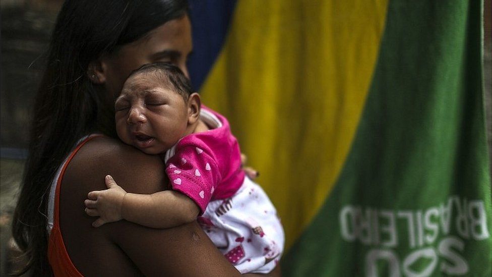
SharecloseShare pageCopy linkAbout sharingImage source, EPAA new outbreak of Zika virus is quite possible, warn researchers, with a single mutation potentially enough to trigger an explosive spread. The disease caused a global medical emergency in 2016, with thousands of babies born brain-damaged after their mums became infected while pregnant.US scientists say the world should be on the lookout for new mutations. Lab work, described in the journal Cell Reports, suggests the virus could easily shift, creating new variants.Recent infection studies suggest those variants may prove effective at transmitting the virus, even in countries which have built up immunity from previous outbreaks of Zika, say the team from the La Jolla Institute for Immunology. Experts said the findings, although theoretical, were interesting – and a reminder that viruses other than Covid could pose a threat. Shapeshifting virusZika is spread by bites from infected Aedes mosquitoes. The insects are found throughout the Americas – except for Canada and Chile, where it is too cold for them to survive – and across Asia. While for most people Zika is a mild illness, with no lasting effects, it can have catastrophic consequences for babies in the womb.If a mother contracts the virus during pregnancy, it can harm the developing baby, causing microcephaly (unusually small head) and damaged brain tissue.Image source, SPLThe Zika virusAlthough the virus is mostly spread by mosquitoes, it can also be sexually transmittedFew people die from Zika and only one in five people infected is thought to develop symptomsThese can include fever, a rash and joint painSince there is no treatment, the only option is to reduce the risk of being bittenScientists have begun work on a Zika vaccine to help protect pregnant womanThe researchers recreated what happens when Zika passes back and forth between mosquitoes and humans, using cells and living mice in their experiments. When Zika passed between mosquito cells and mice in the laboratory, small genetic changes occurred.This meant it was relatively easy for Zika to mutate in a way that allowed the virus to thrive and spread, even in animals that had some previous immunity from a similar mosquito-borne infection called dengue.More investigation Lead investigator Prof Sujan Shresta said: “The Zika variant that we identified had evolved to the point where the cross-protective immunity afforded by prior dengue infection was no longer effective in mice. “Unfortunately for us, if this variant becomes prevalent, we may have the same issues in real life.”Prof Jonathan Ball, an expert in viruses at the University of Nottingham, told the BBC: “We’ve heard so much lately about the rapid evolution and emergence of coronavirus variants, but this is a timely reminder that shapeshifting is a common feature shared by so many viruses.”This work shows just how rapidly a single letter change in the genome sequence of a virus can arise, and the stark impact it can have on the disease capability of a virus. But viruses that share these changes haven’t often been seen in outbreaks and, as the authors point out, these intriguing insights require more thorough investigation.”Dr Clare Taylor, from the Society for Applied Microbiology, said: “Although these findings were seen in laboratory experiments and therefore have limitations, it does show that there is potential for variants of concern to arise during the normal Zika transmission cycle and reminds us that monitoring is important to follow viruses as they evolve.”She said it might be possible to predict which variants could cause significant issues in the future and intervene early.Prof Paul Hunter, Professor in Medicine at the University of East Anglia, said past infection with Zika might still offer some protection against new variants – as has been seen with Covid. More on this storyIndia’s Kanpur reports latest Zika virus outbreakZika vaccine for pregnant women soughtRelated Internet LinksCell ReportsThe BBC is not responsible for the content of external sites.

Read more →