Australia lifts ban on former UK residents giving blood

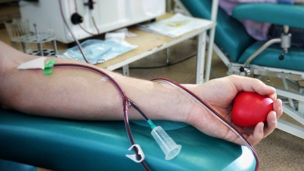
SharecloseShare pageCopy linkAbout sharingImage source, Getty ImagesAn Australian rule banning many former UK residents from giving blood over fears they could spread Creutzfeldt-Jakob disease (vCJD) has been scrapped.For two decades anyone who lived in the UK during its “mad cow disease” crisis has been barred from donating.In rare cases, the fatal illness has been spread through blood transfusions. But citing a review of epidemiological data and expert advice, Australia’s health regulator said the cohort would no longer be excluded.People who lived in the UK between 1980 and 1996 will soon be able to roll up their sleeves to give blood or plasma.Bovine Spongiform Encephalopathy (BSE) is estimated to affected 180,000 cattle during the outbreak. Its human form – vCJD – has been attributed to 178 deaths. It’s thought that one in 2,000 people in the UK is a carrier of the disease. But it appears that relatively few who catch the infectious agent that causes the disease then go on to develop symptoms.’Mad cow disease’: What is BSE?Australia’s blood donation service, called Lifeblood, hopes the long-awaited move will unlock new donors at a time when high demand is straining stocks.”It’s the number one query that we’ve had for change in Australia over the last few years and certainly, anecdotally, lots and lots of people are telling us that this change will enable them to donate,” executive director Cath Stone said.”We are optimistic that we’ll see tens of thousands of new people.”Lifeblood is working to update their screening processes to accommodate the change, but those who want to donate will be able to by the end of the year.With a weekly need for 33,000 donations, the organisation is hoping barriers to donation for men who have sex with men will also soon be removed.Australia was not alone in banning donations from former UK residents – others included France, the US and Canada. But other countries have begun to lift or relax the restrictions, after Ireland lifted its ban in 2019.You may also be interested in:This video can not be playedTo play this video you need to enable JavaScript in your browser.

Read more →

Exercise is good for joints with wear-and-tear arthritis

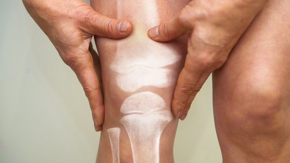
SharecloseShare pageCopy linkAbout sharingImage source, Getty ImagesExercise is good for people with wear-and-tear joint arthritis and should be a “core treatment”, new draft guidelines for the NHS advise. It may hurt to begin with, but can then ease pain and help individuals with osteoarthritis stay supple, healthy and slim, says the National Institute for Health and Care Excellence (NICE). Scans aren’t needed to diagnose it and strong painkillers are not recommended. There is no evidence flushing out the joint helps either. Wear and tearOsteoarthritis is very common – about 7.4 million people in England over the age of 45 have it.It can happen when the joints become damaged with age and injury. Being overweight is another risk factor. Some people have mild symptoms. For others the pain, stiffness and swelling can be severe. That can make exercising difficult, but according to the new NICE guidelines, physical activity should be the main treatment – not painkillers. The charity Arthritis Action said it hoped the guidelines would reassure people with osteoarthritis that exercise is a good intervention.Spokeswoman Dr Wendy Holden said: “Many wrongly believe that exercise can harm the joints, so this guidance is very important and will hopefully empower patients, and give them more confidence to make healthy lifestyle changes that will really help improve their pain and quality of life.”What helpsExercise builds muscles and can help people maintain a healthy weight, which is important for managing osteoarthritis. If there is pain, consider using an anti-inflammatory cream or gel or taking an ibuprofen or similar type of non-steroidal anti-inflammatory (NSAID) medicine, but not a strong opioid, says the advice. Arthritis charities are concerned that too many patients are put on painkillers rather than getting the right type of help – whether that is exercise therapy or a timely hip or knee replacement.A backlog of operations during the pandemic means many who need surgery are still waiting. NHS waiting-list backlog will take year to clearMost hip and knee replacements last longer than thoughtNICE says a joint replacement may be the right option for some and that hospitals should not be turning away people who could benefit, but are overweight. Obesity is not a bar to surgery, but losing weight by following a healthy diet and exercising will reduce strain on the joints. Tracey Loftis, from the charity Versus Arthritis, said: “Our own research into the support given to people with osteoarthritis showed that far too many do not have their conditions regularly reviewed by healthcare professionals, and even fewer had the opportunity to access physical activity support. “The lack of alternatives means that, in many cases, people are stuck on painkillers that are not helping them to live a life free from pain.”Dr Paul Chrisp, from NICE, said: “Muscle strengthening and aerobic exercise can have an impact on not just managing the condition, but also providing people with an improved quality of life. “Beginning that journey can be uncomfortable for some people at first, and they should be supported and provided with enough information to help them to manage their condition over a long period of time.”We have taken the decision to not recommend some painkillers, such as paracetamol and some opioids for osteoarthritis. “This is because new evidence has shown there was little or no benefit. In the case of strong opioids, there was evidence that they can cause harm in the longer term, including possible addiction.”The guidelines do not cover other types of arthritis, such as rheumatoid arthritis. More on this story’The pain is so bad I have to sit down regularly’Microphones ‘diagnose noisy arthritic knees’Around the BBCBBC Two – Trust Me, I’m a Doctor – Can I avoid, or treat, arthritis?Related Internet LinksOsteoarthritis – NHSThe BBC is not responsible for the content of external sites.

Read more →

Barkha Dutt: 'My father did not have to die'

SharecloseShare pageCopy linkAbout sharingImage source, Barkha DuttLast summer hundreds of thousands of people died when an unprepared India was hit by a catastrophic second wave of Covid-19 and its health care system buckled. Journalist Barkha Dutt, who was chronicling the pandemic, lost her father to the virus in April 2021. Here she writes on her loss, and other daughters who suffered the same fate. It’s been a year since I have been able to hear music; a year since the death of my father to Covid at the peak of India’s insatiable second wave.So, a few days ago, the faint strains of a familiar tune felt like a jolt to the system. Guantanamera Guajira Guantanamera…My hand trembled as I heard the chords of the Cuban folk song that has been variously sung to invoke romance, patriotism, protest and change.Inside I was shaking. Memory can be a beast. For my sister and I, this was Papa’s Song that marked the milestones of our lives, played out on scratchy cassettes when we were kids, remastered for an eight-track system in our teens, graduating to CDs when we went to college and finally heard on loop at his desktop. Here, he was surrounded by grandchildren, dogs, meccano sets, and odd looking wires – bit parts of kettles, speakers, coffee makers – machines he was repairing for friends, sometimes opened up for the sheer joy of tinkering with them. Image source, BARKHA DUTTSP Dutt – ‘Speedy’ to friends and family – was one of the hundreds of thousands of Indians taken by a virus that pummelled our health system into submission. In the wasteland of a nation’s grief, April was indeed the cruellest month as oxygen ran out, hospitals shut their gates to patients who died on the streets, vaccines were delayed and elections were mockingly on schedule. We could only turn to each other, daughters desperate to save fathers, as institutions collapsed. In Mumbai, Samridhi Saxena, reached out in the hope that I could help her father with an oxygen cylinder. He was struggling with a rare neurodegenerative disease. From Patna, Manisha called to say her 53-year-old father may die because the hospital where he was admitted no longer had oxygen. “Oxygen, oxygen, can you get me oxygen?”In Bangalore, 21-year-old Bharini asked me: “How strong am I expected to be?” She lost her biological parents in an accident, now her adoptive mother had died from Covid. Journalist Stutee Ghosh and I mirrored each other’s gnawing guilt – that somehow we were letting down our fathers; but also the guilt of knowing that even in our worst moment, we were better off than hundreds of thousands of other Indians, just because our dads were in hospital and not stranded on the road. Her father, like mine, didn’t make it. And over the weeks, her grief mutated into rage at “the indignity in death for so many who are dying and not even being counted by the government”. Image source, EPAWe were bereft. As daughters. And as citizens. Before my father died, we always valorised our mother, Prabha Dutt, who we lost when I was 13 years old to a brain haemorrhage. As India’s first woman war correspondent, who died at 40, she was the stuff of legend. It was easy to take the person in front of us – our dad – for granted. It took losing him to Covid for me to realise he was the epicentre of my existence. How India failed to prevent a deadly second waveIn April 2021, when the news first came that my father had Covid, I was at a crematorium in Mumbai, fighting back tears, as an elderly man in a wheelchair waved goodbye to his wife. Covid was already my whole life. I travelled more than 30,000km (18,641 miles) by road across India, through the first wave and then again thousands of kilometres by both air and road through the more lethal second spell. I’d been the chronicler of death and despair; but now the news had come home. I am forever haunted by my decision to take my dad to hospital in a private ambulance. Image source, ReutersIt was a ramshackle van with no paramedics, no stretchers and a crew of one – the driver. Eventually the single oxygen cylinder placed under the hard vinyl bench on which my father now lay like a helpless child, did not work. By the time we reached the hospital – slowed down, like scores of other ambulances, by random police checkpoints – his condition had worsened, the cylinder had failed to administer high flow oxygen. When we took his body from the hospital to the cremation ground, the irony was staggering. Now that he was dead, we’d managed to organise a professional ambulance. The stretcher in the hearse was shiny brown, the leather new, the height of the seat adjustable. And there was my father, in a beige zip bag, as if he were a frozen vegetable. Today we are keen to forget, desperate to block out triggers of sorrow. But tying up an open gash doesn’t heal it. Should India be bracing for a fourth Covid wave?The collective memories of a wounded nation have long shadows. I still cannot bring myself to step into my father’s room, where over his bed three circular tracks were built from the roof for his collection of self-made trains. At heart, my father was a man of science. He was the brilliant student who made the first toy rocket at school. As a young adult he built a vending machine for Cadbury chocolates. Later, he made shoe shine machines eponymously named ‘Speedshine’.Science, he liked to say, taught us that “what cannot be cured, must be endured”. When he tested positive, he repeated his favourite phrase. But he was wrong. Had India rolled out its vaccines sooner, things might well have been different. The government’s first order for vaccines was placed on 10 January 2021 for a little over 10 million doses, not even enough to administer the first dose to Delhi’s adult population. My father, like hundreds of thousands of Indians, should not have had to endure what he did.My father did not have to die. Barkha Dutt is an award-winning TV journalist, anchor and author, most recently, of Humans of Covid: To Hell and BackThis video can not be playedTo play this video you need to enable JavaScript in your browser.More on this storyShould India be bracing for a fourth Covid wave?

Read more →

High-frequency spinal cord stimulation shows improved longer lasting pain relief

Spinal cord stimulation (SCS) for chronic pain involves delivering low levels of electricity directly into the spinal cord using an implanted device, which modifies or blocks nerve activity to minimize the sensation of pain reaching the brain. The approach is most often used after nonsurgical pain treatment options have failed to provide sufficient relief.
The underlying mechanisms of how SCS works are not fully understood, but in a new paper published in the April 28, 2022 online issue of the journal Bioelectronic Medicine, a research team led by scientists at University of California San Diego School of Medicine report high-frequency SCS proved more effective at improving perceived pain reduction (PPR) than low-frequency SCS in patients studied, and that there was some variation in PPR between male and female patients.
Low-frequency SCS (50 Hz) was originally approved by the U.S. Food and Drug Administration (FDA) as a treatment for intractable back and leg pain in 1989. In 2015, the FDA approved high-frequency SCS (10,000 Hz), which delivers electrical stimulation pulses that are shorter in duration, lower in amplitude and do not induce paresthesia, the abnormal sensation of tingling or prickling.
The newly published retrospective study examined 237 patients who had received SCS treatment between 2004 and 2020: 94 patients (40 females, 54 males) who received HF-SCS and 143 patients (70 females and 73 males) who received LF-SCS. At three and six months post-implantation, the researchers found that PPR across all patients improved compared to baseline, but HF-SCS produced greater PPR than LF-SCS. HF-SCS was also associated with less subsequent use of opioids to mitigate pain.
However, there were differences in the findings between sexes: Male PPR, for example, was significantly better for HF-SCS at three and six months when compared to LF-SCS, while this was only true for females at the 6 month time point. LF-SCS males used more opioids post-implantation and at six months while females used more opioids post-implantation, at three, six and tended to use more opiates at the 12-month time-point.”Our work was sparked by a growing literature that demonstrate sex specific immune pathways differentially contribute to chronic pain processes,” said senior author Imanuel Lerman, MD, an associate professor of anesthesiology, pain management specialist at UC San Diego Health, and an affiliate of the Qualcomm Institute. “The observed parameter-specific (high versus low frequency) sex-based differences in spinal cord stimulation efficacy and opiate use are definitely intriguing.
“It’s a first step in the right direction, but clearly more work needs to be done to carefully characterize sex specific pain regulatory pathways that may prove responsive to specific types of neuromodulation and or pharmaceutical therapies.”
Co-authors include: Rosalynn R.Z. Conic, Zabrina Reyes and Sopyda Yin, all at UC San Diego; Jacob Caylor, UC San Diego and Northwest Pain Care, Spokane, WA; Christina L. Cui, Duke University; and Eric Nelson, Pacific Western University of Health Sciences.
Story Source:
Materials provided by University of California – San Diego. Original written by Scott La Fee. Note: Content may be edited for style and length.

Read more →

Unlocked enzyme structure shows how strigolactone hormone controls plant growth

As sessile organisms, plants have to continually adapt their growth and architecture to the ever-changing environment. To do so, plants have evolved distinct molecular mechanisms to sense and respond to the environment and integrate the signals from outside with endogenous developmental programs.
New research from Nitzan Shabek’s laboratory at the UC Davis College of Biological Sciences, published in Nature Plants, unravels the underlying mechanism of protein targeting and destruction in a specific plant hormone signaling pathway.
“Our lab aims at deciphering sensing mechanisms in plants and understanding how specific enzymes function can be regulated at the molecular levels” said Shabek, assistant professor of biochemistry and structural biology in the Department of Plant Biology. “We have been studying a new plant hormone signal, strigolactone, that governs numerous processes of growth and development including branching and root architecture.”
The work stems from a study by Shabek, published in Nature in 2018, unravelling molecular and structural changes in an enzyme, MAX2 (or D3) ubiquitin ligase. MAX2 was found in locked or unlocked forms that can recruit a strigolactone sensor, D14, and target for destruction a DNA transcriptional repressor complex, D53. Ubiquitins are small proteins, found in all eukaryotes, that “tag” other proteins for destruction within a cell.
To find the key to unlock MAX2 and to better understand its molecular dynamics in plants, postdoctoral fellows Lior Tal and Malathy Palayam, with junior specialist Aleczander Young, used an approach that integrated advanced structural biology, biochemistry, and plant genetics.
“We leveraged structure-guided approaches to systemically mutate MAX2 enzyme in Arabidopsis and created a MAX2 stuck in an unlocked form,” said Shabek, “some of these mutations were made by guiding CRISPR/Cas9 genome editing thus providing us a discovery platform to study and analyze the different signaling outputs and illuminate the role of MAX2 dynamics.”
Regulating a massive gene network
They found that in the unlocked conformation, MAX2 can target the repressor proteins and biochemically decorate them with small ubiquitin proteins, tagging them for destruction. Removing these repressors allows other genes to be expressed — activating a massive gene network that governs shoot branching, root architecture, leaf senescence, and symbiosis with fungi, Shabek said.
Sending these repressors to the proteasome disposal complexes requires the enzyme to relock again. The team also showed that MAX2 not only target the repressors proteins, but once it is locked the strigolactone sensor itself gets destroyed, returning the system to its original state.
Finally, the study uncovered the key to the lock, an organic acid metabolite that can directly trigger the conformational switch.
“Beyond the implication in plants signaling, this is the first work that placed a primary metabolite as a direct new regulator of this type of ubiquitin ligase enzymes and will open new avenues of study in this direction,” Shabek said.
Additional coauthors on the paper are specialist Mily Ron and Professor Anne Britt, Department of Plant Biology. The study was supported by NSF CAREER and EAGER grants to Shabek. X-ray crystallography data was obtained at the Advanced Light Source, Lawrence Berkeley National Laboratory, a U.S. Department of Energy user facility.
Story Source:
Materials provided by University of California – Davis. Original written by Andy Fell. Note: Content may be edited for style and length.

Read more →

Neural pathway key to sensation of pleasant touch identified

Studying mice, scientists at Washington University School of Medicine in St. Louis have identified a neural circuit and a neuropeptide — a chemical messenger that carries signals between nerve cells — that transmit the sensation known as pleasant touch from the skin to the brain.
Such touch — delivered by hugs, holding hands or caressing, for example — triggers a psychological boost known to be important to emotional well-being and healthy development. Identifying the neuropeptide and circuit that direct the sensation of pleasant touch eventually may help scientists better understand and treat disorders characterized by touch avoidance and impaired social development, including autism spectrum disorder.
The study is published April 28 in the journal Science.
“Pleasant touch sensation is very important in all mammals,” said principal investigator Zhou-Feng Chen, PhD, director of the Center for the Study of Itch & Sensory Disorders at Washington University. “A major way babies are nurtured is through touch. Holding the hand of a dying person is a very powerful, comforting force. Animals groom each other. People hug and shake hands. Massage therapy reduces pain and stress and can provide benefits for patients with psychiatric disorders. In these experiments with mice, we have identified a key neuropeptide and a hard-wired neural pathway dedicated to this sensation.”
Chen’s team found that when they bred mice without the neuropeptide, called prokinecticin 2 (PROK2), such mice could not sense pleasant touch signals but continued to react normally to itchy and other stimuli.
“This is important because now that we know which neuropeptide and receptor transmit only pleasant touch sensations, it may be possible to enhance pleasant touch signals without interfering with other circuits, which is crucial because pleasant touch boosts several hormones in the brain that are essential for social interactions and mental health,” he explained.

Read more →

Atherosclerosis: How diseased blood vessels communicate with the brain

LMU scientists have been able to demonstrate for the first time that nerve signals are exchanged between arteries and the brain in atherosclerosis.
Laboratories worldwide are carrying out research into the disease atherosclerosis. However, their focus is on atherosclerotic plaques — deposits of cholesterol, fibrous tissue and immune cells that form on the inner layer of arteries. These plaques progressively constrict the lumen of the arteries, such that less oxygen can get to the body tissue. Heart attacks, strokes and peripheral occlusive disease (smoker’s leg) are among the known consequences.
“In recent decades, nobody has asked whether there is a direct connection between the artery and the brain — the obvious reason being that atherosclerotic plaques are not innervated,” says Dr. Sarajo K. Mohanta from the LMU Institute for Cardiovascular Prevention. But it is precisely such a connection that he has now managed to demonstrate together with Professor Andreas Habenicht, also from the LMU Institute for Cardiovascular Prevention, Prof. Christian Weber, director of the institute, and an international team. Crucial results were obtained by Professor Daniela Carnevale and Professor Giuseppe Lembo from the Department of Angiocardioneurology and Translational Medicine, IRCCS Neuromed, Sapienza University of Rome. Parts of the study were funded by LMU’s Cluster of Excellence SyNergy and the Collaborative Research Center 1123.
In Nature, the researchers report their findings about signals that are conveyed from the arteries containing plaques via nerves to the brain. After processing of the signals in the brain has taken place, signals make their way back to the blood vessel.
A completely new understanding of atherosclerosis
Some background information: The walls of arteries are made up of three components, an outer layer, a middle layer, and an inner layer. Plaques are found in the inner layer. They are not innervated by nerve fibers — a fact that has long been known. “As such, it did not occur to anyone to investigate whether the peripheral nervous system comes into contact with arteries in the case of atherosclerosis,” says Habenicht.
Since 2004, his research group has been investigating what happens on the outer wall of arteries in patients burdened with atherosclerosis. “After all, atherosclerosis is more than just a plaque, rather it is a chronic inflammatory disease of the entire artery — and relevant to our findings the outer layer of it,” adds Mohanta, who was the lead scientist in charge of the project.
The peripheral nervous system responds to such inflammation. Habenicht’s team discovered that molecular sensors known as receptors play a key role. Receptors are located in the outer layer of the vessels. They recognize where plaques are located and where vessels are inflamed by identifying the inflammatory messengers of the inflammation. Then they translate the inflammatory signals into electrical signals via nerves to the brain. The brain processes the signals and sends a stress signal back to the inflamed blood vessel. This negatively influences the inflammation, and the atherosclerosis gets worse.
Long-term prospects for treating causes of atherosclerosis
This previously unknown electrical circuit between the arteries and the brain is potentially of enormous significance. In an animal experiment, Carnevale severed the electrical connection between a diseased artery and the brain. Eight months later, she compared treated mice with mice that had not had this procedure. In the rodents that had had the experimental therapy, atherosclerosis was in fact less developed than in the control mice. “In the long term, we hope to be finally able to treat the causes of atherosclerosis,” says Mohanta, “although that may well be some way off yet.”
As their next step, the scientists want to find out how exactly the peripheral nervous system is organized — and what role other receptors play. There are also many signs that the interface between brain and diseased blood vessels is regulated by stress. Accordingly, Habenicht is planning to investigate neurobiological aspects: Which cells in the brain respond to signals from diseased blood vessels? And with which regions of the brain are these cells connected in turn?

Read more →

Genetic links revealed between severe COVID-19 and other diseases

A new analysis of data from the Veterans Affairs Million Veteran Program has uncovered genetic links between COVID-19 severity and certain medical conditions that are known risk factors for severe COVID-19. Anurag Verma of the Corporal Michael Crescenz VA Medical Center in Philadelphia, Pennsylvania, US, and colleagues present these findings on April 28 in the open-access journal PLOS Genetics.
Some people with COVID-19 experience the disease more severely than others. Previous research has identified certain variants in specific human genes that are associated with a person experiencing more severe COVID-19. Some of these variants may also be associated with other medical conditions that may already be well understood; identifying these shared variants could improve understanding of COVID-19 and illuminate potential new paths for treatment.
To identify shared variants, Verma and colleagues used an unprecedented dataset of genotypic information linked to electronic health record data (EHR) for more than 650,000 U.S. veterans. They conducted a type of analysis known as a phenome-wide association study (PheWAS) to examine links between variants often found in Veterans who experienced severe COVID-19 and variants associated with a broad selection of medical conditions.
The analysis revealed that certain variants associated with COVID-19 are also associated with known risk factors for COVID-19. Particularly strong links were found for variants associated with venous embolism and thrombosis, as well as type 2 diabetes and ischemic heart disease — two known COVID-19 risk factors.
The analysis also found genetic links between severe COVID-19 and neutropenia for Veterans of African and Hispanic ancestry; these links did not appear for those of European ancestry.
Among respiratory conditions, idiopathic pulmonary fibrosis and chronic alveolar lung disease shared genetic links with severe COVID-19, but other respiratory infections and chronic obstructive pulmonary disease (COPD) did not. Some variants associated with severe COVID-19 were also associated with reduced risk of autoimmune conditions, such as psoriasis and lupus. These findings highlight the need to carefully weigh various aspects of the immune system when developing new treatments.
Despite some limitations of the PheWAS method, these findings could help deepen understanding of COVID-19 and guide development of new treatments.
Verma concludes, “The study demonstrates the value and impact of large biobanks linking genetic variations with EHR data in public health response to the current and future pandemics. MVP is one of the most diverse cohorts in the US. We had a unique opportunity to scan thousands of conditions documented before the COVID-19 pandemic. We gained insights into the genetic architecture of COVID-19 risk factors and disease complication.”
“One thing that stood out to us was the high number of immune-mediated conditions that shared genetic architecture with severe manifestations of COVID-19,” coauthor Katherine Liao adds. “The nature of the associations brought to light how the SARS-CoV2 virus pushes on a pressure point in the human immune system and its constant balancing act of fighting infection while maintaining enough control so that it does not also become an autoimmune process, attacking self.”
Story Source:
Materials provided by PLOS. Note: Content may be edited for style and length.

Read more →

Higher COVID-19 death rates in the southern U.S. due to behavior differences, study finds

During the pre-Omicron phases of the COVID-19 pandemic, regions of the U.S. had markedly different mortality rates, primarily due to differences in mask use, school attendance, social distancing, and other behaviors. Had the entire country reacted to the pandemic as the Northeast region, more than 316,000 deaths might have been avoided, 62% of those avoidable deaths being in the South.
The study, by Georgetown University’s School of Nursing & Health Studies researchers, appeared April 28, 2022, in PLOS ONE.
Excess mortality, which helps account for avoidable deaths from a new disease or situation, is defined by the difference between total current deaths and deaths expected based on earlier time period, usually the previous decade or so. The U.S. Centers for Disease Control and Prevention (CDC) calculates these numbers weekly. For this study, the CDC excess mortality data were analyzed for the period between January 3, 2020, to September 26, 2021. For regional comparison purposes, areas of the country were broken down into the Northeast, Midwest, South and West.
“Our goal was to carefully examine regional differences in COVID-19 death rates based on reliable statistical data,” says Michael Stoto, Ph.D., professor of Health Systems Administration and Population Health at the School of Nursing & Health Studies and corresponding author of the study. “Our study is the first to quantify avoidable deaths and confirm that both COVID-19 deaths and avoidable deaths disproportionately occurred in the South.”
The investigators found that regional differences in COVID-19 mortality rates have persisted throughout the pandemic. The southern part of the United States has had higher mortality rates than the rest of the U.S. since the start of summer in 2020. Since October 2020, 48% of COVID-19 deaths were in the South, which makes up 38% of the population, pointing to disproportionate outcomes regionally.
The researchers also determined that between January 2020 and September 2021 there were 895,693 excess deaths associated with COVID-19, which is 26% more than reported by other experts who track disease. Although the official total neared on one million deaths in the U.S due to COVID-19 by late April 2022, based on this undercount the scientists believe that threshold was actually passed at the beginning of 2022.
These estimates of undercounts are important because most studies have looked at excess mortality at the state and county level in the U.S., but because of small population sizes, the studies have not examined patterns over time. Some earlier studies explored the relationship between COVID-19 mortality and age, education, and other factors as well as vaccine uptake, party affiliation, and other factors. But most studies have used reported COVID-19 deaths rather than excess deaths, as compared to what Dr. Stoto and collaborators have done, and may not be as statistically reliable.
“This is one of a series of planned studies to look carefully at the response to COVID-19 in the U.S. and other countries and to learn from the experience in order to strengthen preparedness for future potential outbreaks,” says Stoto. “Our team has also looked at testing and surveillance, and other COVID-19 metrics to understand how communities have come together to effectively deal with the pandemic.”
In addition to Stoto, the other authors from Georgetown include Samantha Schlageter, who conducted this work as an undergraduate in the Health Care Management & Policy program at the School of Nursing & Health Studies (NHS), and John D. Kraemer, an associate professor in Department of Health Systems Administration at NHS.

Read more →

Mother and child vulnerable to endocrine disruptor exposure

They can be found in cosmetics, plastic containers, furniture, toys, or baby bottles. Endocrine disruptors, molecules disrupting our hormones, are everywhere in our daily lives. However, their alarming effects on human health are little known to the public. A growing number of studies have demonstrated that exposure to these environmental contaminants during pregnancy can lead to medium and long-term health problems for both the mother and the fetus.
This is what a group of researchers from the Institut national de la recherche scientifique (INRS), the Université TELUQ and Queen’s University have shown by conducting an exhaustive review of the literature on more than a dozen of the most common endocrine disruptors (EDs), as well as several whose effects are less well-known. They focused on substances that have been shown to affect the reproductive system, metabolism, and mammary gland development during pregnancy. Their work is reported in an article published in April in the special issue of the journal Environmental Research.
“The idea was to show that, during the pregnancy period, there is not only one person who is affected by endocrine disruptors, but two! We wanted to highlight the co-sensitivity of the mother and her child to these environmental contaminants that are everywhere,” says Professor Isabelle Plante, lead author of the study and researcher in environmental toxicology at INRS. She is also co-director of the Intersectoral Centre for Endocrine Disruptor Analysis (ICEDA).
Defeating the placenta’s defence barriers
Pregnancy is a complex process that involves major physiological changes for the mother and the child. This period of life is regulated by various hormones and signalling pathways. It is therefore a window of opportunity that is particularly sensitive to disruption by external contaminants.
By compiling existing studies, researchers highlighted the role of the placenta and its vulnerability to EDs. The placenta is the regulator of maternal physiology and fetal development during pregnancy. It produces hormones necessary for pregnancy. Any alteration in its functioning affects the health of the mother and her child in the short, medium, and even long term. A malfunctioning placenta can lead to health complications later in life, such as diabetes, obesity, or other chronic diseases.

Read more →